
This is the final part of our article on Chronic Headache (CH) management using medical massage. This part is dedicated to treating the secondary symptoms associated with CH: tension in the scalp using the scalpotherapy and cluster headache using the eyes treatment. For the Parts I to IV please visit #2;#3; #4 2012 issues of JMS and #1 issue of 2013.
SCALPOTHERAPY
As we discussed in Part III of this article (see Issue #4, 2012, JMS), tension in the scalp develops as a result of the irritation or compression of the greater and/or minor occipital nerves. Aside from actual pain, patients feel tension in their scalp on varying parts of their head. This is primarily responsible for the “head squeeze” sensation that many patients complain about.
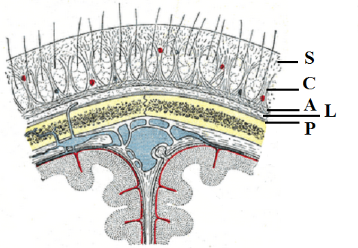
As we also discussed in Part III the major contributor to tension in the scalp is a layer of loose connective tissue (see Fig. 1), which consists from fibrotic string that runs between the cranial aponeurosis and the periosteum, that covers the skull bone.
S – skin
C – connective tissue
A – aponeurosis
L – loose connective tissue
P – periosteum of the cranial bones
In such a case the scalp becomes glued to the skull bones and it additionally contributes to the intensity of CH and to the cervical muscles’ tension. Additional tension in the cervical muscle furthermore irritates the occipital nerves and it triggers a vicious cycle with the patient being caught in the middle. This is why elimination of tension in the scalp, if it is present, is such an important part of the management of CH.
The primal goal of the scalpotherapy is to stretch the fibrotic fibers of loose connective tissue and restore mobility of the scalp along the skull bones. The practitioner should apply scalpotherapy in the areas where tension is present. The evaluation of the scalp which we discussed in Part III of this article is the foundation of the therapy. If the evaluation showed that the patient does not have any areas of tension in the scalp, do not include scalpotherapy into the protocol. The protocol of Scalpotherapy presented below is supposed to be Step 6 in the MEDICAL MASSAGE PROTOCOL for CH management, which we discussed in the previous issue of JMS (#1, 2013).
SCALPOTHERAPY
The duration of scalpotherapy will depend upon the number of painful areas detected in the scalp. The practitioner should spend about 1-2 minutes on each area of tension.
a. Raking effleurage
Pressure: Below the pain threshold.
Begin with raking effleurage along the scalp. The base of the hand provides stability for the strokes and allows the freeing of the fingers for friction. All fingers are bent and spread out. The fingertips are the main contact areas and they conduct circular friction while the hands are moving along the scalp. Try to access the scalp between the hairs without pulling on them.
b. Scalp stretching using the thumb
Pressure: At the level of the pain threshold (first sensation of discomfort).
Place the thumb in the previously detected sensitive area of the scalp. Apply vertical pressure compressing the scalp against the skull. Now without releasing the vertical pressure, add a slow horizontal push, trying to form a fold of skin in front of the thumb.
The practitioner should apply these strokes in a ray-like pattern outward, from the center of the original aching spot. The practitioner may place the thumbs on top of each other to reinforce the strokes and to reduce pressure on the thumb.
c. Scalp stretching between two thumbs
Pressure: At the level of the pain threshold (first sensation of discomfort).
Place the thumbs on opposite sides of the painful point on the scalp. Apply vertical pressure and stretch the scalp, pushing the thumbs in opposite directions.
d. Scalp stretching using crisscrossing thumbs
Pressure: At the level of the pain threshold (first sensation of discomfort).
The application of this stretching technique is similar to the previous one, except that the thumbs push the scalp in a crossing direction.
e. Vertical compression and circular friction
Pressure: Above the pain threshold.
In the painful area, apply repetitive vertical compressions. Reinforce the thumb with the other thumb if needed. Now combine compression with a circular friction stroke of the widest possible radius allowable by the scalp at any given point, without sliding over the skin itself but rather displacing the skin over the underlying bone.
There are four special points in the scalp which should be examined and treated in every patient who suffers from headaches. These points are the upper and lower Grinstein’s points. They are located on both sides of the middle line of the skull, on top of the head, in the areas shown in the video. Use the same combination of compression and circular friction in these areas
EYES TREATMENT (ET)
ET must be part of the treatment of every patient who has Cluster Headache or pain in and around the eye socket. As we discussed in Part III of this article (see issue #4, JMS 2012), the orbital part of the face is richly innervated, especially the extraocular muscles. Their tension is an additional factor, which greatly contributes to the intensity of pain during a Cluster Headache.
Considering the complex anatomical arrangement of soft tissue inside of the socket, the treatment is a complicated issue. To make treatment quick and efficient we have developed the protocol of PIR for the extraocular muscles, and the results from its clinical application have shown that this is an excellent tool to control the intensity and duration of Cluster Headaches.
The protocol of PIR for the extraocular muscles should not be used alone. If the Cluster Headache is a result of Greater Occipital Nerve Neuralgia, then add the treatment of the eyes at the end of the MEDICAL MASSAGE PROTOCOL which we discussed in Part IV (see issue #1, JMS, 2013). If the Cluster Headache is a result of Trigeminal Nerve Neuralgia, add ET at the end of the MEDICAL MASSAGE PROTOCOL addressing Trigeminal Nerve Neuralgia.
Why is it that the protocol for ET is so effective in controlling Cluster Headache? As mentioned above, the orbital area is very richly innervated. Radiation of pain into, or pain having its origin in this area both greatly affect the orbital soft tissue, including the extraocular musculature. One of the first reactions of these muscles to chronic pain is protective tension, which the patient feels as a very heavy, pulsating and ready-to-explode sensation within the orbit and behind the eyeball. Usually any movement of the eyes increases the intensity of the pain, which may also trigger nausea and vomiting.
There exists no other treatment to address extraocular muscles except the protocol of Postisometric Muscular Relaxation discussed below. The PIR protocol uses reflex mechanism to reduce the tension in these muscles. It is safe and highly effective.
During this treatment, the practitioner must work around the patient’s eyes. Thus, the practitioner must explain to the patient the entire procedure in detail and must be certain that the patient is comfortable with this proposed treatment. Contact lenses must be removed before the treatment is begun. The treatment should be conducted within the patient’s comfort level, and he or she must keep the eyes closed. The PIR protocol to reduce tension in the extraorbital musculature should be conducted on two levels.
PROTOCOL OF ET
Step 1.
Duration: 1 min
Pressure: Below the pain threshold.
Start with bi-manual stretching effleurage strokes along the upper orbital edge. Afterwards, use small circular friction strokes along the upper orbital edge. Be sure to press the fingertips against the bone.
For the last part of this step, apply small circular friction below the upper orbital edge. Notice that the practitioner carefully tries to get the fingertips below and behind the upper orbital edge, i.e., between the orbital edge and the eyeball itself.
Depending upon the size of the practitioner’s hands, he or she may choose to use the fingertips of the 5th finger, seeing as this maneuver calls for a very small contact area. The patient must inform the practitioner about any pain he or she experiences during the therapy. This is a main factor which defines the intensity of the pressure applied.
Step 2.
Pressure: Below the pain threshold.
The patient lies comfortably on their back with the eyes closed. Place your right index finger on the medial side of the right eyeball, and your left index finger on the lateral side of the left eyeball.
From the initial position of the eyes looking straight forward, ask the patient to roll the eyeballs to the left while both fingers resist this movement. Hold counter-resistance for 10 to 15 seconds, and afterwards, ask the patient to relax the extraocular muscles without opening the eyes.
For the next part, ask the patient to inhale and, during a prolonged exhalation, to roll the eyeballs all the way in the opposite direction, i.e., to the right. The patient should return the eyes to their neutral position after each prolonged exhalation, inhale, and during the next prolonged exhalation should repeat the rolling of the eyes all the way to the right. This free (i.e., non-resisted) roll to the right is to be repeated by the patient 3 times.
After the last roll to the right, the practitioner asks the patient to keep their eyes all way to to the right and places the same fingers as before on the medial (for the right eye) and lateral (for the left eye) surfaces of the eyeballs. From this new position (eyeballs looking to the right), the practitioner asks the patient to roll both eyeballs to the left while he or she resists the eyes’ movement for 10 to 15 seconds as before.
At the end of this second resistance, ask the patient to return the eyes to their neutral position again and to repeat 3 passive rolls of the eyeballs all the way to the right during the prolonged exhalation, as before.
White arrows indicate the direction of the eyes’ movement against the resistance elicited by the practitioner.
Step 3.
Pressure: Below the pain threshold.
This video repeats the same protocol of the eye treatment, but in the opposite direction: rolling of the eyes to the right against the practitioner’s resistance on two levels, with 3 free rolls to the left after each level of the roll-against-resistance. Notice the position of the index fingers.
White arrows indicate the direction of the eyes’ movement against the resistance elicited by the practitioner.
Step 4.
Pressure: Below the pain threshold.
Start with an application of repetitive vertical compressions on the eyeballs during the patient’s prolonged exhalation. Notice that both fingers shape themselves to the contour of the eyeballs. Fit 3 to 4 vertical compressions into each patient’s prolonged exhalation.
The next part is the application of repetitive compression in the space between the eyeballs and the upper orbital edge. Notice the change of applied pressure compared to the first part of this step. At the end of the video, the vertical compression on the eyeballs are shown from a different camera angle.
Step 5.
Duration: 2 min
Pressure: At the level of the pain threshold (first sensation of discomfort).
Repeat Step 1, but now the pressure applied should be made to reach the pain threshold.
With the publication of this article, we conclude our presentation of medical massage treatment on Chronic Headache and Migraine-Type Headache. We have used the protocol we discussed in Issues #4 2012 and #1 2013 of JMS on a daily basis in our clinic. Its clinical effectiveness still fascinates us. Since Chronic Headaches are such a widespread pathological condition and medical massage plays such a decisive role in successful therapy, we highly encourage our readers to start offering it to patients desperately looking for help. A simple solution is available for them from the hands of the correctly trained practitioner.
Category: Medical Massage