
STIMULATION OF CELLULAR FUNCTIONS BY MASSAGE THERAPY
We continue to publish articles which review various mechanisms responsible for the clinical effects of Massage Therapy (MT). These articles are united by a common theme: How Massage Heals the Body. If you haven’t read the previous articles yet, please do so, since they will equip you with priceless data that will help you understand what you are actually doing while applying MT.
Another important aspect of this data is that it will help you build bridges with other health professions. If you want to establish a solid referral base, you have to speak with physicians, chiropractors or physical therapists using the same language and concepts. These articles give you this ability. Readers may find previous articles which cover other aspects of MT’s healing impact:
HOW MASSAGE THERAPY HEALS THE BODY. PART I: Piezoelectrical Phenomenon in the Soft Tissue
HOW MASSAGE THERAPY HEALS THE BODY. PART II: Streaming Potentials
HOW MASSAGE THERAPY HEALS THE BODY. PART III: Vasodilation Mechanisms 2
HOW MASSAGE THERAPY HEALS THE BODY. PART IV
HOW MASSAGE HEALS THE BODY: CLINICAL APPLICATION OF GATE CONTROL AND NEUROMATRIX THEORIES OF PAIN
This article addresses the fact that MT deeply impacts cellular function, and if therapists understand the events which are triggered by MT even at the cellular level, they will be able to optimize treatment and better help patients.
We think everyone agrees that MT is a way to deliver mechanical force to the soft tissues, and the pressure itself is a healing factor. From this perspective, medical science has accumulated a lot of data which illustrate profound changes in the function of the cells when they function under the application of direct pressure. However, to fully understand and appreciate this data, which has great practical value for therapists, we need to quickly review some very basic principles of cellular biology.
CELLULAR BIOLOGY
Every cell is composed of several main components: the cellular membrane and cytoskeleton, the cytoplasm with organelles, and the nucleus with the nucleolus (see Fig. 1).
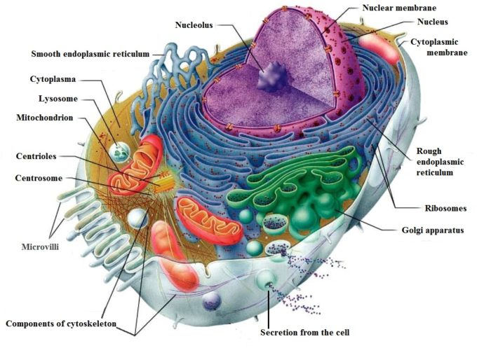
The structure and functions of all cellular components are well known, and they are not the subject of our discussion. However, findings about the cytoskeleton and its relation to external mechanical force applied to the cell are breakthroughs in the understanding of the therapeutic effect of the mechanical stimulation of living cells, including MT.
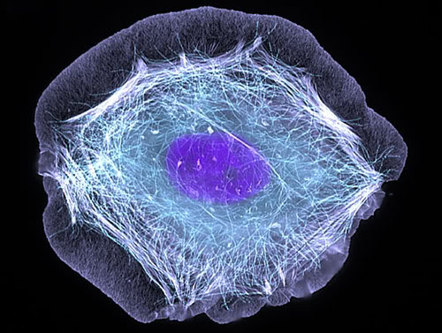
The cytoskeleton is a complex system of fibrillar structures located in the cytoplasm, and it connects the cytoplasmic membrane to the nucleus. It can be compared to the human skeleton, which provides a frame for our body and its dynamic support. Thus, the cytoskeleton gives the cell its form. For example, a fibroblast (see Fig. 4), which is a major repair cell of the body, has its shape because the fibrils of the cytoskeleton are arranged in this particular way. Fig. 2 presents an electron microphotograph of the cytoskeleton of a human keratinocyte (the most abundant cell in our epidermis). Notice that the fibrils of the cytoskeleton run between the cytoplasmic membrane and nuclear membrane.
In contrast with the body skeleton, which is a rigid structure, the cytoskeleton of the cell is very dynamic. It constantly moves, since it must transport organelles around the inner cell, excrete waste and bring nutrition in. The cytoskeleton can very quickly and completely resynthesize and renew itself, and it consists of 3 types of fibrils: microtubules, actin filaments and intermediate filaments.
From the time of its discovery at the beginning of the 20th century, the cytoskeleton was always viewed as a purely mechanical structure that provides the shape of the cell and participates in cell motility, migration (e.g., leukocytes, natural killer cells, etc.), and it is the key component of cell division.
However, modern experimental equipment allowed scientists to set up and conduct more detailed studies of the cytoskeleton. Thanks to these studies, we now know more important functions of the cytoskeleton:
- Controls functions of the cytoplasmic membrane which surrounds each cell.
- Connects the cytoplasmic membrane to the nucleus.
- Regulates cellular functions: synthesis of proteins, hormones, enzymes and other substances which are generated within the cells and secreted from the cell.
- Indirectly controls DNA and individual genes’ expression (i.e., function).
Of course, we are interested in all these scientific data from the MT perspective, since bodywork delivers the external mechanical stimulation to the soft tissues.
Control Over Cytoplasmic Membrane
Therapists work in the soft tissues with repetitive application of strokes which deliver mechanical force with fluctuating pressure. Every time the soft tissues are compressed, the pressure between cells, which compose the different layers of the soft tissue, also increases and fluctuates. These fluctuations are detected by the cytoplasmic membrane of each individual cell and they stimulate cell function.
A cytoplasmic membrane surrounds each cell, and via this membrane, the cells actively interact with the environment. It has a thickness of 7.5 nm, or about 1/3,400,000 of an inch, and is made up of a double layer of phospholipids with integrated proteins. Fig. 3 shows the structure of the cytoplasmic membrane.
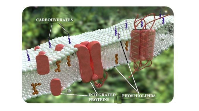
The integrated proteins which are embedded in the cytoplasmic membrane work as gates that connect the external environment and the inner part of the cell. Any molecules that the cell needs for its normal functions pass into the cell through these gates. They also allow the newly synthesized proteins or waste to be secreted or removed from the cell.
The cytoplasmic proteins occupy approximately 50% of the membrane’s mass and form a net of receptors over its entire outer surface. Thus, any outside molecule must first interact with these receptors to be recognized. Only after this occurs will the gate open and let the molecule in.
Mechanical stimuli (e.g., in the form of massage strokes) affect the protein receptors located on the cytoplasmic membrane of all cells which compose the soft tissue therapists work with. When activated, these receptors immediately convert mechanical energy applied from outside (e.g., by MT strokes) into chemical stimuli that are conducted inside the cell, stimulating or inhibiting its functions (Jain et al., 1990; Komuro et al., 1991).
Not all receptors in the cytoplasmic membrane act as mechanoreceptors. One particular family of proteins, called integrins, reacts to the mechanical stimulation (Wang et al., 1993). Thus, for our discussion, the integrins are extremely important and we will trace the events in the cells starting with the activation of those receptors.
Since the fibrils of the cytoskeleton are deeply anchored in the cytoplasmic membrane, the cytoskeleton controls the function, arrangement and even number of mechanoreceptors which are working in the membrane. Thus, by regulating the number and function of cytoplasmic receptors, the cytoskeleton manages the metabolism of the whole cell (Gataullin and Zaripov, 1988; Jain et al., 1990).
Connection of the Cytoplasmic Membrane to the Nucleoskeleton
Now let’s see how mechanical stimuli applied to the soft tissue and individual cells can alter the most sacred part of life: the function of the cell nucleus. As we know, the nucleus stores the vital information of all living objects – DNA. The nucleus is surrounded by its own nuclear membrane, which has pores that open into the cytoplasm and into the cellular structure called the endoplasmic reticulum (see Fig. 1). The endoplasmic reticulum is the part of the cell where everything the cell produces (proteins, hormones, enzymes, etc.) is synthesized before it is unloaded from the cell. The nucleus also has its own nucleoskeleton which has a striking similarity to the cell’s cytoskeleton (McKeon et al., 1986).
One of the most important functions of the cytoskeleton is its ability to conduct information generated by the mechanical stimulation of receptors in the cytoplasmic membrane directly into the nucleus (Maniotis et al., 1997). Such mechanical stimulation has an effect on cell growth, life span, function and even the expression of individual genes (Wang et al., 1993).
Control of DNA and Regulation of Cellular Function
Let’s look at the fascinating process of MT stimulating the cellular function using the example of the human fibroblast, which undergoes repetitive mechanical stimulation during cross-fiber friction. Fig. 4 shows a human fibroblast (FB) under an electron microscope.
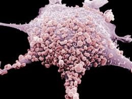
FBs are therapists’ best friends and allies in the patient’s body. To correctly use and activate this major healing force, therapists must know what fibroblasts do and how they operate.
To deal with the consequences of any trauma and inflammation in the soft tissue or inner organs, the body employs FBs. They are everywhere, and they concentrate in the injured or inflamed area to restore function.
The FBs participate in healing by producing a special protein called pro-collagen. The genetic code for the synthesis of pro-collagen is stored within the nucleus of each FB. After pro-collagen is synthesized within the FB, it is unloaded from the cell into the extracellular matrix and it matures there into the collagen fibers which the body uses to heal injured or inflamed tissues or organs. A scar on your finger after you cut it with a knife, healing of tendinitis, filling the damaged part of the brain after a stroke, etc. – all of that is the result of FBs producing and depositing pro-collagen.
The FBs are attracted to the injured or inflamed areas by the histamine released from the affected tissues as a result of their damage. However, when FBs arrive at the damaged or inflamed areas, the mechanical pressure on the cytoplasmic membrane of the FB becomes one of the major driving forces for the stimulation of pro-collagen synthesis inside FBs. This mechanical pressure comes from the edema in the affected tissues, but it can be enhanced by MT, which stimulates the function of FBs by additionally increasing pressure within the tissue.
Fig. 5 summarizes data from various scientific sources on how mechanical stimulation of the soft tissues in the form of MT activates a major healing force in the body – FBs. Please also refer to Fig. 1 for any additional info.
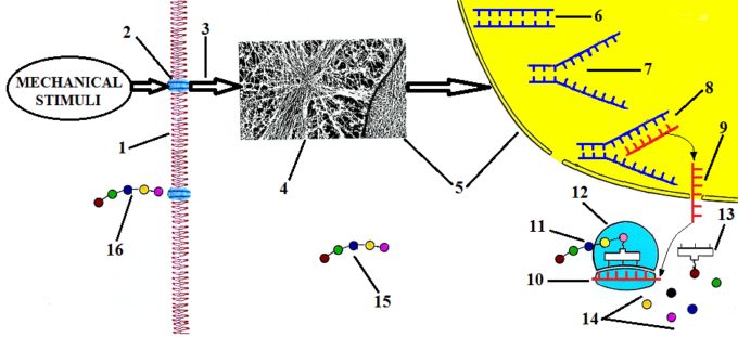
The external mechanical stimuli in the form of repetitive friction additionally increase interstitial pressure, and this increase is immediately detected by integrin receptors (Fig. 5: ‘2’) located in the cytoplasmic membrane (Fig. 5: ‘1’) of the FB. Integrin receptors convert mechanical stimuli applied to the membrane into chemical signals (Fig. 5: ‘3’). These signals trigger the reorientation of the cytoskeleton’s fibers (Fig. 5: ‘4’) in the direction of the force applied by the therapist.
The reorientation of cytoskeleton fibers increases pressure inside the cell in the direction of the applied force, and since the cytoskeleton is connected to the cell’s nucleus (Fig. 5: ‘5’), it causes its distortion. Inside the nucleus, the coil of DNA is stored (Fig. 5: ‘6’), and this is where the code for the sequencing of pro-collagen is located.
Increased pressure inside the nucleus and its distortion are a major trigger for the activation of DNA synthesis and its replication (Curtis and Seehar, 1978; Somjen et al., 1980; Brunette, 1984). The replication of DNA (Fig. 5: ‘7’) is the splitting of its double coil into two identical sequences.
As soon as the DNA splits, the so-called Messenger-RNA or M-RNA (Fig. 5: ‘8’) starts to be synthesized on the available arm of DNA, reading the genetic code which contains the correct sequence of pro-collagen. As soon as M-RNA finishes reading the DNA sequence, it carries the correct data about pro-collagen out of the nucleus, leaving it via pores in the nuclear membrane (Fig. 5: ‘9’). These pores open directly into the Rough Endoplasmic Reticulum (see Fig. 1). Here, M-RNA associates with ribosomes (Fig. 5: ‘12’) which are pro-collagen synthesizing stations.
After newly synthesized M-RNA attaches itself to each ribosome (Fig. 5: ‘10’), so-called Transport-RNA or T-RNA (Fig. 5: ‘13’) starts to catch individual amino acids (Fig. 5: ‘14’) which are floating within the Rough Endoplasmic Reticulum and transport each amino acid to the ribosome/M-RNA complex. Only those T-RNAs which bring the ‘correct’ amino acid to the sequence are allowed to attach to the newly synthesized molecule of pro-collagen (Fig. 5: ‘11’).
After a molecule of pro-collagen is fully synthesized in the Rough Endoplasmic Reticulum it travels through the cytoplasm (Fig. 5: ‘15’) until it reaches the cytoplasmic membrane, where it is secreted out of the FB into the extracellular matrix (Fig. 5: ‘16’). After pro-collagen is unloaded from the FB, it matures into collagen fibers which the body uses to heal injured or inflamed tissues and organs.
Thus, mechanical stimuli activate the FBs and stimulate pro-collagen synthesis, which leads to increased collagen and extracellular matrix production at the injured site. Collagen is the most abundant protein of any living organism and it comprises approximately 1/3 of all proteins in the body (Geneser, 1986).
Here are scientific data which support this critically important clinical effect of MT:
- Direct mechanical pressure on human fibroblasts causes cellular growth to increase 1.7 times and protein synthesis to increase by up to 48% within 24 hours after the mechanical stimulation (Jain et al., 1990).
- Davidson et al. (1997) examined the effect of soft tissue mobilization (intense cross-friction) on experimental tendinitis caused by the injection of the enzyme collagenase into the Achilles tendon of 20 rats. The results obtained from experimental and control groups were examined with light microscopy, electron microscopy, immunoelectron microscopy and by gait analysis.
The authors found that the mean increase in the FB count in the experimental group was 15 ± 11, versus 3 ± 3 in the control group. All FBs in the experimental group (with cross-fiber friction) exhibited a highly developed rough endoplasmic reticulum, which is clear evidence of the stimulation of collagen production. Thus, the mechanical stimuli, cross-friction in this case, stimulate FBs which are working locally and attract the neighboring FBs to the site of inflammation. The clinical outcome of this stimulation is an increase in collagen production and healing of affected tendons.
- Gehlsen et al. (1999) also examined the effect of soft tissue mobilization with different amounts of applied pressure on the FB count. The authors used the same experimental model of tendinitis in the Achilles tendon. They found that the application of strong pressure stimulated the healing process in the tendon much faster (FB count = 375) compared to the treatment with light (FB count = 190) or moderate (FB count = 250) pressure.
For the first time in modern literature, the authors of this study established a clear connection between mechanical stimuli, their therapeutic effect on somatic pathology in the form of soft tissue mobilization, and their effect on cellular functions.
REVIEW OF OTHER SCIENTIFIC DATA
Events we described above are not limited to the stimulation of the FBs’ function only. Direct mechanical stimulation of the soft tissue has a profound impact on other structural components of the body. Here are some examples:
- Mechanical pressure stimulates the formation of new capillaries. This is especially important in the injured areas where capillaries were damaged by traumatic impact. Endothelial cells form the walls of capillaries, and purely mechanical pressure stimulates their growth and correct arrangement.
In 1989, a very interesting study (Shirinsky et al., 1989) confirmed this fact. The authors isolated human endothelial cells in vitro, i.e., in the Petri dish, free from any hormonal or nervous system influence, and applied mechanical pressure to the cell culture, deforming the cytoplasmic membranes. The authors registered the rapid growth and elongation of endothelial cells within 3 hours (!) of applied pressure. However, the most astonishing phenomenon the authors registered 48 hours after the mechanical stimulation of the endothelial cells was that the culture of the cells became uniformly oriented along the axis of the initial application of pressure, which coincided with the future orientation of the new capillary. All of that happened in the Petri dish just after mechanical stimulation of the endothelial cells!
This information has great practical value since if therapists don’t apply pressure in the correct direction and at the correct angle, MT won’t be able to stimulate formation of the new capillary network in the damaged soft tissues.
- It is a commonly accepted fact that passive stretching helps a muscle to relax. This is completely true if the muscle carries tension or spasm. However, passive stretching of the skeletal muscle within the physiological range greatly stimulates healthy muscle performance.
Chen and Grinnell, in a 1995 experimental study, found that stretching the skeletal muscle of a frog in the physiological range (up to 2 mm) more than doubled the spontaneous and evoked release of the neurotransmitter acetylcholine from the motor nerve terminal. Acetylcholine is used in the neuromuscular junctions as a neurotransmitter which is responsible for the transfer of electric impulses from the nerve to the muscle the nerve innervates.
The authors concluded that the increase in pressure in the skeletal muscle during the passive stretch activates integrin receptors in the cytoplasmic membrane. Additionally, it mobilizes Ca+2 which assists in the release of extra acetylcholine. The more acetylcholine is available in the neuromuscular junction, the more responsive the muscle is to motor commands from the CNS.
This study validated the fact that light stretching of the skeletal muscles at the end of massage treatment will stimulate the muscle tone and muscle performance. It is even more important to the professional athlete who uses sports massage to enhance performance.
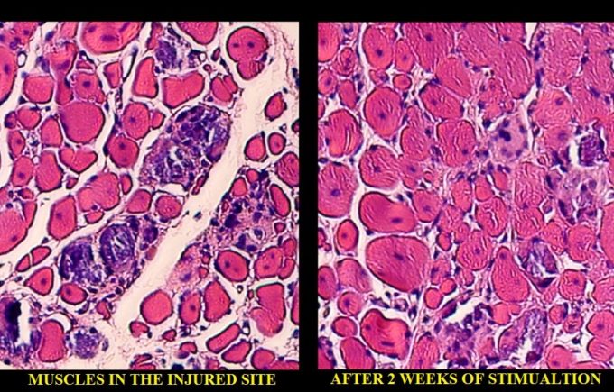
- A recently published study (Cezar et al., 2016) in the very prestigious scientific publication Proceedings of the National Academy of Sciences illustrates how mechanical stimulation of the injured skeletal muscle helps its regeneration.
The authors created local ischemia in the tibialis anterior muscle of rats and after complete loss of function was registered, the animals’ legs were mechanically stimulated by an implanted magnetic device (first group) or an externally placed air cuff which compressed the muscle with kneading-type strokes (second group).
After two weeks of stimulation, the authors registered a 2.5-fold increase in muscle regeneration and reduced fibrosis in the injured muscles compared to no-treatment controls. Fig. 6 illustrates the authors’ findings.

Fig. 6. Histological sample of the muscle after local ischemia and after 2 weeks of mechanical stimulation by kneading-type strokes. On the left you see histological changes in the tibialis anterior muscle after local ischemia was triggered there. The red-colored areas are muscle cells, and the white-colored areas are the fibrotic tissue developed between the muscle fibers as a result of local ischemia. At this point, the animal had completely lost function of the affected muscle. On the right, you see the same tissue after 2 weeks of mechanical stimulation with kneading-type compressions. The white areas of fibrosis are completely gone, and the normal structure of the muscle is completely restored. It coincided with complete restoration of lost function.
At the end of this review, I would like to quote the conclusion from an article by Maniotis et al. (1997) published in Proceedings of the National Academy of Sciences of the USA:
“… direct mechanical linkages throughout living cells raise the possibility that regulatory information, in the form of mechanical stress or vibration (emphasized by R. T.), may be rapidly transferred from these cell surface receptors to distinct structures in the cell and nucleus, including ion channels, nuclear pores, nucleoli, chromosomes, and perhaps even individual genes, independent of ongoing chemical signaling mechanisms.”
REFERENCES
- Brunette DM. Mechanical stretching increases the number of epithelial cells synthesizing DNA in culture. J Cell Sci. 1984;69(1):35-45. doi:10.1242/jcs.69.1.35
- Cezar CA, Roche ET, Vandenburgh HH, Duda GN, Walsh CJ, Mooney DJ. Biologic-free mechanically induced muscle regeneration. Proc Natl Acad Sci U S A. 2016;113(6):1534-1539. doi:10.1073/pnas.1517517113
- Chen BM, Grinnell AD. Integrins and modulation of transmitter release from motor nerve terminals by stretch. Science. 1995;269(5230):1578-1580. doi:10.1126/science.7667637
- Curtis ASG, Seehar GM. The control of cell division by tension or diffusion. Nature. 1978;274(5666):52-53. doi:10.1038/274052a0
- Davidson CJ, Ganion LR, Gehlsen GM, Verhoestra B, Roepke JE, Sevier TL. Rat tendon morphologic and functional changes resulting from soft tissue mobilization. Med Sci Sports Exerc. 1997;29(3):313-319. doi:10.1097/00005768-199703000-00005
- Gataullin RR, Zaripov AT. The role of cytoskeleton in mechanoreceptor activity of Pacinian corpuscles. In: Hník P, Soukup T, Vejsada R, Zelená J, eds. Mechanoreceptors: Development, Structure and Function. Springer; 1988:209-211. doi:10.1007/978-1-4899-0812-4_38
- Gehlsen GM, Ganion LR, Helfst R. Fibroblast responses to variation in soft tissue mobilization pressure. Med Sci Sports Exerc. 1999;31(4):531-535. doi:10.1097/00005768-199904000-00006
- Geneser F. Textbook of Histology. Munksgaard; 1986.
- Jain M, Berg R, Tandon G. Mechanical stress and cellular metabolism in living soft tissue composites. Biomaterials. 1990;11(7):465-472. doi:10.1016/0142-9612(90)90059-Y
- Komuro I, Katoh Y, Kaida T, et al. Mechanical loading stimulates cell hypertrophy and specific gene expression in cultured rat cardiac myocytes: possible role of protein kinase C activation. J Biol Chem. 1991;266(2):1265-1268. doi:10.1016/s0021-9258(17)35310-3
- Maniotis AJ, Chen CS, Ingber DE. Demonstration of mechanical connections between integrins, cytoskeletal filaments, and nucleoplasm that stabilize nuclear structure. Proc Natl Acad Sci U S A. 1997;94(3):849-854. doi:10.1073/pnas.94.3.849
- Shirinsky VP, Antonov AS, Birukov KG, et al. Mechano-chemical control of human endothelium orientation and size. J Cell Biol. 1989;109(1):331-339. doi:10.1083/jcb.109.1.331
- Wang N, Butler JP, Ingber DE. Mechanotransduction across the cell surface and through the cytoskeleton. Science. 1993;260(5111):1124-1127. doi:10.1126/science.7684161
Category: Medical Massage
Tags: JMS 2017 Issue #2
