
My colleague B. Prilutsky suggested that the Journal of Massage Science should publish an article on Periosteal Massage (PM). Once, we addressed that subject in Massage & Bodywork Magazine, but it was many years ago and massage science has developed further since then.
The basic principles of Periosteal Massage were developed in Germany by two physicians, Dr. Vogler, MD, and Dr. Krauss, MD. After years of clinical observations, Dr. Vogler and Dr. Krauss concluded that the periosteum, besides playing a critical role in the normal functioning of our skeleton, is greatly affected by chronic somatic and visceral disorders forming so-called periosteal reflex zones. They in turn start to further complicate the initial somatic and visceral disorders. Thus, Dr. Vogler and Dr. Krauss developed PM as a clinical tool to restore normal function to the periosteum, eliminate periosteal reflex zones and help patients to recover sooner. In Part I we are going to discuss the periosteum and its important functions in the human body.
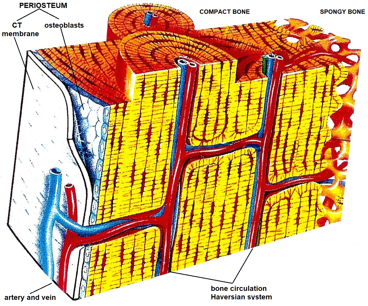
There are five types of soft tissues: skin, fascia, skeletal muscles, tendons and ligaments, and periosteum. The periosteum is a thin membrane (2-3 millimeters) that consists of two layers: outer and inner. The outer layer is a connective tissue membrane similar to the strong fascia. The inner layer is one single layer of cells called osteoblasts. The periosteum covers all bones, and it borders the cartilage which covers joint surfaces. Fig. 1 illustrates the anatomy of the periosteum.
Although the periosteum looks like an ordinary, let’s say, less important type of soft tissue, the periosteum is everything for the bones. Without the periosteum, we are not going to have a stable skeleton and are simply unable to survive. Let’s first enumerate the critically important functions of the periosteum:
- Bone Circulation
- Bone Innervation
- Support of Locomotion
- Bone Remodeling
Let’s review these important functions of the periosteum in detail.
BONE CIRCULATION
The entire bone circulation consumes approximately 10% of cardiac output, and it works under high pressure since the blood must enter resistant tissue such as bone. The bone circulation is called the Haversian system. This network of arteries and veins spreads throughout the entire periosteum, which covers the bones, and enters the inner bone via the so-called nutrient foramen. Fig. 1 illustrates bone circulation and the role that the periosteum plays in it.
BONE INNERVATION
As with all tissues and organs, the periosteum has sophisticated innervation. In reality, when we are talking about a bone’s innervation, we are actually talking about the innervation of the periosteum, which covers all bones. When we fracture any bone, the horrible pain we experience is not coming from the fractured bone but rather from the damaged periosteum. Thus, all nociceptors as well as all other sensory receptors are located in the periosteum but not in the bone itself.
Similar to the skin, skeletal muscles and other tissues and organs, there is clearly an established pattern of the periosteum’s innervation by different segments of the spinal cord. This pattern of innervation is called the sclerotomes map, and it was published in 1944 by Dr. V.T. Inman and Dr. J.B. Saunders. Fig. 2 illustrates the sclerotomes map (Inman and Saunders, 1944).
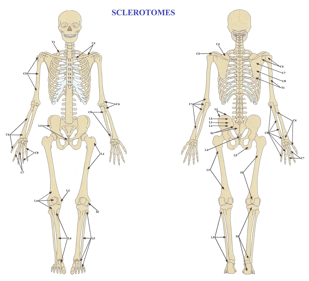
From the clinical perspective, these maps are a very important somatic evaluation tool, and each therapist must use them daily. We will discuss this important issue in the next parts of the article.
SUPPORT OF LOCOMOTION
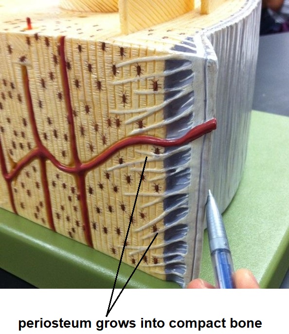
All the soft tissues which support the body’s dynamic and static functions insert into the periosteum. This includes tendons that support muscle contractions and ROM in the joints, ligaments that provide joint stability, and the joint capsules which support their normal functions, all inserted into the periosteum, which itself is very firmly attached to the bones. Thus, the periosteum is a key stabilization component of our locomotion. Its trauma or even mild inflammation deeply affects the function of the affected segment(s), affecting range of motion and eventually the formation of different adhesions. Fig. 3 illustrates how fibers of the periosteum grow into the bone for better stabilization of the muscles, ligaments, etc.
BONE REMODELING
Fractured bone can’t repair itself and reconnect fragments. This critically important healing mechanism is completely under the control of the periosteum. The cells which form the single inner layer of the periosteum are responsible for new bone formation and fracture union.
These cells (see Fig. 1), which are called osteoblasts, are immediately activated when the periosteum is damaged during a bone fracture or bone inflammation. As soon as osteoblasts are active, they start to produce pro-collagen which they unload in the area of bone trauma. Within the line of fracture, the pro-collagen matures into collagen fibers, and later calcium and phosphorus are deposited there, triggering collagen ossification, which finishes fracture healing. That is why we need stabilization of the fractures and complete rest to let the periosteum finish its job and make us mobile again after severe trauma.
The bone remodeling function of the periosteum has its own drawbacks. The formation of bone osteophytes that accompany Osteoarthritis is an example of the negative side of the remodeling function of the periosteum. We will discuss this issue in detail in Part II of this article.
Thus, as you see, the bones of our skeleton are dead if the periosteum is not present, and the function of the skeleton is greatly affected if the periosteum is damaged or malfunctioning.
REFERENCES
- Inman VT, Saunders JBDM. Referred pain from skeletal structures. J Nerv Ment Dis. 1944;99(5):660-667. doi:10.1097/00005053-194405000-00023
- Vogler P, Krauss H. Periostbehandlung. Leipzig; 1953.
Category: Medical Massage
Tags: JMS 2022 Issue #1
