
The article in the July/August issue of Massage & Bodywork Magazine written by Til Luchau is about the iliopsoas’s anatomy, role and treatment options. The issue of iliopsoas therapy requires a separate discussion, since it is greatly misrepresented and unfortunately misused, especially among massage therapists. This is why we can’t stay away from this extremely important topic.
From the very beginning, the author acknowledges the controversy about the iliopsoas’s role and, what is more important, about the treatment which is commonly used. Here are quotes from the article:
“…is also clear: direct myofascial work with the psoas is itself controversial”
and even more striking:
“…the potential for client injury [bold by JMS] from intense abdominal work is real”
However, despite the controversy and even the blunt acknowledgment that the client can be injured, the author continues to suggest direct intra-abdominal treatment of the iliopsoas muscle. According to the article, the author based his views on:
“…my in-depth readings and study of many others’ reports, opinions and approaches.”
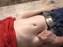
Now let’s see what the author and “others” (BTW, we would like to see references for ‘others’) missed during their in-depth reading. We will use pictures from the Internet which are very close to the pictures in the article. In the article, the author recommends that therapists (see Fig. 1):
“…gently with your client’s breath… sink into the client’s abdomen, patiently making your way to the bottom toward the back and the center [bold by JMS. We will refer to these recommendations later in the review] of the body.”
While the therapist does all of that, the author would like readers to imagine the viscera (i.e., the omentum, intestinal loops and mesentery) as balloons that will gently slip away, letting the therapist touch the psoas muscle without obstruction and work with it directly while the happy viscera are patiently waiting until the important work is finished.
For the second part of the ‘therapy’, the article suggests applying the same pressure while the client slowly slides the leg up and down along the table, as shown in Fig. 2.
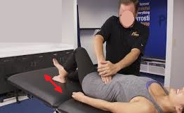
Arrow – directions of leg movements to activate the right iliopsoas muscle
Now let’s see what actually happens in the abdomen while such ‘therapy’ is performed. First of all, as the readers know, the psoas muscle is located so deep that it forms the posterior abdominal wall. Thus, all our abdominal organs are located on and rely on both psoas muscles as support structures.
Fig. 3 below illustrates the anterior view of the abdomen after the skin and abdominal muscles are removed. As you can see, directly under the peritoneum is the omentum, which is an apron-shaped connective tissue structure that contains a great amount of fat and acts as an intestinal protector against any direct abdominal trauma. The omentum has a rich blood supply.
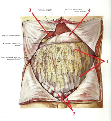
1 – omentum
2 – intestinal loops
3 – liver with gallbladder
4 – stomach
Let’s now remove the omentum (see Fig. 4). Readers can see loops of the small intestine as well as the large intestine, and on the right side, blue dashed lines indicate the lateral and medial edges of the right psoas muscle located under the intestine.
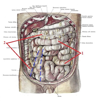
1 – small intestine
2 – large intestine
Blue dashed lines – edges of the right iliopsoas muscle
Our next step is removing the loops of the small intestine (see Fig. 5), and now readers may observe the visible fascial fibers which cover the right psoas muscle. We emphasized the borders of the muscle with blue lines. As you can see, the psoas muscle continues under the ascending colon and cecum (blue dashed lines).
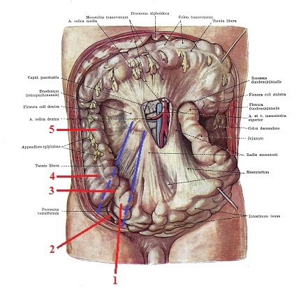
1 – jejunum
2 – appendix
3 – ileocecal area
4 – cecum
5 – ascending colon
Blue lines – borders of the right psoas muscle
In the lower left corner of the picture, you’ll see the ileocecal junction where the ileocecal valve is located. Here, the small intestine transitions into the large intestine, and the ileocecal valve prevents the intestinal contents from moving back into the small intestine.
For the valve to work properly, the cecum and the end of the ileum must be stable. This is why this part of the intestinal tract has a very short mesentery and very low mobility. As you can also see, the ileocecal part of the GI tract lies directly on the right psoas muscle (dashed blue lines).
This is a critical mistake the author, other educators and therapists make. Following the recommendations from the article, the therapist would pin down the ileocecal part of the GI tract, including the valve, against the psoas muscle, at least bruising the intestinal walls and, in some cases, severely traumatizing them.
Let’s finally remove the large intestine, and only now can we see both psoas muscles and the neighboring anatomical structures (see Fig. 6). Please notice how many layers of abdominal structures must be removed before the therapist can finally come into direct contact with the psoas muscle!
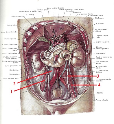
1 – right psoas muscle
2 – vena cava inferior
3 – left ureter
4 – left psoas muscle
Please pay attention to the left side, where the left ureter crosses the anterior surface of the left psoas muscle exactly where the article suggests therapists work, and also notice that the vena cava inferior is normally shifted to the right and is partly located on the anterior surface of the right psoas muscle.
The notion that, as the therapist gently sinks into the abdomen, the viscera peacefully slip away, exposing the iliopsoas muscle, is absurd. The omentum, intestinal loops and/or mesentery don’t have anywhere to go to escape direct intra-abdominal submergence.
It doesn’t matter what the author thinks; he will pin down these structures against the psoas, and the traumatic impact will increase greatly when the therapist asks the client to slide his or her leg up and down the table.
In such a case, as a result of contraction of the iliopsoas, the abdominal organs will be additionally compressed and traumatized between the author’s fingertips and the contracting iliopsoas muscle. Should we also mention the left ureter, which is on the anterior surface of the psoas muscle, and what will happen to it when the therapist works on the left psoas muscle?
Of course, the author and “other” unnamed sources have never observed or conducted abdominal surgery. Let us share with the author and readers our personal experience. First of all, intestinal peristalsis never stops, and it is a very gentle process. The video below illustrates the waves of intestinal peristalsis with their sounds recorded during endoscopic laparoscopy.
The intestinal walls are very delicate, sensitive and easy to damage. This is why, when a surgeon touches the intestine to work with it, he or she always uses gauze (see Fig. 7).
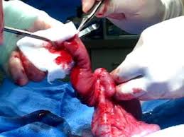
If the surgeon simply squeezes the intestinal loop he or she holds, it immediately stops its peristaltic waves. As soon as the pressure is eased, the peristalsis restarts. This is why young surgeons are constantly reminded by their teachers not to bruise intestinal walls. Imagine for a second what damage to the local peristalsis the therapy suggested in the article does! Direct trauma to the intestinal loop can cause severe consequences for intestinal function, such as colitis, invagination, etc., which appear later.
One of the main arguments the author uses is gentle pressure during the therapy. However, the pictures in the article show significant submergence into the abdomen, which is accompanied by hand movement and becomes even more aggressive when the patient starts to move his lower extremity while pressure is still maintained.
The funny thing about all of this is that the author himself is not 100% sure his recommendations will never hurt the patient. Here is a quote from his article:
“This [i.e., iliopsoas treatment by JMS] … is unlikely [bold by JMS] to damage or disrupt the surrounding structures.”
Since the author uses the word “unlikely,” it is obvious that he does not want to give a 100% guarantee of the safety of the therapy. If there is a 1% chance that massage therapy can worsen the patient’s health – and in the case of Mr. Luchau’s recommendations, this chance rises dramatically – the author does not have the moral right to promote such ‘therapy’ among practitioners.
Unfortunately, the reality of the recommended therapy is much grimmer than the author imagines. The intestine can be easily damaged even by very small trauma. For example, a simple fall from a bicycle without any other significant trauma (Vijayan and Toe, 2013) or even a minor collision during a soccer game (Sandiford et al., 2006) can perforate or transect the intestinal walls. These are just two examples from similar publications on this subject in the medical literature. Fig. 8 illustrates a complete tear of the intestine observed during surgery after relatively mild trauma.
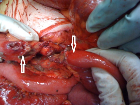
The two white arrows indicate both ends of the severed jejunum
Vijayan and Toe (2013) described the reported case as “jejunal transection, presumably by direct compression against the lumbar spine.” We mentioned the author’s guidelines above for submergence into the abdomen: “patiently making your way to the bottom… toward the back and the center of the body.”
Thus, the article directs therapists to compress the intestinal loops against the psoas muscle, as indicated by the words “toward the back,” and after that, the article recommends switching the direction of pressure “to the center of the body,” compressing the intestinal loops and the psoas muscle against the lumbar spine. This is exactly the mechanism of compression described by Vijayan and Toe (2013)!
We would like to emphasize that the clinical cases described above (Sandiford et al., 2006; Vijayan and Toe, 2013) occurred when the abdominal muscles were able to protect the intestine, since they will always react before any impact reaches the abdominal organs. In the article, the author insists on abdominal breathing and relaxation to remove the abdominal muscles’ protection, which will intensify the intestinal trauma.
Of course, we are not saying that every treatment promoted in the article ends with perforation of the intestinal loops. However, we guarantee that therapists who follow these recommendations are bruising and traumatizing the lower GI tract. The more therapists follow this absurd procedure, the greater the chance that one day a therapist will indeed perforate the intestine.
We have always been puzzled about where this intra-abdominal psoas treatment came from. Who was that original “genius” who came up with this absurd idea? Our research and communications with other equally surprised somatic practitioners revealed that J. Travell and D. Simons’s textbook Trigger Point Manual (1983) is to blame.
Technically speaking, it isn’t the book or its authors but an incorrect interpretation of information from it. In Volume II of Trigger Point Manual, in the chapter dedicated to the Iliopsoas muscle, Dr. Travell and Dr. Simons indeed showed the examination of the tension in the iliopsoas muscle (see Fig. 9). In the same way, many educators commonly suggest that massage therapists actively treat psoas tension.
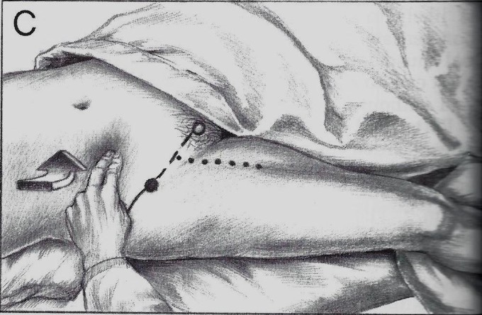
Travell and Simons showed the correct examination procedure for iliopsoas tension, but it was never intended as a treatment! There is no mention of direct abdominal compression as a treatment option in their textbook. Somehow, this legitimate diagnostic test became a treatment procedure, despite the pure absurdity of this idea.
There is no doubt that direct work with the psoas will be helpful for the psoas. However, if the basic rule of medicine, “Do No Harm!”, is still relevant for massage therapists, Mr. Luchau’s approach is unacceptable and his statement,
“Advanced Myofascial Techniques trainings…we see clear benefits that are not easily accomplished by other means”
is an educational disaster.
As long as massage therapy as a profession continues to live in the bubble of unscientific claims (like this one) which are far away from medical science, while whining that no one in traditional medicine would like to listen – nobody will listen!
There are other means to reduce tension in the iliopsoas muscle which are equally effective. There is a direct approach to the iliopsoas muscle above and below the inguinal ligament, completely away from the abdominal organs, and direct work on the iliac portion of the iliopsoas when the patient is positioned on the side. There are also indirect therapies like Postisometric Muscular Relaxation. The combination of these approaches gives the same results without injuring the omentum, intestine or mesentery.
Massage therapy is a relatively safe treatment option with few side effects. Direct intra-abdominal work on the iliopsoas muscle is one of the instances when therapists are hurting patients. The intestinal damage can be acute, but pathological changes in the function of the lower GI tract may also occur later in life following the ‘healing’ effect of intra-abdominal work.
Massage therapy is very flexible in its application. One therapist prefers to use Neuromuscular Therapy, while another uses Myofascial Release. In the case of direct treatment of the iliopsoas muscle, there is no compromise since the patient’s health is at stake. Don’t listen to anyone who tells you that the iliopsoas must be treated through the abdominal organs! From a medical point of view, this is completely absurd!
REFERENCES
- Kiss F, Szentágothai J. Atlas Anatomiae Corporis Humani. Medicina; 1960.
- Sandiford NA, Sutcliffe RP, Khawaja HT. Jejunal transection after blunt abdominal trauma: a report of two cases. Emerg Med J. 2006;23(10):e55. doi:10.1136/emj.2006.038604
- Travell JG, Simons DG. Myofascial Pain and Dysfunction: The Trigger Point Manual. Williams & Wilkins; 1983.
- Vijayan R, Toe K. Delayed complete small-bowel and mesenteric transection following seemingly minor blunt abdominal trauma. BMJ Case Rep. 2013;2013:bcr2013008817. doi:10.1136/bcr-2013-008817
Category: Medical Massage
Tags: JMS 2015 Issue #4
