
Thank you to everyone who shared their expertise in response to our original post. Just as a reminder, the question was what two local conditions can explain the skin reaction to the Compression Test presented in the video.
As correctly mentioned by many therapists, it is true that Thoracic Outlet Syndrome can cause similar reactions on the hand. However, the patient must have numbness and/or burning pain on the entire arm and hand if Thoracic Outlet Syndrome (TOS) is to blame. If the patient has Anterior Scalene Muscle Syndrome, as a lighter form of TOS, the sensory abnormalities must be present as well, but in a lesser form, for example, tingling. The patient didn’t exhibit these symptoms; otherwise, we would have mentioned them in the original post.
Those of you who mentioned Carpal Tunnel Syndrome were absolutely correct, but it was only one of two pathological scenarios presented in the video. There were a couple of therapists who mentioned Median AND Ulnar Nerve Neuralgias. That is actually the correct answer. The patient exhibits Carpal Tunnel and Guyon’s Canal Syndromes at the same time. The most interesting fact is that the ulnar nerve in Guyon’s Canal was affected more than the median nerve in the Carpal Tunnel.
Please pay attention to the very beginning of the video before testing. The local vasoconstriction is clearly visible along the hypothenar with the hand hanging in extension under its own weight without a Compression Test even applied. Also, the application of a Compression Test over the carpal tunnel triggered more pronounced vasoconstriction on the hypothenar (ulnar nerve distribution) compared to the thenar and the palm (median nerve distribution).
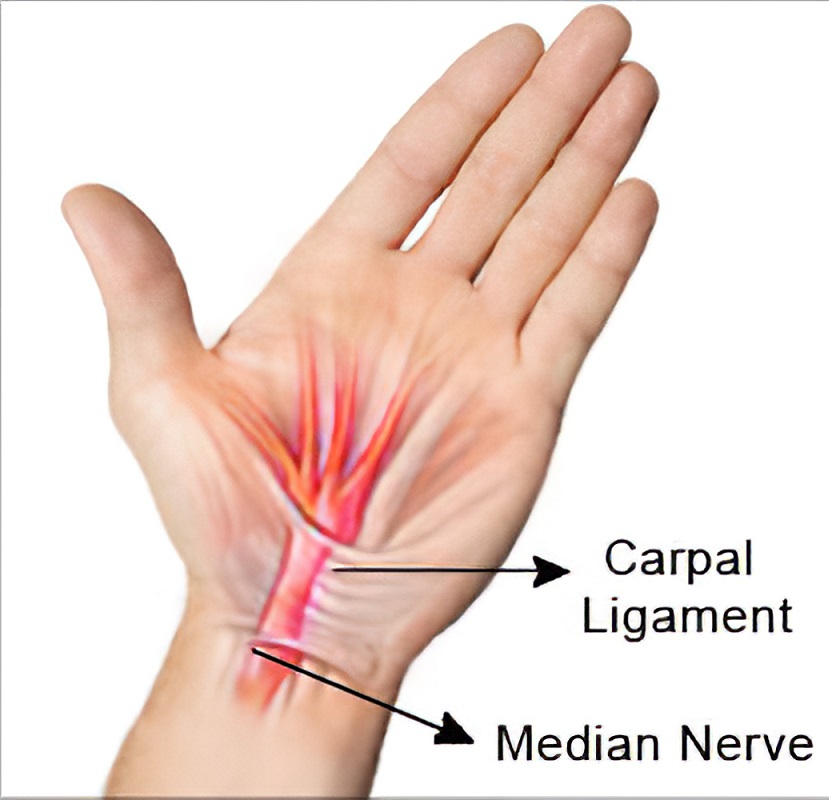
Here is an anatomical situation in the wrist that therapists should consider. The Transverse Carpal Ligament (TCL) forms the roof of the carpal tunnel and lets the flexor tendons and the median nerve pass under it on their way to the palm and fingers. In other words, the carpal tunnel is an entrance gate to the palm. The TCL laterally originates from the scaphoid and trapezium carpal bones, runs across the palmar wrist and inserts into the hamatum and pisiform bones medially. Fig. 1 illustrates the position of the transverse carpal ligament and its relation to the median nerve.
On the ulnar side of the wrist joint, medially and more superficially to the TCL, is the Volar Carpal Ligament (VCL), which originates from the hook of the hamatum bone and inserts into the pisiform bone, forming the so-called Guyon’s Canal. Thus, the walls of this very narrow and short, but clinically important, canal are formed laterally by the hook of the hamatum, medially by the pisiform bone and on the bottom by the TCL, and its roof is the VCL. Fig. 2 illustrates the anatomy of Guyon’s Canal.
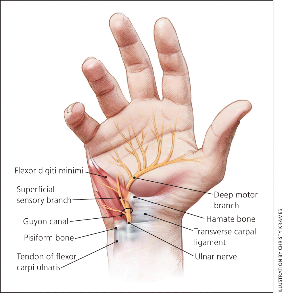
Guyon’s canal contains the ulnar nerve and artery. Technically speaking, some fibers of the TCL contribute to the more superficially positioned VCL. Thus, tension and scarification of the TCL may also affect the VCL, and vice versa. In such cases, therapists will observe a mixed clinical picture of Median and Ulnar Nerve Neuralgias at the same time.
The Compression Test presented in the video additionally stressed the TCL to examine the degree of pressure formed in the carpal canal. The test indeed indicated median nerve entrapment within the carpal canal, since new sensations on the palm were reported by the patient and visible signs of vasoconstriction appeared on the thenar (they are clearly visible in the video). However, vasoconstriction was much more prominent on the hypothenar, which is the ulnar nerve distribution.
Thus, we are dealing with Median Nerve Neuralgia and Ulnar Nerve Neuralgia at the same time, with the ulnar nerve more compressed within Guyon’s Canal than the median nerve within the Carpal Canal.
It is obvious that these changes are work-related. Repetitive motions and the pressure generated during massage strokes are the initial triggers. Many therapists use the pisiform bone as an additional pressure tool during deep strokes. Another aspect is scarification of the TCL, which secondarily decreases the cross section of Guyon’s Canal due to the medial displacement of the pisiform bone, and it compresses the ulnar nerve first, since this canal is very narrow compared to the carpal canal.
Let’s look at both canals in close proximity. Fig. 3 illustrates the relations between the Carpal and Guyon’s Canals.
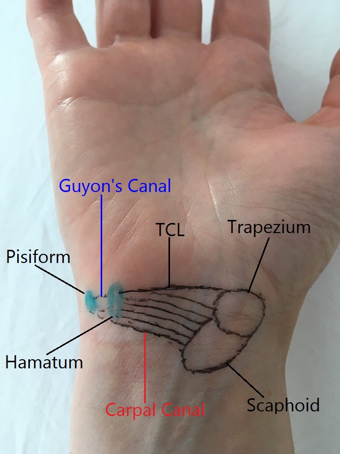
From the treatment perspective, both canals must be decompressed separately, with Guyon’s canal as a priority in this clinical case. Since Guyon’s canal is very narrow, the therapist is forced to work with precision at the insertions of the VCL into the hamatum and pisiform bones, decrease adhesions between the skin with subcutaneous tissues and the VCL, and finally stretch Guyon’s Canal itself, separately from the Carpal Canal, which should be worked on in the second part of the session. Remember that, unlike the Carpal Canal, Guyon’s Canal is superficial and shallow, with both the ulnar nerve and artery at the bottom of it against the carpal bones. Be very careful with the degree of applied pressure. While working in the canal and on its walls, ask the patient to report any new sensations on the hypothenar, pinky and half of the 4th finger. If sensations appear, immediately decrease the amount of applied pressure.
Fig. 4 illustrates the position of the finger to work on the lateral wall of Guyon’s Canal (hook of hamatum).
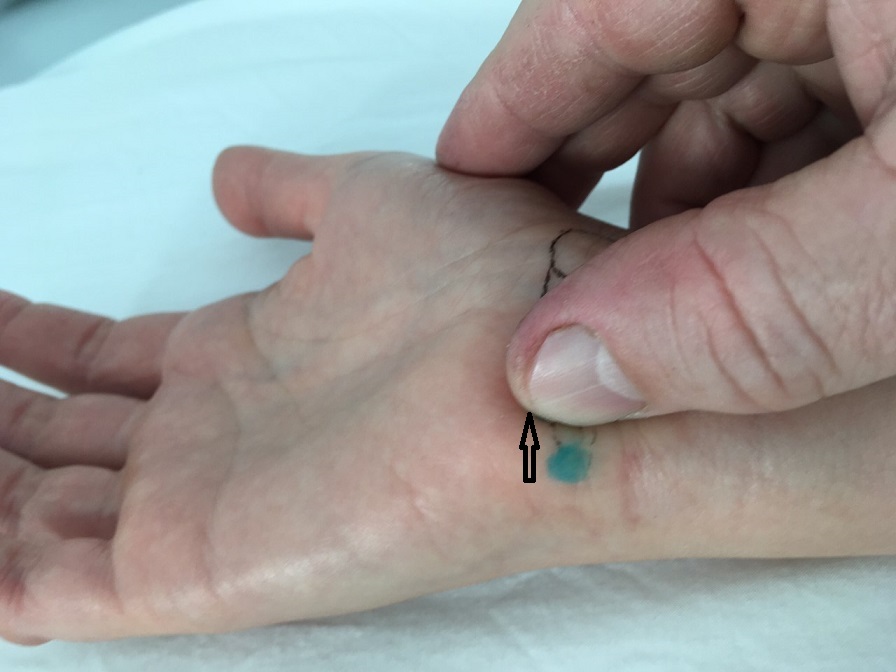
The arrow shows the direction of pressure
Fig. 5 illustrates the position of the finger to work on the medial wall of Guyon’s Canal (pisiform bone).
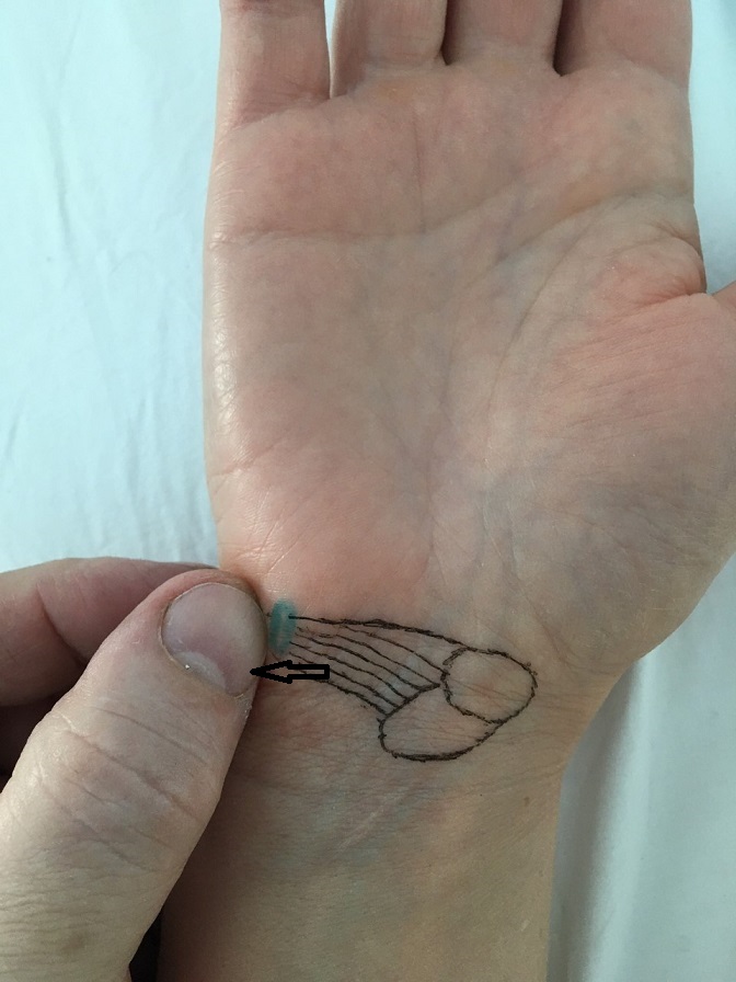
The arrow shows the direction of pressure
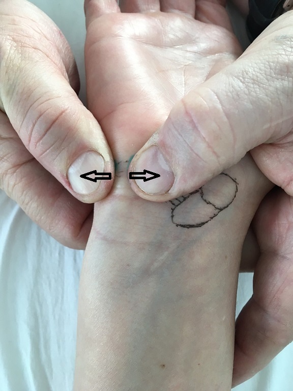
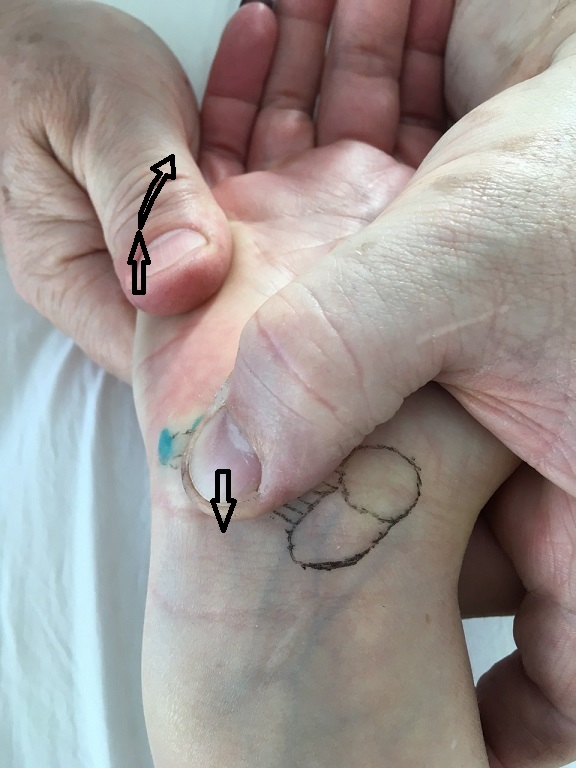
Finally, Fig. 6 illustrates manual decompression of Guyon’s Canal in the perpendicular direction and Fig. 7 in the longitudinal direction. Please note that stretching along the canal requires a combination of forces: stabilization of the hamatum bone with one finger and wrist extension with simultaneous radial deviation.
Category: Blog
Tags: JMS 2020 Issue #3
