
This is our third article on the subject of Pediatric Massage (see the two previous issues of JMS). The first two articles were dedicated to Infant Massage of prematurely born but overall healthy children. The goal of massage therapy in these cases was to stimulate the child’s development and help preterm infants close the gap with full-term infants as soon as possible.
However, there is another aspect of Infant Massage: How to work with those children who were born with various pathological conditions ranging from CNS abnormalities to torticollis (i.e., lateral tilt of the head due to flexion contracture of the cervical musculature). Reading various articles on Infant Massage recently published in professional literature leaves the impression that practitioners are taught by various sources to use basic sets of stress-reduction massage strokes on each baby without any differentiation between a healthy child and a baby with various pathological conditions.
In clinical reality, the application of correct and scientifically sound MEDICAL MASSAGE PROTOCOLS to help children born with various pathological conditions is a separate and extremely valuable branch of Pediatric Massage. Of course, we are unable to address all infant abnormalities in just one article. This is why we decided to discuss here the protocol of medical massage for infants with abnormalities in CNS function that have developed as a result of Perinatal Asphyxia (PA), or lack of oxygen at birth.
There are various causes of PA: abnormalities of the placenta and umbilical cord, premature delivery, birth trauma during labor, infections, etc. PA occurs in 2 to 10 per 1000 newborns born at term, and this number rises more significantly for preterm infants (Barkovich & Truwit, 1990).
Even a short decrease in blood perfusion to the brain or a failure of respiration greatly affects the function of infants’ CNS. In cases of severe PA, the damage to the brain is irreversible, and for these patients, medical massage only helps to maintain quality of life. However, mild to moderate cases of PA are much more common, and early medical massage rehabilitation plays a critical role in the infants’ recovery. When conducted correctly and on time, medical massage helps to increase brain perfusion and save neurons that may have a chance to recover. As stated by Prof. A.M. Aksenova, MD (2003):
“Application of proper rehabilitation during phase of reversible changes after PA allows to save larger number of neurons affected by oxygen insufficiency. This why therapeutic modalities to enhance the blood supply to the brain and spinal cord must be used since infant’s first days of life.”
Prof. A.M. Aksenova, MD
We would like to share with our readers the results of an extensive clinical study conducted by Prof. A.M. Aksenova, MD, in Russia. She is one of the most respected world authorities in the field of medical massage and pediatric massage in particular. This publication is based on the treatment of 700 newborns who experienced PA of various degrees. We don’t want to overwhelm the readers with the statistical analysis and data the authors obtained before and after the applied treatment, but we think it will be beneficial to our readers if we discuss the actual protocol Prof. Aksenova developed as a result of this study.
The method of Deep Reflex Massage (DRM) was developed by Prof. Aksenova for infants experiencing even short intervals of PA and is accepted as a standard treatment procedure in many pediatric hospitals. The goal of DRM is to increase blood circulation through the brain and spinal cord by using intense reflex stimulation of the soft tissues.
The basic physiological fact that any stimulation of the peripheral sensory receptors requires the formation of a reflex motor response to the stimulated tissue is the theoretical foundation of DRM. The formation of a motor response requires activation of CNS function, and it can be done only by increasing the blood supply to the CNS and activating local metabolism (e.g., glucose metabolism in the brain). As Prof. Aksenova’s measurements showed, the application of regular full-body massage for infants with PA significantly increased blood circulation through the brain and spinal cord. Based on these initial results, Prof. A.M. Aksenova experimented with different massage protocols while examining blood circulation through the brain. The DRM developed by the author showed the greatest stimulation of blood circulation through the CNS.
DEEP REFLEX MASSAGE FOR INFANTS AFTER PA
The initial course of DRM consists of 3-5 sessions, two times per week, in the hospital, starting on the first day of life, and 10-15 sessions, 2-3 times per week, conducted after the child is discharged from the hospital. The duration of each session is 10-20 minutes, depending on the size of the infant’s body. The entire course of 10-15 sessions can be repeated after 2 months, if needed.
PLAN OF SESSION
The protocol of DRM is a very important factor in the overall success of the therapy. The sequence of the treatment consists of the consecutive application of massage to the following areas: anterior neck, lateral neck (alternating right and left sides), posterior neck, upper shoulders, both temporal areas, head, back and gluteal areas. The practitioner must start with neck massage and spend more time on the neck and head.
Another critically important aspect is the set of massage techniques. Various types of kneading make up 80% of the techniques used during each session. These techniques require lifting, stretching, compressing and rolling soft tissue between the fingers and against bone structures. Scrolling, double scrolling, transverse kneading, simple kneading, etc., are used. Considering that the massage areas are small, the practitioner should use pinching (or three-finger) prehension while working with the tissues. The readers may find a detailed description and video presentation of all kneading techniques in the Therapeutic Part of our Video Library.
DRM doesn’t mean the application of pinpoint deep pressure. The author uses the term ‘deep’ to emphasize the necessity to address deeper layers of the soft tissue. Only in this case does DRM have a stimulating impact on the circulation in the CNS.
STEP-BY-STEP PROTOCOL OF DRM
Step 1.
Place the infant on their back. Start with bimanual light effleurage strokes on the anterior neck starting from the origin of the sternocleidomastoid muscle (i.e., the mastoid processes behind both ears) down along the muscle, following lymph drainage. Use both thumbs, keep them flat and direct pressure forward to the clavicles. When you reach the supraclavicular area, move the thumbs laterally along both clavicles while applying pressure on both trapezius muscles using the thenar area of each hand until you reach both acromioclavicular joints.
Step 2.
Apply kneading along the sternocleidomastoid muscles as well as moderate friction at their origin at the mastoid processes. The contact areas are the palmar surfaces of the distal phalanges of the index or index and third fingers. Be sure to stretch the muscle fibers between the hands and alternate this with careful compression of the muscle tissue against the underlying cervical vertebrae. While working on the sternocleidomastoid muscle, turn the child’s head to the same side.
Finish this step with the application of careful spiral friction along the pathways of the major nerves and carotid arteries on the anterior neck. With the fingertips of the index or index and third fingers, apply careful friction along the anterior edges of both sternocleidomastoid muscles.
Step 3.
Using the thumbs placed flat along the clavicles, work in both supraclavicular areas using friction and gentle manual vibration in interruptive mode. Slowly increase the pressure, trying to submerge into the supraclavicular areas and even reach the first rib, which is at the bottom of the supraclavicular fossa.
Step 4.
Alternate work on each lateral surface of the neck. Turn the child’s head to the opposite side and use the same combination of techniques.
Step 5.
Work on the posterior neck while the child is placed on their side. Use a small pillow to support the head. Use the same combination of techniques and give attention to the suboccipital area and occipital ridge. While working in the suboccipital area, apply circular friction and several repetitive compressions using the fingertip of the 5th finger in the area of the vertebral artery.
Both vertebral arteries are hidden in the cervical vertebrae, and they are directly accessible only at the vertebral artery point, which is located between the occipital ridge and the transverse process of the first cervical vertebra, the most prominent bone on the lateral surface of the neck just below the skull.
The vertebral artery point is very easy to find, because it is located inferior to the occipital ridge, just in front of the free edge of the upper portion of the trapezius muscle (see Fig. 1).
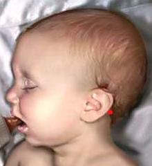
Red dot – location of the point
Work in this very small area has a great impact on the increase of blood perfusion in the pool of the basilar artery (the basilar artery is located on the pons and is formed by the union of the two vertebral arteries).
When working on the cervical paravertebral muscles, use kneading between the fingers as well as kneading against bony structures.
Step 6.
Work on the scalp of both temporal areas first and later on the other parts of the scalp. Start with raking effleurage using the fingertips, moving them between the hairs in a spiral motion. Later, switch to careful friction, placing the fingers flat. Don’t apply significant pressure, especially vertical pressure. Remember the location of cranial fontanelles. Try to move the fingers with the scalp, applying pressure horizontally along the skull. Vasodilation in the scalp triggers an increase in brain circulation through reflex pathways. This reflex reaction is a well-established physiological fact.
Step 7.
Briefly work on the entire back and gluteal areas at the end of the session.
If you are working in a pediatric hospital or have friends and family members who recently had a child with PA, use this protocol to help infants recover from the consequences of this dangerous condition. Also, educate other health practitioners (pediatricians, nurses) about the clinical benefits of Pediatric Massage.
REFERENCES
- Aksenova AM, Aksenova NI. Method of deep reflex massage of infants with perinatal disorders of the central nervous system. Medical Gymnastics & Massage. 2003;3(6):9-11.
- Aksenova AM, Reznikov KM, Trofimova OV. Effects of deep reflex-muscular massage and exercise on regulatory processes in the body. Klin Med (Mosk). 1997;75(7):50-52.
- Barkovich AJ, Truwit CL. Brain damage from perinatal asphyxia: correlation of MR findings with gestational age. AJNR Am J Neuroradiol. 1990;11(6):1087-1096.
Category: Pediatric Massage
Tags: JMS 2011 Issue #2
