
In the Mar/Apr, May/June, July/Aug, and Sept/Oct 2009 issues of JMS, we published a four-part article on Trigger Point Therapy (TPT). Since then, we have read several articles on this subject and participated in and observed many discussions in professional forums on the same subject. This personal experience convinced us to revisit the issue of trigger points and TPT in a follow-up article.
While working on the original article, we were sure that the science of TPT was well-known and an already settled issue. We saw that the purpose of our original article was to put the main theoretical and clinical components under one roof. To our great surprise, participating in professional forums and reading recent articles revealed that TPT remains a most controversial, misunderstood and misrepresented issue.
We also realized that our attempts to express scientifically and clinically based opinions in the majority of cases were seen as just our personal opinion/theory. This is why we decided to address the TPT issue one more time from a scientific point of view and update the original article.
First, we would like to summarize the most common misunderstandings and simple mistakes that the issue of TPT has generated.
- A trigger point is a local pathology without any involvement of the nervous system.
- There is no scientific data which supports the theory that trigger points can be the result of any neurological abnormalities or reflex zone formation.
- The best way to eliminate the trigger point is through the application of mild to moderate pressure.
- Ischemic compression as a part of TPT is useless because it is not logical to treat the trigger point where circulation is already compromised by additional oxygen deprivation of the affected tissue.
Some of these ideas started to circulate in the massage community after a publication by Dr. J. Muscolino (2008) on this subject. We greatly respect this author. He is in the minority of educators who really know what they write and teach. However, we completely disagree with his views on the subject of TPT.
Recently, the situation became even more complicated when other practitioners and educators contributed to the confusion with their own ideas rather than scientifically based information. Frequently, statements are made without any data or references, and as soon as authors are confronted about the origin of their ideas, we hear similar replies: “I practice TPT for 20 years”, “I wrote books on TPT”, “I have taught TPT for 15 years”, “200 MD’s were impressed with my treatment” and so on. In other words, “I can’t be wrong because I am right.”
We think that at this point the American massage therapy industry lacks a unified view on the majority of important subjects. A great number of educators espouse their own ideas and agendas without even bothering to examine scientific data that already exists but in many cases undermines their personally developed concepts and system of beliefs. Some educators mislead readers by manipulating existing data (Good/Bad Apples in the Nov/Dec 2010 issue of JMS). The sad part is that many educators do not even agree with each other and teach completely opposite approaches, which dramatically increases the confusion. AMTA is just an observer of this contradictory state without the means or desire for leadership.
As a result of all these events, the massage therapy practitioner trying to break into the medical field frequently relies on personal opinions and anecdotal experiences rather than on firm scientific data. We agree with the fact that every practitioner is entitled to their own opinion. However, this freedom stops at the moment the practitioner starts to work on patients rather than clients. At this moment, the personal views become invalid if they are not supported by scientific data. This is why we decided to revisit the issue of TPT as one of the most misrepresented issues. Although this article is long, we encourage the reader to finish it because we believe our views will benefit everyone in the profession.
A trigger point is a local pathology without any involvement of the nervous system
There is a belief that a trigger point is a local pathology without any involvement of the nervous system. This belief system absolutely correctly describes the chain of events that are responsible for the formation of a trigger point: excessive calcium release, formation of a taut band, shortage of ATP, local ischemia and so on. However, supporters of this view fail to provide any reasonable explanation for what actually triggers these events.
Yes, it is correct that a trigger point could be the result of trauma, and this is one of the possible causes. Yes, a trigger point could form in muscles that are overused, especially if they are worked a lot in the isometric regime. For example, trigger points form in the trapezius muscle of a computer programmer. In these two cases, the trigger points are indeed a local pathology as a result of direct trauma or chronic overload, and they develop secondary to overused muscles.
However, this theory fails to explain the commonly observed clinical facts of active trigger point formation in the muscles innervated by an irritated or compressed spinal or peripheral nerve. Why, for example, does the patient with acute disk bulging or herniation almost immediately exhibit active trigger points in the piriformis muscle or in the peroneal group? Any practitioner who works on patients with an irritated or compressed brachial plexus finds trigger points in various locations on the upper extremity, and the location of these trigger points will coincide with the irritated part of the brachial plexus. We can go on and on with these examples.
The readers may ask: Is there any merit in this discussion? What is the difference if the nervous system plays or does not play a role in the formation of a trigger point? The answer to what look like purely theoretical questions is very simple: The success of your entire practice is at stake!
Those who read various professional forums may regularly see pleas for help from practitioners who are desperately trying to eliminate a ‘stubborn’ trigger point without any success. Why is that? The answer is very simple: Those practitioners approached the active trigger point as a local pathology, exactly as proponents of this theory recommend, instead of finding the real cause of the problem. In this case, it was mild irritation of the nerve that supplies the muscle harboring active trigger points. The practitioner is knocking on the same but unfortunately wrong door, and this is the reason for the failure. Trigger points are the result of many different factors, and the treatment protocol should consider and address the original cause; otherwise, the practitioner shouldn’t expect quick and sustained results.
Considering that the majority of authors use their personal experience or beliefs as a justification for this and similar concepts, we decided to present our personal experience as well. We conducted very basic research that examined the results from two clinics, one in Phoenix, Ariz., and another in Los Angeles, Calif. Evaluation of our patients’ initial clinical symptoms and outcomes of the therapy for the month of August 2011 allowed us to conclude that 86% of our patients (320 total treatments) had somatic abnormalities accompanied by active trigger points in the skeletal muscles. Of this 86%, approximately 70% were cases in which active trigger points were a complication of spinal or peripheral nerve entrapment. Our treatment protocols initially targeted the area of irritation, and later, local therapy in the area of the active trigger point was added. We aren’t claiming statistical proof of this data. This is our personal clinical observation.
Conclusion: active trigger points are the result of local abnormalities as well as involvement of the nervous system. We personally think that in clinical practice, the majority of patients who have active trigger points have nervous system involvement, including viscerosomatic reflexes in cases of inner organ disorders, and that to successfully eliminate this pathology, local treatment isn’t enough. It is a recipe for failure.
There is no scientific data which supports the theory that trigger points can be the result of any neurological abnormalities or reflex zone formation
This is the most bizarre statement we have ever read. Even from a historical point of view, the proponents of this statement are wrong. They attribute the concept of trigger points solely to the works of J. Travell, MD. It is a great misunderstanding to see Dr. Travell as the author of the trigger point concept. A great number of scientists contributed to the development of the trigger point concept way before Dr. Travell was even born:
- Dr. R. Froriep (1843) was the first scientist who studied trigger points;
- Dr. H. Nothnagel (1877) proposed the original theory of the neurological origin of trigger points in the skeletal muscles;
- Dr. H. Schade (1921) developed the concept of myogelosis as the outcome of the chronic presence of a trigger point;
- Dr. J. Mackenzie (1923) proved that changes in the motor activity of the patient’s nervous system are key components in the development of hypertonic muscular abnormalities, especially trigger points. Thus, Dr. Mackenzie was the first to formulate the concept of reflex zones in the skeletal muscles, including trigger points.
As you can see, trigger points have been intensely studied since the end of the 19th century, and these scientific studies greatly influenced Dr. Travell’s and Dr. Simons’s work.
Supporters of the local theory of trigger point formation widely cite selective quotes from Dr. Simons’ earlier articles. It seems they use outdated data to reinforce their concept, while Dr. Simons moved with the times and concluded that the nervous system is a major factor in the formation of trigger points. He endorsed the works of the German scientist Dr. S. Mense and even coauthored an article on this subject (Hoheisel et al., 1993).
According to this experimental study, the triggering of muscle pain leads to the projection of these signals within the dorsal horns of the spinal cord, which in turn trigger referred pain and the formation of trigger points in completely new areas. Thus, the nervous system is directly responsible for trigger point formation in the skeletal muscles or, in other words, in the areas of reflex zones.
Recently, some authors have started to return to the original theory that active trigger points are mostly the result of nervous system abnormalities. This theory was originally proposed by Dr. Nothnagel in 1877 and was later replaced by Travell’s and Simons’ works, which concentrated on the explanation of the local events within the trigger point.
We are sure that it will surprise supporters of the strictly local theory of trigger point formation that overall frustration with the unsuccessful treatment of trigger points has forced some scientists to abandon the local theory completely in favor of the original neurological theory (Quintner and Cohen, 1994). We are sure that such an opposite extreme is as incorrect a view as the local theory, and that both mechanisms are equally responsible for the trigger point formation. In many cases, both mechanisms even contribute to the clinical picture in the same patient.
Another example of why supporters of the local theory are wrong is the fact that active trigger points are formed in the skeletal muscles as a result of various visceral abnormalities. This is one of the greatest contributions of Dr. Travell to medicine. She and Dr. Simons later showed without any doubt that active trigger points form in the skeletal muscles (e.g., pectoralis major and minor, trapezius, etc.) of patients with chronic and acute cardiac disorders. As the original works of Dr. Travell showed, the deactivation of active trigger points in the chest and upper shoulder muscles significantly decreased the intensity of the chest pain even in patients with acute myocardial infarction (i.e., heart attack) (Rinzler et al., 1954). Additionally, active trigger points in the skeletal muscles do not easily disappear even after cardiac circulation is restored and the patient survives the heart attack (Landmann, 1949).
Considering that the formation of active trigger points in cases of visceral abnormalities is an undisputed, established medical fact, the local origin of trigger points alone simply does not make common sense. For the active trigger points to form in the pectoralis muscles immediately during a heart attack, the nervous system must be involved. This is a perfect example of reflex zone formation, and the supporters of the local theory usually do not mention this fact because it undermines the entire ‘local only cause’ concept.
Conclusion: claims that there is no scientific data that supports the nervous system as one of the leading causes of trigger point formation mean only one thing. The supporters of the ‘local only cause’ concept do not know or do not want to acknowledge all the scientific data that completely rejects a local-only origin of trigger point formation. We can’t even believe that we are discussing this issue in the 21st century!
Even our short review of articles shows how wrong proponents of the local theory are and how confusing this subject is for the practitioners who waste their time on useless treatments trying to eliminate active trigger points with a local-only treatment approach. The scientific and clinical data show that trigger points cannot be addressed as local pathology only. Such views will complicate and derail medical massage treatment and prolong patient suffering.
We treat patients in our clinics who initially received incorrect somatic rehabilitation based on the local treatments applied by chiropractors, physical therapists and massage practitioners, and in many cases, the lack of clinical success forced patients to undergo unnecessary surgeries, which could have been prevented if the correct MEDICAL MASSAGE PROTOCOL had been chosen.
The best way to eliminate the trigger point is the application of mild to moderate pressure
The supporters of this idea can be split into two camps:
- Those who advocate that sustained mild pressure must be used because active trigger points are the result of psychological conflict and such gentle pressure is a comforting factor, which helps the clients to balance their emotional state and thus eliminate active trigger points.
- Those who advocate that moderate repetitive pumping strokes must be used to mechanically enhance arterial blood supply and venous drainage.
Both groups oppose the application of ischemic compression: The first one on spiritual grounds, while the second one bases its logic on the fact that an active trigger point is an area of ischemia and that it does not make any sense to impose additional ischemia on already ischemic tissue. We will address the issue of ischemic compression below.
Ironically, both groups are correct in their own beliefs, but their supporters fail to see the bigger picture. The body is a very complicated but efficient machine, and it always will defy those who use a simplistic approach.
Yes, active trigger points always will be exaggerated by emotional stress and, in these cases, the mild pressure is a correct clinical tool to address the tissue as a first component of TPT. It helps the patient to adapt to and be comfortable with the practitioner while accomplishing the very important goal of desensitizing the area of the active trigger point by applying massage therapy in the inhibitory regime. It allows the elevation of the threshold of peripheral receptors, especially pain receptors, in the area of future TPT.
Yes, pumping strokes in the area of the active trigger point are an equally important tool. Such strokes, in combination with the subsequent application of permanent fixed vibration, help to trigger local vasodilation using mechanical force, which prepares the active trigger point for the application of ischemic compression. This is one example of why the application of ischemic compression alone is not enough, but an integrative approach to TPT is the only correct clinical solution.
However, both groups make the grave mistake of singling out each of these tools from the context of TPT and using them as independent therapies. Such a one-sided, simplistic approach to TPT confuses the practitioner and is directly responsible for the unsuccessful cases of TPT so frequently reported in the posts in various forums.
Conclusion: The ultimate success of TPT lies in an integrative combination of treatment tools, which is the only way to eliminate active trigger points with stable clinical results.
The ischemic compression part of TPT is useless because it isn’t logical to treat a trigger point where circulation is already compromised with additional oxygen deprivation of the affected tissue
As we discussed above, the supporters of this concept base their ideas on what seems to be reasonable logic: The active trigger point is an area of ischemia, and it makes no sense to impose additional ischemia on the tissue with already compromised circulation.
To support this assumption, the Second Edition of the Trigger Point Manual by Travell and Simons is cited:
“deep-stroking massage as being … probably the most effective way to inactivate central TrPs when using a direct manual approach.”
First of all, this was the personal opinion of Dr. Simons. When this quote is read carefully, the word ‘probably’ does not reflect the firm belief and conviction even from Dr. Simons himself. The fact that this single quote is a personal belief is supported by the fact that Dr. Simons did not measure the clinical outcomes of deep-stroking massage alone against ischemic compression. Without such a study to confirm it one way or another, such a quote remains a personal opinion and this is exactly why Dr. Simons uses the word ‘probably’.
Those who are familiar with his publications have witnessed many times that he was very forceful in expressing his opinions (usually in the form of letters to the editors of scientific journals) when he was sure of his views. Here is one of the examples: Simons (2007).
The Trigger Point Manual is definitely a remarkable book. However, we would like anyone who has access to it or owns this publication to open the book and look at the treatment options the Manual offers. The readers will find that this book offers the following treatments: injection, muscle energy techniques, cold and stretch approach (in the first edition) and lifestyle modifications. Massage as a treatment option is mentioned one time, at the beginning of Volume I in the general chapter where all possible treatment options are reviewed.
Thus, neither Travell nor Simons considered massage a valuable clinical tool. Otherwise, massage options, including deep stroking, would be offered to the reader as a treatment option in each chapter. So why is that? Both volumes are written by physicians for physicians and, to a lesser degree, for physical therapists. Massage practitioners were not the original target audience. Plus, neither Travell nor Simons personally used or studied massage therapy as a main treatment tool for their patients.
Now we would like to take our readers, especially those who believe that ischemic compression is an outdated and even harmful tool, on a short journey into the most scientifically trusted source of the modern understanding of circulation in the skeletal muscles, written by Dr. R.J. Korthuis: Skeletal Muscle Circulation (2011).
Let’s look at Fig. 1 from this great book, which we highly recommend to those interested in this subject.
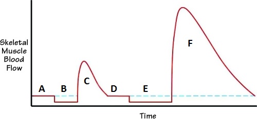
Let’s read this diagram.
A – baseline of circulation in the skeletal muscles before occlusion
B – occlusion of circulation less than 1 minute
C – reflex vasodilation after occlusion
D – restoration of the circulation to the baseline
E – occlusion of circulation more than 1 minute
F – reflex vasodilation after occlusion
Please pay attention to the difference in the peaks of reflex vasodilation (C and F) in regard to their intensity (height of the vasodilatory wave) and duration (time of the vasodilatory wave) after occlusion of less than 1 minute (B) and of more than 1 minute (E).
Here is a quote from the book (Korthuis, 2011) that explains Fig. 1.
“When the arterial inflow to skeletal muscle is suddenly occluded, blood flow decreases (B). Upon removal of the occlusion, skeletal muscle blood flow is markedly enhanced (C). The magnitude of this reactive hyperemia, both in terms of peak response and time to recover to baseline (D), varies directly with the time of occlusion. When the occlusion period exceeds 1 minute (E), maximal peak responses are achieved (F), but the time for blood flow to return to control levels is progressively increased.”
If anyone still has doubts, here is the final scientific medical data, but we are not done yet. We think it is extremely important to our profession to finally put this issue to rest. We recognize that no matter how many references we use to support the scientific foundation of TPT, this article will have limited influence on the followers of the deep-stroking concept. Some of them exhibited very strong convictions. This is why, instead of going back and forth with references, we decided to use visual proof of the effectiveness of ischemic compression and illustrate it with a couple of very informative tests. This discussion has become so complicated by conflicting opinions that we think that it is better to actually see how everything works rather than read conflicting opinions.
Test 1. Reflex Vasodilation in the Normal Tissue
As we discussed above, the supporters of deep-stroking massage treatment alone deny the fact that reflex vasodilation is a result of ischemic compression. Using a simple application of Test 1, we can visually prove the existence of such a phenomenon in normal, unaffected tissue. It is sad that this basic physiological mechanism requires extended discussion and proof for the practitioner who is confused by inaccurately presented information.
Please pinch the skin on your inner forearm for one minute. The pressure should be significant but just below the pain threshold. In such a case, you are creating ischemic compression in the skin on the forearm. After you release the compression, the immediate picture you will see is a white spot that matches the compressed area. This is local ischemia as a result of skin compression. Soon you will see that this white spot changes color to pink or red. This is reflex hyperemia or direct evidence of vasodilation as a result of ischemic compression. However, this vasodilation is not the result of the mechanical effect of effleurage/friction strokes (you just compressed the skin). This vasodilation is a direct result of oxygen deprivation in the compressed tissue. In other words, cutting the blood supply to the compressed tissue triggers reflex vasodilation, because the body will do anything to restore oxygenation of the tissue that has experienced even a short period of hypoxia.
To do that, the reserve capillaries must be open, and this allows an extra amount of oxygenated blood to rush into the compressed tissue that you observe as skin redness. In a minute, the skin color is restored, which means that the reserve capillaries are now closed and normal circulation is restored. This exact chain of events happens in the area of the active trigger point in the skeletal muscle, and this is why ischemic compression is an important tool which, of course, should be combined with proper preparation of the soft tissues in the form of effleurage and deep pumping strokes. Thus, by finishing this simple experiment, you prove to yourself that temporarily applied local hypoxia from ischemic compression triggers local vasodilation using reflex mechanisms (e.g., axon reflex; see the Vasodilation article in this issue of JMS).
Test 2. Dermographism Test (DT)
Our opponents may have a reasonable counterargument that Test 1 does not prove anything because it was used on tissue (skin in our test) that initially had normal circulation, while the trigger point is an area of local ischemia where there is insufficient local circulation. Such final doubts can be easily put at rest by using the more sophisticated Dermographism Test, which we would like to discuss here as well.
DT is a much more complicated test and diagnostic tool. To fully understand the value of DT, we need to discuss first what dermographism means and how it looks in a healthy person (see Video 1).
DT on the inner forearm of a healthy person
The best way to trigger DT is to quickly stroke the skin with your fingernail (see video above). In the video above, you can see that immediately after we stroked the skin, a white line appeared along the stimulated part. This line is called white dermographism, and it is the result of reflex vasoconstriction as a response to skin stimulation. Almost immediately, this line started to turn pink and later red, and this new reaction to the initial skin stimulation is called red dermographism. The red dermographism is reflex vasodilation, which normally appears after initial vasoconstriction. The red line slowly fades away, and the original color of the skin in the stimulated area is eventually restored. This is the normal chain of events triggered by the skin stimulation. There are two possible pathological outcomes of DT: long-lasting white dermographism and excessive red dermographism.
Long-lasting white dermographism is a sign of increased activity of the sympathetic nervous system in the stimulated area. In such a case, the white line does not turn red quickly enough. The excessive red dermographism is a sign of overactivity of the parasympathetic nervous system. In these cases, the white line turns red within the normal time frame, but the red line increases to the point that the stimulated area sometimes becomes edematous.
For our discussion, the case of long-lasting white dermographism is critically important. As we just mentioned, in the case of long-lasting white dermographism, the white line, which forms on the skin after its stimulation, doesn’t turn red and in some cases continues to increase in size. You may see this phenomenon in patients with peripheral nerve neuralgia, for example, on the back of the leg in cases of Sciatic Nerve Neuralgia.
We may use the patient with long-lasting white dermographism as an exact analogy of what is going on with the circulation in the area of an active trigger point in the skeletal muscles. In both scenarios (i.e., white dermographism and active trigger point), there is local vasoconstriction as a result of increased activity of the sympathetic nervous system.
To demonstrate the phenomenon of long-lasting white dermographism, we filmed DT on one of our patients with Sciatic Nerve Neuralgia (see video below). Despite the very visible white dermographism reaction, it was difficult for us to capture it on video. This is why we ask you to pay attention to the slightest difference in skin color in the presented video.
Long-lasting white dermographism on the posterior leg of a patient with Sciatic Nerve Neuralgia
At the beginning of the video, you may see that we stroked the skin on the posterior leg three times and got white lines that did not turn red and even started to widen. In the next part of the video, we compressed the skin in one area of the white line between the thumb and index finger. We kept the skin compressed just below the pain threshold for half a minute. In the final part of the video, we released the compression while the white lines above the compressed area were still clearly visible. At this moment, you may observe that the compressed skin starts to turn pink (white arrow), while the rest of the white line does not change color at all.
This test recreates the exact chain of events during the application of ischemic compression in the area of an active trigger point. By compressing the skin with visible signs of local vasoconstriction, we additionally deprived the compressed areas of oxygen. After compression stopped, reflex vasodilation was triggered (white arrow) as a response to temporary hypoxia caused by our compression. At the same time, the other parts of the white line did not change their color because the vasoconstriction was still raging there.
There is no doubt that if one strokes the area of the white line, as suggested by proponents of deep-stroking massage only, he or she will get the same vasodilation effect, while other parts of the white line are still affected by vasoconstriction. However, our goal was to prove that ischemic compression in an area with already compromised circulation was able to trigger reflex vasodilation as a result of pure compression.
The mechanisms which are responsible for vasodilation as a result of deep stroking and as a result of ischemic compression are quite different. In the first case, the practitioner obtains vasodilation as a result of a mechanical effect, while in the second case, the reflex mechanism of vasodilation is used. This is why deep stroking and ischemic compression should never be measured against each other. They are two equally important components (plus muscle energy techniques) of a scientifically sound protocol of TPT. As we discussed in the Sept/Oct 2009 issue of JMS, the application of ischemic compression in combination with deep pumping strokes and permanent fixed vibration allows optimized treatment by using local and reflex mechanisms of vasodilation during the same treatment session.
Conclusion: We hope that the scientific data presented in this article has finally put the four concepts we mentioned at the beginning of the article to rest. For the most stubborn believers, this or any other source of scientific information will not change their beliefs, and we are powerless in those cases. For the rest of the readers, we would like to emphasize that fragmentation of the profession is a dead end. The clinically effective practitioner must see the larger and more integrative picture for the clients’ and patients’ health benefits; he or she must base his or her practice on scientific data rather than on personal opinions and always challenge any new concept before applying it to the client or patient.
REFERENCES
- Froriep R. Ein Beitrag zur Pathologie und Therapie des Rheumatismus. Weimar; 1843.
- Hoheisel U, Mense S, Simons DG, Yu XM. Appearance of new receptive fields in rat dorsal horn neurons following noxious stimulation of skeletal muscle: a model for referral of muscle pain? Neurosci Lett. 1993;153(1):9-12. doi:10.1016/0304-3940(93)90064-R
- Korthuis RJ. Skeletal Muscle Circulation. Morgan & Claypool Life Sciences; 2011. doi:10.4199/C00035ED1V01Y201106ISP023
- Landmann HR. Trigger areas as cause of persistent chest and shoulder pain in myocardial infarction or angina pectoris. J Kans Med Soc. 1949;50:69-71.
- Mackenzie J. Angina Pectoris. Henry Frowde & Hodder & Stoughton; 1923.
- Muscolino J. Understanding and working with myofascial trigger points. Massage Ther J. 2008;(Spring):1-5.
- Nothnagel H. On neuritis in relation to its diagnosis and pathology. In: Volkmann R, ed. Clinical Lectures on Subjects Connected With Medicine, Surgery and Obstetrics by Various German Authors. New Sydenham Society; 1877:201-236.
- Quintner JL, Cohen ML. Referred pain of peripheral nerve origin: an alternative to the myofascial pain construct. Clin J Pain. 1994;10(3):243-251. doi:10.1097/00002508-199409000-00012
- Rinzler SH, Stein I, Bakst H, Weinstein J, Gittler R, Travell J. Blocking effect of ethyl chloride spray on cardiac pain induced by ergonovine. Proc Soc Exp Biol Med. 1954;85(2):329-333. doi:10.3181/00379727-85-20871
- Schade H. Untersuchungen in der Erkaltungstrade: III. Über den Rheumatismus, insbesondere den Muskelrheumatismus (Myogelose). Munch Med Wochenschr. 1921;68:95-99.
- Simons DG. Cardiology and myofascial trigger points: Janet G. Travell’s contribution. Tex Heart Inst J. 2003;30(1):3-7.
- Simons G. Letter to the editor. J Man Manip Ther. 2007;15(3):E65-E67. doi:10.1179/jmt.2007.15.3.65E
- Travell JG, Simons DG. Myofascial Pain and Dysfunction: The Trigger Point Manual. Williams & Wilkins; 1983.
Category: Medical Massage
Tags: JMS 2011 Issue #4
