
Two weeks ago, we had an unusual patient in our office. The patient was in her early fifties and very fit. Four years ago, she did a lot of step aerobics and started to have pain on the bottom of her right foot. She continued to exercise until she started to feel pain in her left foot. From that point, her pain continued to increase despite all therapies she tried.
She worked with 4 different podiatrists, had three steroid injections, and tried acupuncture and stretching. Three (!) custom orthotics were made for her at a total cost of $1500 and they only made her foot pain worse.
Her recent foot X-ray ruled out a calcaneal spur on either foot. Currently, she stays away from exercise, and she feels pain all day long. The pain is mostly located in the balls of her feet while she is standing, or she feels it throbbing at rest if she has spent a lot of time on her feet.
The initial evaluation showed that she indeed had a classic picture of Plantar Fasciitis. The puzzling part was the fact that for so long, and despite all the correct treatment options, the clinical picture didn’t improve but got worse.
Examination of the foot itself at least partly explained this. The picture below shows two right feet.

The left picture shows the plantar fascia (red arrow) of the average foot when the great toe is passively extended. The right picture shows our patient’s foot. The difference is striking: the plantar fascia (red arrow) in our patient is under such tension that it is almost entirely exposed as a cord directly under the skin. The tension in this cord was so high that one could snap it back and forth like a string.
It is impossible to say at this point if it was her inborn foot structure or if tension developed as a pathology later after years of pain and unsuccessful therapies.
Surprisingly, she had only moderate pain and tension when direct pressure was applied along the plantar fascia. The mild symptoms didn’t fit into the picture of acute Plantar Fasciitis, which was supposed to be the cause of her pain. Such a moderate clinical picture in combination with unsuccessful therapies made us curious about the real trigger of her pain. A detailed examination of her feet revealed two important findings, presented in the pictures below:
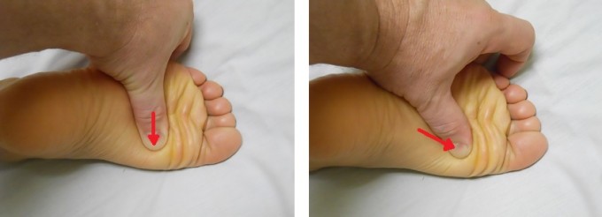
- As we mentioned above, the examination of the plantar fascia revealed moderate tension all the way to the heads of the metatarsal bones, which form the ball of the foot (left picture) when strictly vertical pressure (red arrow) was applied to the fascia at its insertion to the head of a metatarsal bone.

However, when the angle of pressure was changed (red arrow) to the direction of the metatarsal heads (right picture), the patient practically jumped off the table and she shouted that this was exactly the pain she felt; no one until then had been able to find its exact location.
This finding means that the plantar fascia had lost its elasticity and by itself doesn’t trigger pain sensations, because it is partly scarred. The pain she felt in the ball of the foot came from the periosteal trigger points that had developed in the heads of metatarsal bones and that was the real cause of her pains.
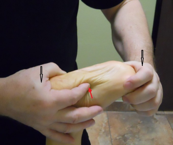
- Direct application of pressure along the plantar fascia didn’t trigger severe foot pain. As shown in the first picture, great toe extension exposed the plantar fascia. Black arrows indicate the points of pressure. Out of professional curiosity, we decided to examine the part of the plantar fascia which is normally inaccessible. Using the tip of the index finger (picture below) we got on the inner surface of the plantar fascia and applied very moderate pressure vertically. This examination triggered another scream of pain from the patient.

This finding means that while the outer layer of the plantar fascia completely lost its elasticity, the inner layer of collagen fibers is still functioning. The pain which comes from the foot itself and heel is the result of this inner tension.
We recommended that she stop wearing orthotics, since, according to the patient, they hurt her feet more than they helped. We asked her to buy Dr. Scholl’s gel inserts for $10, gave her a set of home stretches, forbade ice application which she constantly used as recommended by her podiatrists, and encouraged her to do Epsom salt foot baths two times per day.
With regard to the treatment protocol, we used the standard protocol for Plantar Fasciitis from the Medical Massage Library, but we modified it to be more effective for this particular patient. We concentrated on the part of the plantar fascia just at its insertion into the metatarsal heads and gradually applied the Periosteal Massage on the metatarsal heads (see picture above for the direction of friction and compressions).
Another modification was work on the inner layer of the plantar fascia. We used the same trick we applied during the examination. Using the tip of the index finger, we penetrated as far as possible under the fascia from the medial edge of the foot along the inner foot arch and worked along it.
The switch to gel inserts gave the patient relief just after the first session. We used 5 sessions of medical massage before giving her a week’s break. We saw her today. For the first time in 4 years, she got overall pain relief of approximately 70%. Next week, we plan another push of 5-6 sessions to make the results clinically stable.
LESSONS
- Pay attention, but never use someone else’s ideas as a guide. If the patient is in your office, it means one thing: people before you failed. Don’t follow their ideas even if they have more credentials than you. Otherwise, your therapy will be just another failure in a line of previous failures. Find out what was done to exclude it from your examination and therapy
- Spend time examining the area and tissues. You may be surprised how much they have changed, especially if the patient has chronic somatic abnormalities
- Stop extensively recommending ice therapy in cases of chronic somatic pathologies. It is true that ice blocks the pain-analyzing system, but it also blocks the results of your therapy. Showers, hot baths and Jacuzzis followed by passive stretching are the only correct supportive therapy at home.
Ice is supposed to be used in cases of acute trauma (first 24-48 hours), acute inflammation (e.g., rheumatoid arthritis) and severe nerve pain (i.e., neuralgia).
Category: Blog , Medical Massage
Tags: JMS 2015 Issue #1
