
This clinical case is exceptionally well thought through, with beautifully orchestrated treatment. Gerry Ivanov, LMT, from Switzerland, was able to solve an extremely difficult and seemingly unsolvable clinical case by integrating various techniques and modalities of somatic rehabilitation and attacking the patient’s difficult situation from several directions. Also, notice how cleverly and patiently he increased pressure on the patient’s body, slowly building up the clinical response.
Although Medical Massage was the cornerstone of the entire treatment process, we decided to name this clinical case ‘Integrative Therapy’ rather than ‘Medical Massage’.
Gerry greatly illustrated for us how important correct integration of techniques and modalities is if therapists would like to reach stable clinical results in a relatively short time.
INTEGRATIVE THERAPY vs. SEVERE SPONDYLOSIS AND FUNCTION LOSS
The patient is a 61-year-old male who owns a home-based business. For almost three years now, he has had acute pain in the lower back, which greatly affects his range of motion and everyday activities. During these years, the patient was unsuccessfully treated with medications and physical therapy in several hospitals and clinics in Moscow and Switzerland.
EVALUATION
Complaints During First Visit:
Intense bilateral pain in the lower back, general weakness in both legs, especially the right, numbness in the left hip and anterior-lateral thigh. His sleep pattern is greatly affected, and the patient is physically and emotionally exhausted from pain and lack of night sleep.
Clinical Evaluation:
The patient exhibited a complex clinical picture which indicated severe irritation of the L1-L5 lumbar spinal nerves and lumbar plexus. Measurement of his legs indicated that the right lower extremity is 1.6 cm (about half an inch) shorter due to the pelvis tilt.
Examination of the cutaneous (i.e., skin) reflex zones indicated the presence of sensory deficit (paresthesia or ‘pins and needles’) along the left iliotibial band and leg, all the way down to the top of the foot. It indicated irritation of the sciatic and common peroneal nerves on the left side. Dr. Solomon’s compression points to test for possible sciatic nerve irritation were positive on both lower extremities.
Examination of the reflex zones in the skeletal muscles indicated the presence of active trigger points in the quadratus lumborum muscles on both sides and in the gluteus maximus and piriformis muscles.
Further testing indicated the presence of significant muscle weakness in the iliopsoas, gastrocnemius, extensor hallucis longus, and tibialis anterior muscles.
The patient’s biceps femoris was so hypercontracted that it was simply impossible to perform Lasegue’s Test to examine the degree of sciatic nerve inflammation. Tension in the biceps femoris greatly contributed to inflammation and adhesions formed along the sciatic nerve.
Thus, the patient exhibited a motor deficit within the sciatic (common peroneal and tibial nerve) distribution as well as the femoral nerve distribution.
In the MRI report, the radiologist described advanced spondylosis with disc herniation at the L3-L4 level; disc protrusions at L2-L3, L4-L5, and L5-S1; a synovial cyst of the left transverse joint at the L3-L4 level; and multiple Schmorl’s hernias at levels T12-L2. Degeneration of intervertebral disks caused deformation of the dural sac and compression of the cauda equina (or bundle of L1-L5 spinal nerves).
Considering the complexity of the clinical picture and the lack of positive progress with conservative therapy, the only solution offered to the patient was spinal surgery.
TREATMENT
A radiologist’s report as well as our evaluation confirmed a very difficult and complex clinical case. The patient was unsuccessfully treated in various clinics and hospitals, but his treatment history indicated that all therapies were done with very limited sets of clinical tools. Thus, it was obvious that the integration of methods and modalities of somatic rehabilitation was missing.
I decided to give the patient a last chance with conservative therapy and to customize an integrative treatment protocol. My idea was to carefully combine clinical modalities and slowly increase the complexity of the therapy in the affected area while the patient was going through the treatment process.
Here is the treatment protocol I designed for the patient:
- Medical Massage in an inhibitory regime starting from lower extremities. The goal is to release the tension and rebalance the actions of large muscle groups. Strokes were directed laterally and proximally.
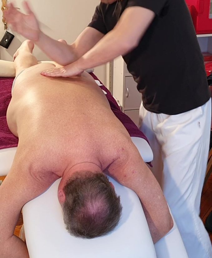
- Acupuncture – L5-S2 at periosteal depth, bilateral, and also in Vastus Lateralis, Iliotibial Tract, Gluteus Maximus, Gluteus Medius, Piriformis, Quadratus Lumborum.
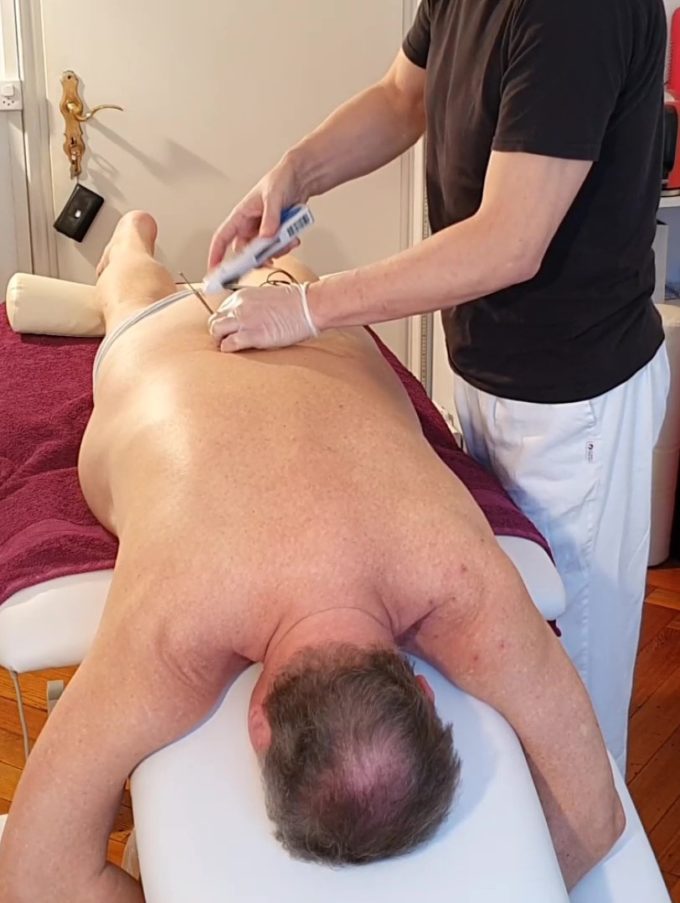
- Trigger Point Therapy massage in Quadratus Lumborum, Piriformis, Gluteus Maximus, Gluteus Medius, Biceps Femoris, Semitendinosus and Adductor muscles.
- Myofascial Release in the thoracic and lumbar areas.
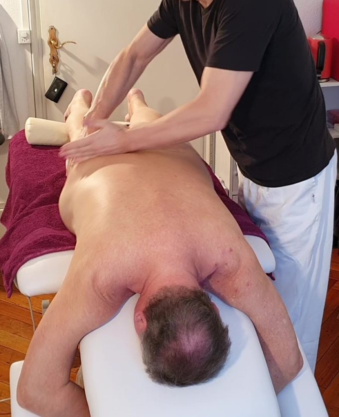
- Radial Shockwave 10 and 15 Hz, 2.5-4 bar administered on Biceps Femoris, Semitendinosus, Adductors group, Gluteal group, and Sacrospinalis.
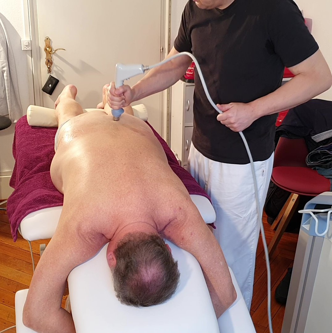
- Sacroiliac and Hip joints stretching.
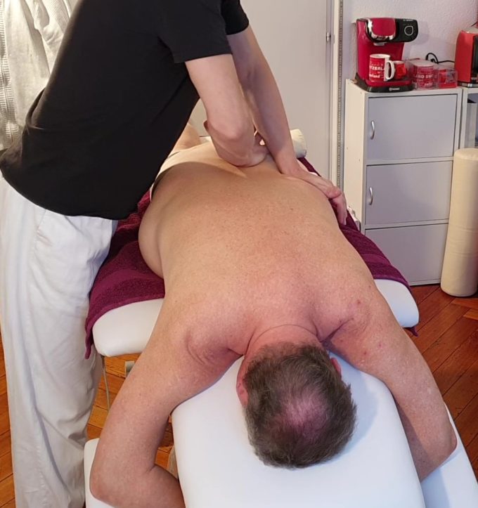
- Stationary and Mobile Cupping paravertebrally along the lumbar and thoracic spine and both legs.
- Electric Vibration along the thoracic and lumbar spine to additionally control the pain-analyzing system.
- Low-level Laser Therapy administered to all branches of the L3-S1 spinal nerves.
- Pressure release in the lower back using PIR (Postisometric Muscular Relaxation) and tractions in the lower back and lower extremities.
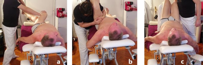
a – PIR for piriformis
b – PIR for hamstring muscles
c – Perl’s Traction
- At the end, I applied bee venom cream to all areas of pain.
I also suggested taking immunotherapy support formulas: INFLAM, ARTH and OSTEO-N.
Even after the first session, the patient reported a short period (7-8 hours) of being pain-free! Very carefully, from session to session, I increased the intensity of the Medical Massage protocols, the power levels of the Radial Shockwave, and the degree of laser energy. I also started gentle massage along the Valleix points associated with the sciatic nerve, from the ischial tuberosity all the way to the popliteal fossa.
These first 7 sessions were spent preparing the patient for the application of the full power of Medical Massage in the form of Segment-Reflex Massage protocol for Thoracic and Lumbosacral Spondylosis. The techniques I used, in sequence:
Pulling and rolling a fold of skin in the lower back in the directions of Connective Tissue Massage to eliminate adhesions below the skin and decompress the superficial fascia. Sawing friction in the cranial direction.
Kneading, stretching and relaxing the paravertebral muscles. Electric vibration. Friction between spinous and transverse processes of the lumbar vertebrae.
Spiral frictions along the sacrum, insertion of gluteal muscles into the sacral edge and SI joint, into the greater trochanter and along the muscle fibers. Kneading of gluteal muscles, vibration and shaking of the pelvis.
Electric vibration with medium intensity, kneading in the inhibitory regime, and stretching along the sciatic nerve. Weakened muscles in the thigh and leg were stimulated with various friction, compression, and percussion techniques.
One of the major problems for this patient was very weak iliopsoas and anterior thigh muscles. Thus, according to Sherrington’s law of reciprocal inhibition I concentrated on the hamstring muscles first to inhibit their tone and as a result to stimulate activity of iliopsoas and quadriceps muscles.
After the 9th session (with two consecutive sessions of SRM), the numbness along the left thigh completely disappeared, the numbness and weakness in the right leg became very mild. The patient regained muscle power and his gait normalized. For the first time, he was able to easily go upstairs. He stopped having pain at night and his sleep pattern completely normalized.
Currently, I continue to work on the patient to eliminate the residuals of spinal nerve compression, but spinal surgery is not even an option anymore.
ABOUT THE AUTHOR
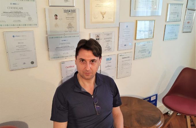
Gergan (Gerry) Ivanov, LMT
Gergan (Gerry) Ivanov graduated from the National Sports Academy in Sofia, Bulgaria and in 2007 he obtained a Master of Science Degree from University Paisii Hilendarski Plovdiv, Bulgaria. Presently he studies at The French Institute of Micro-immunotherapy.
Gerry is certified in the application of a great number of therapeutic modalities from basic massage therapy and Sports Massage to Medical Massage methods and techniques as well as laser therapy, shock wave therapy, acupuncture, reflexology, nutrition, phototherapy, etc.
Gerry worked in clinics and hospitals in Bulgaria, France, and the Netherlands. He currently lives and works in Geneva, Switzerland. He is a member of the International Society of Physical Medicine and Rehabilitation.
Category: Case Studies
Tags: JMS 2020 Issue #2
