
We thank everyone who commented on the original post. Those of you who mentioned Cauda Equina Syndrome were absolutely correct, and we appreciate your clinical expertise.
The patient is a 52-year-old male who had no previous health issues and owns a small windshield replacement company. As we mentioned in the original post, he felt extreme electric-shock pain in the right lower back and the entire lower extremity after he lifted a heavy windshield. Excruciating pain of high intensity brought him to his knees and caused a complete drop of the right foot and a sudden urge to urinate, and he was almost immediately taken to the emergency room.
This patient presented the worst-case scenario of disk problems. Instead of disk bulging or even herniation, the body of the L5 disk was sequestered and part of it was slightly displaced to the right, severely compressing the L5 and S1 spinal nerves and creating a very complex clinical picture of Cauda Equina (a.k.a. Horse’s Tail) Syndrome.
In these cases, besides severe lower back and lower extremity pain, the patients complain about abnormalities in the function of the pelvic organs (delayed or frequent urination, constipation, erectile dysfunction, etc.). Another aspect of Cauda Equina Syndrome is the frequent presence of vasomotor abnormalities. It is the cause of the patient’s peripheral edema in the right leg and foot, which was presented in the original post.
The presence of peripheral edema on the affected side means that compression of the spinal nerves is so severe that it blocks the two-way traffic of signals between the CNS and the tissues and organs. Compression and inflammation blocked the ascending sensory flow to the CNS, and it triggered the severe pain and dysfunctions that the patient felt (pain, numbness, burning, etc.). The same compression affected the descending motor output from the CNS to the skeletal and smooth muscles, and they lost their normal tone.
The decrease in the descending flow of motor signals weakened the skeletal muscles, and it impaired the normal function of the muscle pump responsible for proper drainage. At the same time, altered motor output weakened the smooth muscles in the walls of the veins as well, causing their dilation. Failure of the muscle pump and dilation of the veins are the two main mechanisms of this patient’s peripheral edema. The same impact on smooth muscle originally relaxed the sphincter of the urinary bladder, causing urination.
Cauda Equina Syndrome requires immediate surgical decompression due to the possible failure of the pelvic organs. The problem was that the patient did not have medical insurance, and in the emergency room he agreed only to an epidural injection, which brought him relief. While he was trying to figure out medical insurance issues, he came to our clinic to get some additional help.
The complexity and intensity of the clinical symptoms required a gradual multi-disciplinary approach to his rehabilitation. While the patient continued to take his medications, we extremely carefully started therapy with a gentle application of the Medical Massage protocol for Lumbalgia without provoking even the slightest discomfort. Initially, we concentrated on the inhibitory regime of therapy to balance the autonomic nervous system and suppress overactivity of the pain-analyzing system. Later on, we added more sophisticated modalities, arranging them in order to better use the ‘lateral shift’ principle of Medical Massage. At the same time, the patient started gentle exercises in the swimming pool and passive stretching as homework.
The Medical Massage protocol itself consisted of the gradual addition of new modalities each time the patient was ready for them and decompression of the soft tissues in the middle and lower back and the sacral area on a layer-by-layer basis (skin, superficial fascia, lumbar erectors, middle fascia, QL muscle, deep fascia, rotators and finally periosteum at the insertions into spinous processes, lower ribs, iliac crest, sacrum and SI joint). The local therapy on the foot and leg used the same strategy but was added later, when clear improvements in the symptoms and restoration of function were observed. At the beginning, we only maintained local drainage using LDM.
During the first 2 months (2-3 sessions per week), the patient slowly but steadily improved. At that point, we added acupuncture, gentle manual therapy (without any adjustments) by a DC, a TENS unit along the sciatic nerve, passive stretching at home and, recently, very careful application of the inversion table.
We got so far that the neurosurgeon who recently consulted the patient did not even consider surgery anymore, while continuing to monitor his condition on a monthly basis. Peripheral edema started to dissipate first. Fig. 1 illustrates the patient’s feet before the start of conservative decompression and after 6 weeks of therapy.
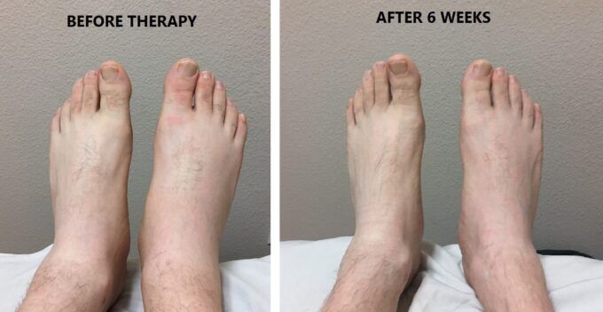
Very light edema in the right foot is still visible, as is weakness of the big toe.
The patient entered our clinic still in pain and requiring support to move about. His right lower extremity was very swollen, and he continued to have to drag his right foot. Unfortunately, we don’t have a video of the patient at the beginning of therapy, since it would have been unethical to ask for his permission at that time. The video below was recorded following 4 weeks of therapy after which the patient showed significant improvement and gladly gave us permission to record his improved but still abnormal gait. Notice that for the patient to walk steadily, he still needs to bend his right knee to elevate the weak foot to avoid stumbling. Peripheral edema in the entire right lower extremity is still visible. We repeated the same short video 3 times.
The video below was recorded this week. The patient’s gait is normalized, and he is no longer bending his knee in an unnecessary way. The patient is able to maintain the transition of his foot from one stroke to the next. There are no visible or palpable signs of peripheral edema. At this point, the patient shows only residual weakness in his right foot’s ability to support his gait.
Currently, the patient doesn’t have any original symptoms besides residual weakness of foot extension and weaker toe elevation. Now he can lift all four toes on the right foot, with the big toe starting to jerk only this week, which is a great sign.
Successfully treating Cauda Equina Syndrome with a combination of somatic modalities, with Medical Massage as the backbone of the therapy, once more proves the exceptional clinical value of Medical Massage Therapy for patients even in dire situations.
As was correctly pointed out in several comments on the original post, the patient was in a very difficult situation. His treatment must be approved and done in cooperation with the patient’s physician, and the therapist must have very intensive training and personal clinical experience. For many patients with Cauda Equina Syndrome, spinal surgery is the only solution. However, if the therapist has enough clinical expertise, it is always worth it to try conservative therapy first.
LESSONS
- You may see smaller peripheral edema in less severe cases of Sciatica. For Sciatica patients, you have to test soft tissues for hidden edema by applying mild compressions along the anterior foot and tibia. If even a small depression stays in the tissue, it is a sign of hidden edema, and, generally speaking, you can’t expect a very quick recovery, since you are dealing with significant irritation/inflammation of the spinal/sciatic nerve. It takes time for the nerve to recover. The presence of any visceral symptoms accompanying lower back pain (for example, constipation) is another red flag of severe or chronic spinal nerve irritation.
- The encouraging factor in this case was the significant relief of symptom intensity by the epidural injection. Usually, it gives very limited help to patients with Cauda Equina Syndrome. Our theory of why conservative therapy worked is as follows: the disk was already compromised, but the brain was able to keep it in a compensatory state – he lifted heavy windshields from his truck – the already compromised disk fragmented by the gravity and compression forces (at that point he entered a decompensation state) – the first reaction of the brain to the alarm from the damaged disk is protection – the brain placed the lumbar erectors, quadratus lumborum, iliopsoas and rotator muscles into additional spasm to prevent any additional displacement of the disk – combination of fragmentation and excessive protective reaction triggered Cauda Equina Syndrome – epidural injection decreased initial protective reaction and the brain re-evaluated the situation and realized that despite fragmentation being present there is no significant shift of the fragmented part of the disk – gradual elimination of protective muscle tension, restoration of normal mobility between layers of the soft tissues and decompression of the spinal segment by the therapy and water exercises eventually released pressure within the L5 segment and freed the spinal nerve.
- At this point, the patient has residual effects of the initial nerve damage, which we hope will fade away. If he continues his homework and stays away from lifting, there is a very good chance that the fragmented part of the disk will heal, but it is going to take time.
- It takes time for the injured nerve to recover and it also takes time for the therapy to work. Both therapist and patient must be patient. Sometimes for several sessions nothing happens, and it seems that therapy has reached a plateau. In reality, the gradual and careful building up of a clinical response pays off by suddenly giving another improvement.
- Accumulation of clinical expertise allows you to move from local therapy where the symptoms are (edema in this case) to the identification and elimination of the actual cause. Otherwise, be very careful with disk abnormalities, especially Cauda Equina Syndrome.
Category: Blog
Tags: JMS 2020 Issue #3
