
This clinical case is unique and extremely complex, and its description is long. However, we ask therapists interested in the clinical application of massage therapy to finish it because nothing can better illustrate the clinical power of Medical Massage, which most patients don’t have access to.
Our clinic works on complex patients referred by medical clinics and hospitals, with no other viable clinical solution offered for many of these patients. Eventually, we helped the majority of them partially or decisively. Having said that, I would like to state that the clinical case you are about to read now, from our perspective, beats the complexity of patients in our clinic.
I tip my hat to our former student Mike Devo for his professional expertise and dedication to his patients. We are happy when our former students fully grasp the clinical flexibility of the Medical Massage concept. After learning all the necessary basics, they start to develop their treatment strategies using the tools and techniques we offered them. They don’t feel afraid to face tremendous professional challenges anymore while delivering stable clinical results, beating all odds. Helping therapists acquire such knowledge and clinical expertise justifies our educational efforts! Patients desperately need more therapists like Mike Devo and our current and former students.
CASE STUDY OF SOMATIC DISASTER DUE TO MULTIPLE OPEN CHEST SURGERIES
I was first introduced to this patient in November of 2019. Her father is a long-time patient of mine, and he was hopeful that I could somehow help her. The patient is a 41-year-old female.
In 2005, at the age of 26, she worked as a firefighter/EMT. On 12/29/2005, she suffered a SCAD event (Spontaneous Coronary Artery Dissection), a tear in the Left Anterior Descending (LAD) coronary artery. The tear clotted as she was misdiagnosed and sent home, but this delay caused a massive Myocardial Infarction (Heart Attack) in the LAD territory. Due to her training as an EMT, she knew that the situation was serious, and she rushed to the hospital. Unfortunately, a non-viable stent was put in the LAD during emergency surgery. That means the stent was defunct upon installation, and there was already a 23-hour delay after the initial episode. In December 2006, one year later, she was diagnosed with Class D End-Stage Heart Failure/Left Ventricular dysfunction and Cardiomyopathy. The Ejection Fraction was approximately 22% (normal cardiac ejection fraction is 60-75%), and she was put on a heart transplant list.
In 2006 the patient began intensive (twice a week) treatment with a PTSD therapist and continued until 2016. That greatly supported the patient through challenging times.
In May of 2007, cardiac catheterization revealed a terminal aneurysm (critical enlargement) in the LAD. EF (ejection fraction) dropped to 16% at that time, and she experienced multi-system organ failure. There was no longer time to wait out the long waiting list for a transplant.
In June of 2007, the first open-chest cardiac surgery was done in lieu of a transplant: Sternotomy and Left Ventricular Resection with Remodel using Ventricular Mannequins. The patient survived! (see procedure video in attachment).
In September of 2007, she was diagnosed with sternal instability with Chronic Non-union. In other words, both fragments of her sternum, which was cut during the cardiac surgery, didn’t unite to heal correctly.
In November of 2007, the first revision of her sternal non-union was done with a repeat sternotomy and bone debridement (removing non-unioned and inflamed sternum bone), bone grafting, and sternal rewiring.
Due to repeated non-union, in May of 2008 a third sternotomy was performed with titanium rigid fixation plates installed to replace the entire sternal body. Despite that, she continued to suffer from anterior chest pain due to sternal instability with chronic bilateral clavicular dislocations. Each episode was excruciatingly painful for her.
In December of 2010, a pacemaker/defibrillator was installed to control her cardiac rhythm.
In January of 2014, the patient got a service dog, Arthur. He helped the client get up from a chair or off the floor. Due to multiple surgeries and chronic pain, the client was last employed part-time in 2008.
In March of 2016, she was diagnosed with Thoracic Outlet Syndrome, which most likely developed due to sternal instability. The surgeon offered a third sternal repair via muscle flap, but this option was denied due to possible side effects for the heart transplant. She was sent to 16 physical therapy (PT) treatments, and after having no significant improvement, she was referred to the thoracic surgeon again.
In June of 2016, the titanium fixation plate which held the sternum was removed. However, the star plate, which replaced the upper part of the sternum (called the manubrium), was left in place since it was wired to her clavicles. At this point, there was still very little fusion along the sternotomy line, and both sternal fragments were held together only by scar tissue and cartilage. Thus, there was open space in the middle of her chest where the sternum body once was, and only skin and scar tissue separated her heart, mediastinum, and lungs from the outside environment. Ten weeks after the surgery, the patient was referred for and completed a new 16-treatment round of physical therapy—no significant improvement.
In 2017, the patient was sent through a third round of PT with the same results.
Out of desperation, the patient found a clinic that specializes in deep tissue therapy, believing that such treatment could solve her chronic somatic pains. While the treatment helped with general pain intensity, the patient started to have acute muscle spasms in the posterior side of the pacer pocket, and it triggered unnecessary pacer activation. Within an hour after pacer activation, her entire upper body on the left side would be “locked” up with tension, sometimes with left clavicle dislocation.
From 2016 to 2019, the client’s Ejection Fraction fluctuated between 30% and 40%. Below 35% is considered active heart failure. As of Feb. 2019, the client’s EF was at 38%, and she was on a heavy daily medication list.
EVALUATION
Complaints
The patient’s initial evaluation took quite a while, and the client was happy I did not run out of the therapy room while she was telling me her medical history! On the contrary, I was excited to work with her and felt we could get her out of the chronic pain situation. Her main concerns were:
- She felt chronic pain, weakness, and numbness in both arms/hands.
- On either side, the clavicles tend to “pop out” at the acromioclavicular (AC) joint. That was excruciatingly painful for her, and these episodes can sometimes last for days.
- The pectorals are tender and swollen from all the trauma incurred. The patient could not lie on her side due to her missing sternum and chest instability.
- Stress from all of the above and lack of sleep.
Clinical Observation
The patient exhibited shallow, frequent breathing patterns. There was bilateral edema in the hands, arms, and pectoral areas. The edema was more pronounced on the left, especially around the “pacemaker pocket.” ROM in both shoulders was very restricted. The presence of edema is a direct sign of drainage failure due to the compression of the venous and lymphatic structures or incorrect innervation of the upper extremities.
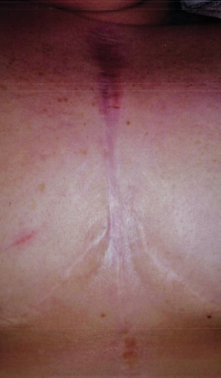
Her posterior and anterior cervical muscles became so weak that she struggled to keep her head up for any length of time. There is a long postsurgical scar in the middle of her chest with overstretched scar tissue. It means that the scar tissue is under significant stress trying to keep the chest stable during each inhalation. Fig. 1 illustrates the postsurgical scar. Notice the absence of the ridged scar, which is now pulled apart by respiratory efforts. Two half-circled scars visible in the picture are the result of breast-reduction surgery done years before the patient’s health issues started.
Palpation
- Wartenberg’s Test, which indicates the degree of brachial plexus compression by the anterior scalene muscle, was negative.
- Wright’s Test, which examines possible compression of the brachial artery and brachial plexus by the pectoralis minor muscle, was positive.
- There was a significant spasm in the Pectoralis Minor and Major muscles with multiple active trigger points.
- Tingling sensations (or Paresthesia) along both thumbs and forefingers indicate irritation of the radial nerve. Referred Pain and itch sensation along the C6-T1 dermatomes revealed ulnar nerve irritation on the left. The Sensory Test was positive for both nerves, indicating significant sensory deficits.
- Lack of pincer grip indicated the presence of a motor deficit.
- Skin discoloration along the area of the missing sternum body indicated proliferation of the scar tissue and its replacement of the normally structured skin along her sternum.
- The patient noticed that her period always worsens the pain and edema.
No issues with low back, hips, or legs. No swelling in legs or feet.
PRE-TREATMENT CONSIDERATIONS
First of all, I needed clearance from the patient’s physician to start medical massage therapy. The patient’s doctor encouraged the attempt since all other treatments had failed.
I reviewed the Medical Massage protocol for Cardiac Disorders from Medical Massage Volume 2 (by Dr. Ross Turchaninov) to get therapy guidelines. The treatment should consider that the stroke speed should be slow to moderate. No Trigger Point Therapy using ischemic compression since the activation of the pain-analyzing system should be avoided. Use gentle kneading and moderate friction instead, especially on the fingers and toes. No Post Isometric Muscular Relaxation or Muscle Energy techniques to prevent an increase in the peripheral vascular resistance. Use passive stretching only. Treating cardiac patients requires the “long game” approach with constant feedback from the patient.
Since her heart was not working at full capacity, I had to consider these recommendations when developing my treatment strategy. Although the client came in with an already established diagnosis of Thoracic Outlet Syndrome (TOS), my evaluation showed that she had a combined clinical picture of Pectoralis Minor Muscle Syndrome and Pectoralis Major Syndrome rather than TOS. Both syndromes will trigger an almost identical clinical picture since the brachial plexus can be equally affected in both conditions. However, the negative Wartenberg’s Test ruled out TOS.
I considered Pectoralis Minor Muscle Syndrome the main problem because the attachments of the Coracoid Ligament contributed to the patient’s history of clavicular dislocations. The tension in the pectoralis minor muscle affects the brachial plexus, and the brain triggers the tension in the pectoralis major muscle as a secondary protective reaction. Combined tension in both pectorals restricts ROM in the shoulder joints. Also, the pectoralis major muscle is the only muscle in the upper limb supplied by all five parts of the brachial plexus.
Thus, the patient exhibited multiple areas of peripheral nerve irritation combined with excessive postsurgical local trauma to the chest. The recurrent surgical chest trauma endured by my patient has wreaked havoc on her somatic and neurological structures.
TREATMENT
My plan for Nov 14, 2019 to Jan 2020
I started the first testing session with Lymphatic Drainage for the upper body using the very gentle Thoracic Pump technique, followed by Supraclavicular Pump techniques bilaterally. I did a visual assessment and took a photo of the sternum. During the 20-minute session, the client’s breathing was good and steady, and overall my first interaction with the patient’s body was positive and pleasant.
For future treatments, I suggested twice-weekly 45-minute sessions. Each session started with LDM to reduce any soft tissue swelling and relax the patient, balance the autonomic nervous system, and maintain proper breathing. In the first few sessions, I started with the posterior neck as the origin of pectoral innervation. I used medium pressure on the trapezius and paravertebral muscles, applying the Relaxation of Cervical Paravertebral Muscles Technique.
After that, I focused on the pectoralis major and minor muscles, working on their attachments using light to medium touch. Extensive scar tissue had formed in the area along the missing sternum and cartilaginous parts of the upper ribs. I used light friction along and across the edges of the scar.
Gradually I would increase time and pressure while working on the pectorals. Considering the complexity of the patient’s situation and the fact that the pain-analyzing system was already compromised, I replaced Trigger Point therapy in active trigger points with slow kneading in the inhibitory regime and careful Aligned and Circular frictions. In the same way, I would also work on the Deltoid, Latissimus, and Intercostal muscles.
Gradually I added work on the anterior neck, including her scalene and Sternocleidomastoid (SCM) muscles and elements of the Carpal Tunnel protocol for hand, wrist, and forearm. I used passive stretching as well.
During the first five weeks, we experienced as many setbacks as advances, including a couple of clavicular “pop-outs.” Several pacemaker activations triggered upper back pain with alternating pain and numbness in either arm/hand. I steadily continued to push forward, constantly correlating the patient’s sensations with the duration and intensity of the therapy.
Not until six weeks did we arrive at the point when the patient felt no pain or numbness in either upper extremity. She started to do short walks now. That was a big step forward for her.
From the very beginning, I told the patient that we were playing the long game here, and she gladly agreed since she started seeing real progress in her condition for the first time.
Jan – March 2020
I re-analyzed our situation and concluded that the next step in the treatment was clavicle stabilization to decrease the painful clavicular “pop-outs.” The star plate, which replaced the sternum’s manubrium, was wired into the clavicles, immobilizing them. I thought that this lack of mobility was the direct cause of clavicular instability. Thus, I saw my immediate goal as addressing the soft tissues around the clavicles’ attachments to reduce tension and make the attachments more flexible and elastic to give clavicles some room to give. I didn’t expect to eliminate the possible “pop-outs,” but I hoped to make them less painful when the clavicle slipped in and out.
While focusing on the Pectoralis protocol, I introduced more scapula passive ROM, including some light traction. The aim was to lengthen the Coracoid ligament attachments. I also added light repetitive frictions at the clavicular insertions. Finally, I added elements of the Medical Massage Protocol for Sternocleidomastoid (SCM) bilaterally to take pressure off the sternal end of both clavicles.
Another aspect I added was working on the posterior cervical muscles. I started careful and gradual passive stretching of the posterior cervical muscles. After that, I used Sherbak’s friction on the spinous processes of C6-T7 to take pressure off the superficial fascia and stimulate and decompress cutaneous branches of C6-T7 spinal nerves. All these curtailed most of the referral of neurological pain to the patient’s back. Next, I worked along T1-T7 intercostal spaces on both sides to decompress intercostal muscles and, consequently, intercostal nerves. I avoided the side position during the treatment due to the thoracic cage instability. This treatment immensely helped the patient restore proper depth and rhythm of respiration. The increase in blood oxygenation gave skeletal muscles enough ATP to fuel more powerful contractions.
Every four sessions, I would add a lower-body stress management massage to encourage full-body homeostasis. During this time frame, the setbacks started to be less frequent. Recovery time after the previous session was quicker. There were two days when the client took nitroglycerin for the cardiac pain. Those episodes did not cause any soft tissue setbacks, however. The patient started to take longer walks since her respiration greatly improved. I felt encouraged to tell her that she would get back into the workforce again one day.
March 17 – June 10, 2020
No appointments due to the COVID-19 pandemic. Due to her already compromised health condition, we stopped treatments out of precaution. During this time, the patient moved into a new apartment and continued lengthy walks with her service dog.
June 11 – Dec 31, 2020
The patient moved further away from my office, so we changed to weekly sessions. Remarkably, after a ten-week absence of therapy, the patient did not have much of a setback. Yes, the pectorals and both arms fatigued quickly, and the referral of nerve pain to the back had returned. However, her latest concern was pain in the “pacemaker pocket.” It felt almost like it had moved or pulled out.
In July, the patient’s “cardiac cocktail” meds were updated, causing a drop in blood pressure, and in the first week of August, she fell when walking her dog. Due to hyperextension trauma, the patient suffered a right wrist fracture and strain of metacarpophalangeal and interphalangeal joints of the third, fourth, and fifth fingers. She wasn’t put in a cast but used a hand brace for four weeks.
When the brace was removed, we added some treatment to address her right wrist and hand to help with the ROM restoration. The hand and wrist have healed nicely in a month with some lingering trigger finger in the fifth finger.
The client had a flare-up of Plantar Fasciitis and mild leg Anterior Compartment Syndrome in October. That was a good sign because this resulted from the increase in the distance the patient had been walking.
As I mentioned above, in 2019, her Ejection Fraction (EF) was approximately 35-40%, but in October 2020, EF rose to 50%, which classifies her as “recovered” with “reduced function” rather than in the “heart failure” classification. This was fantastic news! It was the first time she had reached that EF number since 2005. There was no doubt that her gradual increase in walking played a critical role in the recovery of cardiac function. She was able to do that only after MM could control the intensity of her somatic pain and restore proper respiration.
We continued the same treatment strategy through December 2020. She noticed occasional pain flare-ups and pectoral tension around her menstrual cycle. To my complete surprise, the painful clavicle “pop-outs” were almost completely gone, as were the numbness and weakness in both hands. The strength and endurance of her hands, forearms, and shoulders have increased significantly. The strength in all cervical muscles has improved, allowing the patient to hold her head up for longer lengths of time. The patient was able to make some stencil ornaments for Christmas that year. Even a year ago, that would not have been possible.
Recently the patient started to complain about tension and discomfort in the pacemaker area. It almost feels as though the device shifts inside the pocket. I have introduced light massage with drainage and circular frictions surrounding the “pacer pocket” soft tissues. She has commented that there has been significant improvement in that regard. In 2022 her cardiologist plans a replacement of the old pacemaker, and the new model will be smaller and less invasive to the soft tissues.
In 2021 we reduced our treatment sessions to twice a month and eventually once a month as supportive therapy. The hope for 2022 is that she will be able to return to work—at least a part-time position to start.
Below is a link that describes the LAD remodel procedure: Myocardial reconstruction in ischaemic cardiomyopathy.
P.S. You just finished reading an astonishing clinical case. It is not another successful treatment of Carpal Tunnel Syndrome or Chronic Headache. Using the principles and tools of Medical Massage that SOMI gave him, Mike saved the patient’s life. These are not empty words. Patients around the country need more therapists who can deliver such important and irreplaceable clinical expertise! If Mike slowly and gradually didn’t restore the patient’s somatic functions and via the reflex mechanism of MM therapy didn’t unload cardiac function of already weakened heart transplant it will fail in a short time and a very big chance that the patient’s life was over. Dr. Ross Turchaninov
Letter From the Patient
So to start: my diagnosis when I saw Mike for the first time was Chronic Post-Sternotomy Nonunion. Chronic Clavicular Instability.
The constant clavicular dislocations created chronic inflammation and swelling on and alongside the scar line in the middle of my chest. As a result of these dislocations, it was challenging for me to hold my head up and shoulders back. Movements in the neck and upper body were very restricted and painful. I didn’t have any strength to even use either arm without extreme pain and fatigue. My inhalation and exhalation were greatly limited. I could not walk any distance.
Since Mike conducted medical Massage treatment, I have only had ONE true episode of a complete clavicular dislocation since Nov 2019! Also, I had only one partial dislocation in the last 18 months, which corrected itself within 48hrs.
I have moments of extreme tension and pressure (what I would classify as “pre-dislocation” pain), but this became a rare occurrence with Mike’s treatment plan. Before Medical Massage therapy, typically, I would have had two true clavicular dislocations in a month, which would each render me bedridden for a few days at the minimum. I would have to hug a sternal pillow to apply deep pressure along the scar line to provide enough support in my upper body and chest to pull myself upright to stand or even lift my head.
Now, I walk several times a week, often averaging over 20k/week (in 2021). Now I am without significant incidence of intercostal spasms, sternal pressure, or sternal and clavicular pain – and without any of the symptoms which would have been previously exacerbated by even the minor activity of standing upright for extended periods.
Feeling BETTER AND STRONGER than I have since my first sternotomy in 2007, I have done much more than Mike has advised – specifically during my move in September. I pushed myself, lifted, hoisted, carried heavy boxes in ways that I’m sure are not consistent with continued improvement or healing. I often do too much because I feel so good comparatively, and I forget to be dutiful and careful. Or I do it anyway at the moment because I CAN in that one moment. I am sincerely trying to be more aware of proper body mechanics (as Mike suggested) while limiting over-use. Symptoms of Thoracic Outlet/Pectoralis Minor Syndromes have mainly been resolved.
Julie B.
ABOUT THE AUTHOR

Mike Devo, LMT, CMMP
For 29 years Mike worked as a delivery driver for UPS. This was an extremely physical job, and he had his own share of back, neck, and shoulder issues. He had always been interested in the field of massage therapy as a second career. In 2009 Mike completed massage school while still driving full time. It was during this time that he developed Thoracic Outlet Syndrome. He tried orthopedic physical therapy with no luck, so he sought out help from a chiropractor who also did massage therapy. After one session of releasing the scalene muscles, the TOS was 50% better. After three sessions it was completely gone. That was a huge ah-ha moment for Mike, and from that experience, he made it his goal to pursue a career in Medical Massage.
Mike’s next break came in a chance phone call to Boris Prilutsky, one of the true pioneers of bringing Medical Massage to the United States. This call led to Mike forming The International Medical Massage Association in an effort to bring therapists like Boris and others to Chicago to teach Medical Massage. This proved a little more difficult in reality than on paper. While organizing a successful class with Boris in Chicago was great, it took a lot of time to be a promoter instead of focusing on his real desire to be a therapist. It was through this relationship with Boris that Mike learned of Dr. Ross Turchaninov and his SOMI certification program. This was exactly what he was looking for. Boris and Dr. Ross use many of the same methods they brought with them from Ukraine. It was a natural progression for Mike. He found it to be very humbling to work, learn, and train with such experienced Medical Massage therapists. Mike hopes to use this knowledge and training to spread the word of Medical Massage and its power to heal. Someday Mike hopes to train and teach other therapists to carry on these methods.
Category: Medical Massage
Tags: JMS 2022 Issue #1
