
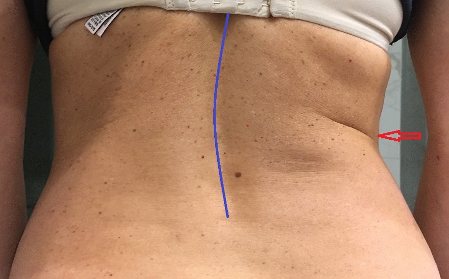
The original question was:
A patient came into our clinic with acute right-lower-back pain that amplifies during rotation, deep breathing, and sneezing. The lower back pain started after an excessive workout in the gym. There are no peripheral neurological symptoms. Lumbar compensatory scoliosis to the left (blue line on the photograph in the picture above), upper body tilt, and right pelvis elevation (red arrow on the photograph) are clearly visible. These structural changes are not improved when the patient is relaxed and lying face-down with a pillow underneath the belly. The patient had never had lumbar curvature before.
Considering the patient’s history and visual observation from the photograph, what likely triggered her acute lower back pain?
Thank you to everyone who posted their answers. It was great that the post generated such a wonderful professional exchange. We will get better together!
Let us summarize the most common suggestions:
- Spasm in Lumbar erectors
An acute spasm in the lumbar erectors may indeed trigger compensatory lumbar scoliosis. However, this scoliosis will more likely correct itself during sleep or while the relaxed patient is positioned on the stomach with a pillow under the belly.
- Spasm in the iliopsoas muscle
Yes, acute tension in the Iliopsoas muscle may create compensatory scoliosis, but the patient will bend slightly forward. Upper torso flexion is a way to decompress tension within the iliopsoas muscle. As seen in the picture, our patient didn’t exhibit a forward tilt.
- Spasm in serratus muscles
All serratus muscles belong to the upper torso muscles and don’t have attachments to the pelvis. In such cases, their tension, while triggering many discomforts, never triggers compensatory lumbar scoliosis. The same is true of rib subluxation.
- Disk pathology
Yes, a disk bulge or herniation will trigger the same clinical picture, and compensatory scoliosis will not correct itself with the patient positioned on the stomach. However, the patient must exhibit some peripheral neurological symptoms. Examples include sensory deficit (tingling or numbness in the lower back or lower extremity) or motor deficit (muscle weakness). Our patient didn’t have that.
- Pelvis rotation and elevation, short lower extremity, incorrect gait, etc.
All of that can be present in the patient, but these symptoms are not the triggers but rather the brain’s compensatory reactions to the trigger. Generally speaking, we should consider postural changes when evaluating a patient, but in many clinical cases, they are purely compensatory reactions used by the patient’s brain to deal with the initial trigger.
- Fascial Tension
Yes, every patient with LB pain will exhibit varying degrees of fascial tension and, in chronic cases, even superficial and/or deep fascia scarification. However, fascial tension is not a trigger but the result of adhesions formed between layers of soft tissues due to the inability of the tissues to freely slide along each other during regular movements.
- Inner organ pathologies
Yes, some patients with kidney colic, for example, may exhibit similar compensatory scoliosis. However, it is a mistake to treat somatic pain from the perspective of the visceral disorder alone. If there is suspicion of a possible visceral disorder, it needs to be examined and addressed medically first. At the same time, if the physician establishes a diagnosis of a chronic visceral disorder, the therapist may address the visceral disorder via reflex therapy or use visceral manipulations after this issue is discussed with the patient’s physician. Our patient had purely somatic pain without a visceral component. She was otherwise healthy.
- Scoliosis
We mentioned in the original post that the patient didn’t have lumbar scoliosis before the pain onset.
The therapists who answered spasm in the right Quadratus Lumborum (QL) muscle were correct.
Here are clues from the original post that therapists may consider when thinking about a possible acute spasm in the QL muscle:
- Lumbar pain formed after gym exercise that increased the respiratory rate.
- In patients with an acute spasm in the QL muscle, deep breathing and sneezing are agony because the QL is the primary muscle of respiration. Any tension there will be aggravated by deep breathing or sneezing.
- The absence of peripheral neurological symptoms makes disk pathology less likely to be the trigger.
- The main indicator of a possible QL muscle spasm is the presence of compensatory scoliosis, which doesn’t correct itself if the patient is comfortably lying on the stomach with a pillow under the belly.
The QL muscle is a main postural muscle that keeps humans upright. Animals that walk on four legs have the QL muscle in rudimentary form. The QL muscle is potent because it consists of three divisions (lumbocostal – connects transverse processes of lumbar vertebrae and the 12th rib; iliocostal – connects iliac crest and the 12th rib; iliolumbal – connects iliac crest and the transverse processes of lumbar vertebrae). This weird anatomy, in which muscle fibers cross each other within the same belly, prevents the QL muscle from relaxing if the muscle is traumatized or overworked. That is why compensatory scoliosis stays even during the patient’s sleep in a comfortable position. It is a clinical characteristic of acute QL spasm.
Patients with a severe QL spasm are in the emergency room, since it creates horrible pain. Interestingly enough, a very severe spasm in the QL muscle passes the stage of compensatory scoliosis, and it creates weird whole-body torsions, which patients cannot correct. Here are pictures of our patients with severe QL spasms. Eliminating a QL spasm of THIS intensity is an uphill battle:
[Image is no longer available]
Of course, the final decision on whether the patient has or doesn’t have tension in the QL muscle must be confirmed by further evaluation, which includes palpation and testing. Indeed, our patient’s palpatory evaluation indicated active trigger points in the lumbar erectors and BOTH upper and lower active trigger points in the right QL muscle. While palpatory examination of the active TPs in the lumbar erectors triggered a moderate ‘jump sign’, it was impossible to touch the lateral aspects of the patient’s right lower back above the iliac crest and below the 12th rib. As soon as these areas were palpated, the patient’s entire right lower and middle back started to cramp.
Here is our patient’s initial clinical picture, the image of the same patient at the end of the 2nd session of the Medical Massage protocol for the QL Muscle Syndrome, and the final picture after the 4th session. You may observe the gradual elimination of compensatory scoliosis and restoration of function and balance in the lower back.
[Image is no longer available]
The treatment strategy included: drainage; addressing connective tissue zones in each layer separately (skin, superficial fascia which covers lumbar erectors, deep fascia which separates lumbar erectors from QL muscle); Trigger Point Therapy separately for erectors and QL muscle; Postisometric Muscular Relaxation separately for the erectors and QL muscle; Periosteal Massage at the insertion of erectors into the sacrum, SI joint, spinous processes of lumbar vertebrae and PM at the insertion of QL muscle into the iliac crest; Homework stretches, light repetitive exercise in the swimming pool. This case is almost two years old, and the patient hasn’t had the same problem since. She regularly does the homework we suggested as a preventive measure.
Category: Blog
Tags: JMS 2022 Issue #2
