
This article, written by Boris Prilutsky, LMT, MA, has a very interesting history. Currently, Boris works with a group of scientists who examine the impact of Medical Massage on patients after concussion and with post-traumatic encephalopathy. At the same time, Boris hosted a professional blog in the online edition of Massage Magazine.
One day he decided to share with therapists some concepts of this therapy and preliminary clinical data obtained so far. To his and our surprise, several therapists who belong to the massage bureaucracy sent letters to the Editor of Massage Magazine, demanding that this information be deleted, since, according to them, it was outside the scope of the profession.
We in JMS regularly witness incredible reluctance on the part of the American massage bureaucracy to embrace, or at least keep an open mind about, Medical Massage. Some even support people who spread completely false fears that the clinical aspects of massage therapy are out of the scope of massage practice. While physicians currently examine and treasure Boris’s expertise in post-concussion treatment with Medical Massage, Massage Magazine gave in to the pressure and ended this important discussion.
We would like readers to judge for themselves if the information Boris shared with therapists is within the scope of massage practice. Here is the first part of his article.
MANAGEMENT OF POST-CONCUSSION SYMPTOMS AND POST-TRAUMATIC ENCEPHALOPATHY WITH MEDICAL MASSAGE. PART I
American football is a huge part of American life. It is impossible to imagine our country without the Super Bowl. However, football also has a very dark side associated with death and irreversible changes in the brain function of players.
Let’s look at the life of an average football player. From a relatively young age, he undergoes body and head collisions of varying intensity. One of the most dangerous consequences occurs during high school and college years when the brain is still going through the final stages of development and at the same time is actively loaded with information.
During these years, young athletes frequently experience so-called Repetitive Head Injury Syndrome (RHIS). In these cases, the young athlete continues to compete after what initially seems like a relatively mild head trauma. While doing so, he sustains repetitive mild concussions, which add up and create a slowly lingering clinical picture of RHIS.
As it was shown by Boden et al. (2007) 39% (!) of high school football players continued to play during the same season while still having residual symptoms of recent concussions. Later, these students, who in reality suffer from RHIS, are diagnosed with attention deficit disorders or tagged as “academically incapable.”
“The effect of multiple concussions over time remains significant and can result in long-term neurologic and functional deficits. These multiple brain insults are termed Repetitive Head Injury Syndrome” (Cifu et al., 2014).
While the professional football player continues his career, the intensity and violence of his head injuries increase. Let me give you one stunning example. Scientists from Boston University examined 79 brains of deceased NFL players from a brain bank and found that 76 (!) exhibited changes associated with Chronic Traumatic Encephalopathy (Breslow, 2014). These brains were donated by relatives or arranged to be donated by players before they died.
Boxing and Ultimate Fighting are even more gravely dangerous to athletes, while less alarming but still hazardous situations occur in ice hockey, soccer and other contact sports.
Finally, we are forever in debt to our veterans who come back home after being severely injured by improvised explosive devices (IEDs) in Afghanistan and Iraq. It was reported that 20% (!) or 233,000 service members who served in Iraq and Afghanistan suffer from Traumatic Brain Injury (Meyer et al., 2010).
CONCUSSION AND ITS CONSEQUENCES
For further discussion, the mechanism of concussions and their impact on the human brain need to be clarified. When we are running, jumping or even walking, neuronal and axonal membranes are stretched within the normal physiological range. A significant blow to the head can produce a sudden strain of neuronal and axonal membranes and a bouncing impact of the brain against the skull. Fig. 1 illustrates the mechanism of concussion when opposite sides of the brain are traumatized after one direct blow to the right temple.
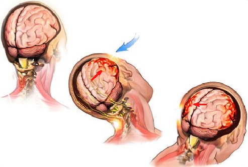
Blue arrow – direction of initial blow to the head
Red arrows – direction of the brain bouncing in the skull with bruising on the opposite sides
We can split the events after a severe concussion or RHIS into three stages:
1. Hypermetabolism Stage
The stress on brain membranes and microbleeding into the brain tissue trigger the release of different neurotransmitters, especially glutamate, and substances called cytokines which accompany any type of inflammation in our body.
The next step is post-traumatic cellular derangement and an increase in cerebrospinal fluid (CSF) secretion. The first outcome of these events is the increase of intracranial pressure, which compromises cerebral circulation. As was shown by Yamakami and McIntosh (1989), cerebral circulation can be reduced to as little as 50% of normal.
Another equally important event in the early stages after a concussion is a dramatic increase in brain metabolism, which requires extra glucose to support it (Giza and Hovda, 2001). However, decreased cerebral circulation is unable to deliver the necessary amount of glucose to the brain cells for use as fuel. Thus, there is a mismatch between the injured brain’s glucose demand and its availability, which triggers what McKee and Daneshvar (2015) called an “energy crisis in the brain.”
2. Hypometabolism Stage
Approximately 2 to 4 weeks after a concussion, brain metabolism slows dramatically because of the negative impact of local inflammation in combination with a lack of proper oxygenation (Bergsneider et al., 2000).
At this point, production of ATP, or fuel, by mitochondria greatly diminishes. As soon as the normal amount of ATP diminishes, it becomes the first step to the irreversible changes in neural connectivity, especially if the individual continues to suffer repetitive concussions.
3. Post-Concussion Encephalopathy
If the patient has experienced repetitive mild concussions, he or she starts to develop Post-Concussion Encephalopathy (movement disorders, memory loss, psychiatric behavioral disorders, chronic headaches, etc.). One of the major mechanisms of these profound changes in the function of the brain is alteration in the function of so-called tau-protein.
Tau-protein is an important part of normal brain function since it supports the function of neurons. This protein forms microtubules which support neurons by transporting necessary proteins, nutrients and ATP molecules along the neuron and transporting waste products back. Without that, the neuron would deteriorate (Ballatore et al., 2007). Fig. 2 illustrates tau-protein in normal and damaged neurons.
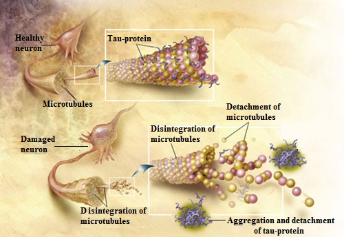
As a result of concussion, especially RHIS, the normal microtubules deteriorate, tau protein accumulates excessive phosphorus and clumps together and eventually detaches from neurons. Then, these wandering tau proteins accumulate in different parts of the brain tissue, disrupting its normal electrical activity and causing all the neurological symptoms associated with RHIS (Gerson et al., 2016). By the way, the same mechanism is responsible for the progression of Alzheimer’s disease.
GLYMPHATIC SYSTEM
Separately, I would like to address the Glymphatic System, which is very important for this article and for post-concussion patients. Since the presence of lymphatic vessels wasn’t detected in the brain, the exact mechanisms of waste removal from the CNS were until recently a great puzzle for scientists. However, thanks to the work of the brilliant Danish neuroscientist Dr. M. Nedergaard, we now know how it works. Dr. Nedergaard named the brain’s main waste removal system the Glymphatic System. I will illustrate this complex issue with the help of the picture from Dr. Nedergaard’s original article (2013).
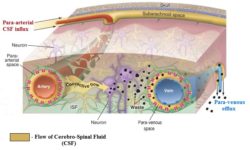
The picture above illustrates a cross section of the brain’s circulatory system. The light brown areas indicate the flow of CSF. At the top of the picture, you see the skull with the subarachnoid space, which contains CSF. As arteries, which bring blood, enter the brain, they also pull along CSF, which flows along the arteries in a so-called para-arterial influx route located in the para-arterial space. Under the pressure generated by arterial pulsation, CSF leaves the para-arterial space and forms the convective flow, which now goes through the brain tissue itself (i.e., brain parenchyma) and it cleans the waste produced by neurons.
Eventually, the convective flow ends up around the neighboring veins in the para-venous spaces which allow the CSF, which now carries waste products, to be drained along the veins via so-called para-venous efflux until CSF with all the waste it carries enters the lymphatic system outside of the brain in the head and neck areas. Thus, the flow of CSF itself is a major waste removal mechanism the brain uses to allow neurons to function properly.
However, Dr. Nedergaard didn’t stop there. Recently, with a group of colleagues (Xie et al., 2013), she discovered another critical piece of information. According to a recent study, more than 60% of the waste formed during normal CNS activity and the neurotoxic waste formed additionally as a result of post-traumatic encephalopathy or Alzheimer’s Disease drains during sleep! As the authors concluded:
“The restorative function of sleep may be a consequence of the enhanced removal of potentially neurotoxic waste products that accumulate in the awake central nervous system.” (Xie et al., 2013).
Finally, there is another recent breakthrough in the work of Louveau et al. (2015) who proved that the drainage to the lymphatic system doesn’t happen only outside of the CNS, but also inside the brain itself. These scientists for the first time detected the presence of lymphatic vessels inside the brain, and in such cases, besides the indirect flow of waste products through the Glymphatic System, we now know that the brain can drain waste directly into the lymphatic system inside the CNS. Fig. 4 illustrates the lymphatic vessels inside the brain.
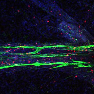
The information about the Glymphatic System is critically important to massage therapists and to the protocol we will discuss in Part II of this article. It scientifically justifies the critical role Medical Massage plays in the treatment of post-concussion symptoms.
APOPTOSIS
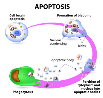
There are a lot of pathological events in cases of RHIS which affect the function of the brain and the brain tries to cope with them to the best of its ability. One of these mechanisms relevant to the Medical Massage practitioner is called apoptosis (AP). AP, or cell death, is executed by the body itself in a programmed fashion. In other words, this protective mechanism allows some cells to die in order to avoid further massive damage. This programmed death is a slow disintegration of the cell.
After the cell gets a signal to deteriorate its inner skeleton (or cytoskeleton), it starts to disintegrate and cells develop areas of blebbing and condensation of the nucleus. Finally, the cells burst into pieces to form apoptotic bodies which are later cleaned by phagocytes during phagocytosis. Fig. 5 illustrates the stages of AP.
The only way to prevent AP and help neurons survive is to increase cerebral circulation and decrease CSF pressure.
As readers will see in Part II of this article the information we discussed above has a direct impact on what the massage therapist must or shouldn’t do while working on patients after concussion or post-traumatic encephalopathy. In the final part of the article, we will go over the MEDICAL MASSAGE PROTOCOL which must be part of rehabilitation therapy.
REFERENCES
- Ballatore C, Lee VMY, Trojanowski JQ. Tau-mediated neurodegeneration in Alzheimer disease and related disorders. Nat Rev Neurosci. 2007;8(9):663-672. doi:10.1038/nrn2194
- Bergsneider M, Hovda DA, Lee SM, et al. Dissociation of cerebral glucose metabolism and level of consciousness during the period of metabolic depression following human traumatic brain injury. J Neurotrauma. 2000;17(5):389-401. doi:10.1089/neu.2000.17.389
- Boden BP, Tacchetti RL, Cantu RC, Knowles SB, Mueller FO. Catastrophic head injuries in high school and college football players. Am J Sports Med. 2007;35(7):1075-1081. doi:10.1177/0363546507299239
- Breslow JM. 76 of 79 deceased NFL players found to have brain disease. FRONTLINE. Published September 30, 2014. Accessed August 16, 2026. https://www.pbs.org/wgbh/frontline/article/76-of-79-deceased-nfl-players-found-to-have-brain-disease/
- Cifu DX, Drake DF, Steinmetz BD. Repetitive head injury syndrome. Medscape; 2014. Accessed August 16, 2026. https://emedicine.medscape.com/article/1163653-overview
- Gerson J, Castillo-Carranza DL, Sengupta U, et al. Tau oligomers derived from traumatic brain injury cause cognitive impairment and accelerate onset of pathology in hTau mice. J Neurotrauma. 2016;33(22):2034-2043. doi:10.1089/neu.2015.4262
- Giza CC, Hovda DA. The neurometabolic cascade of concussion. J Athl Train. 2001;36(3):228-235.
- Louveau A, Smirnov I, Keyes TJ, et al. Structural and functional features of central nervous system lymphatic vessels. Nature. 2015;523(7560):337-341. doi:10.1038/nature14432
- McKee AC, Daneshvar DH. The neuropathology of traumatic brain injury. Handb Clin Neurol. 2015;127:45-66. doi:10.1016/B978-0-444-52892-6.00004-0
- Meyer KS, Marion DW, Coronel H, Jaffee MS. Combat-related traumatic brain injury and its implications to military healthcare. Psychiatr Clin North Am. 2010;33(4):783-796. doi:10.1016/j.psc.2010.08.007
- Nedergaard M. Garbage truck of the brain. Science. 2013;340(6140):1529-1530. doi:10.1126/science.1240514
- Xie L, Kang H, Xu Q, et al. Sleep drives metabolite clearance from the adult brain. Science. 2013;342(6156):373-377. doi:10.1126/science.1241224
- Yamakami I, McIntosh TK. Effects of traumatic brain injury on regional cerebral blood flow in rats as measured with radiolabeled microspheres. J Cereb Blood Flow Metab. 1989;9(1):117-124. doi:10.1038/jcbfm.1989.16
Category: Medical Massage
Tags: JMS 2016 Issue #2
