
We value our readers, their thoughts, opinions and contributions. This article is a great example of that. The idea to write it came to us as a result of an e-mail sent to Journal of Massage Science by Carole Clinch, BA, BPHE, who asked us to comment on the latest breakthrough in pain science.
There are a lot of misleading statements circulating in massage therapy, since the profession tends to exist in its own little bubble, which is frequently detached from modern medical science. We understand that this article contains a lot of scientific data and info, but we think its subject is of great importance, and we encourage readers to read this article all the way through. Otherwise, they will continue to be fed partial truths about pain science.
A SHORT TRIP THROUGH HISTORY
Before we get to the actual topic of the article, we need to take a short trip through history.
- In 1965, Professors R. Melzack and P.D. Wall in the world’s most prestigious scientific publication, Science, published the Gate Control Theory of Pain which emphasized the role of the spinal cord’s gating mechanism in the formation of pain perception by the brain.
- In 1990, Professor R. Melzack recognized that further development of the Gate Control Theory of Pain was needed. He introduced the Neuromatrix Theory of Pain, which showed the role the brain itself independently plays in the formation of pain perception in cases of phantom and chronic pains. Learn more about the Neuromatrix Theory in HOW MASSAGE THERAPY HEALS THE BODY. PART V: PHENOMENON OF PAIN AND MASSAGE THERAPY: NEUROMATRIX THEORY.
- The main concepts of the Neuromatrix Theory of Pain were later picked up by Professor L. Moseley, who deserves credit for spreading this theory in the medical community. However, Prof. L. Moseley generalized the Neuromatrix Theory and used its concepts to explain the formation of pain perception in ALL clinical cases of pain. Such views seemed revolutionary at that time, created waves in the medical community and needed to be validated.
- In 2012, a study was set up to examine the clinical outcome of Prof. L. Moseley’s views on the formation of pain perception to find out if indeed his treatment methods have clinical value when applied to actual patients. Prof. L. Moseley was invited as a co-author of the study which tested his treatment techniques in the clinical setting.
The study examined the clinical effect of the Rubber Hand Illusion, which is the trademark of Prof. Moseley’s work. According to him, this illusion is supposed to have impressive clinical benefits in the control of pain by addressing and altering the brain’s ability to form pain perception. We explain Prof. Moseley’s Rubber Hand Illusion at the end of this article:
The results of the study were published in 2012 (Mohan et al., 2012). Unfortunately, the study showed that the treatment proposed by Prof. Moseley failed miserably in the clinical setting. Apparently, it is one thing to theorize in the lecture room and another to work on actual patients with pain. Here is the reference for this study where the name of Prof. Moseley as a co-author is in bold:
Mohan R, Jensen KB, Petkova VI, Dey A, Barnsley N, Ingvar M, McAuley JH, Moseley GL, Ehrsson HH. (2012). No Pain Relief with the Rubber Hand Illusion. PLoS One. 7(12):e52400.
And a final quote from the article:
“In two experiments, undertaken by independent research teams and both powered to detect a clinically relevant effect, we failed [bold by JMS] to detect any modulation of pain thresholds, nor of pain evoked by individually calibrated high and low painful stimuli, in association with the rubber hand illusion.”
MASSAGE THERAPY AND ‘NEW’ PAIN SCIENCE
Massage therapy, alongside other health professions, initially embraced the ‘new’ pain concept. However, this initial enthusiasm quickly faded for other health practitioners as soon as the independent study on the effect of the Rubber Hand Illusion, which we have referenced above, was published. This study confirmed that there are no widespread clinical benefits of pain management via the brain-only treatment approach, as suggested by Prof. Moseley. Despite this evidence, a vocal group of massage therapists continued to follow the same views and considered the ‘new’ concept of pain a breakthrough in pain science.
We observed that firsthand, since several years ago we were actively engaged in the discussion on this subject. To settle the ridiculousness of some therapists’ claims, we contacted Prof. R. Melzack and asked for clarifications. Seeing direct danger to his legacy, Prof. Melzack gave an interview to the Journal of Massage Science although he had retired from science and the last interview he gave was almost 10 years ago.
We published this interview in JMS Persons of the Month 2013 Issue #4.
To our complete surprise, the same therapists claimed that we had misled Prof. Melzack with our questions. To this day, it is still puzzling to us how we were able to mislead a world-famous scientist with a set of simple questions.
So, where was the problem to start with? Prof. Melzack was the author of both theories and although they address different aspects of pain perception, the author insisted that they are two equally important components.
However, Prof. Moseley concentrated on the Neuromatrix Theory and suggested that ONLY the brain creates and modifies pain perception. For some of his followers, the presence of the Gate Control Theory of Pain, which emphasized the critical importance of a gating mechanism in the spinal cord, didn’t fit into this scenario. From this perspective, the only way to make things work in their favor was to proclaim that, by introducing the Neuromatrix Theory of Pain, Prof. Melzack had denied his own Gate Control Theory of Pain, although in all publications he insisted that they complemented each other.
Before we move forward, we would like readers to pause for a second and read the following: If the Rubber Hand Illusion, which was the trademark of the so-called ‘new’ pain science, failed in the clinical setting and that failure was confirmed by an independent study, how did it happen that the brain-only approach to somatic therapy still flourishes in the massage therapy community? Would you buy a car with new-concept brakes if they failed in a crash test? We don’t think so, but this is exactly what continues to happen in the massage therapy profession.
The new study published in the Journal of Clinical Investigation in May of 2017 puts everything in the correct perspective, and that is the main subject of this article.
THE IRREFUTABLE REBUTTAL TO THE ‘NEW’ PAIN SCIENCE
Before we get to the actual results of this breakthrough study of pain, we need to quickly review the basics of neuroanatomy so readers can fully understand the value of the information the study uncovered. Fig. 1 illustrates the general view of the spinal segment.
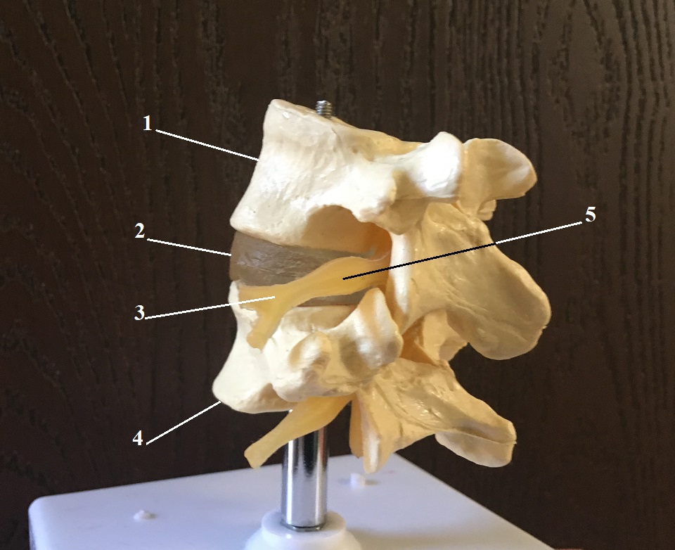
| 1 – L4 lumbar vertebra |
| 2 – intervertebral disk |
| 3 – L4 spinal nerve |
| 4 – L5 lumbar vertebra |
| 5 – dorsal root ganglion (DRG) |
Fig. 2 illustrates the basic arrangement of the nervous system at the level of the spinal cord.
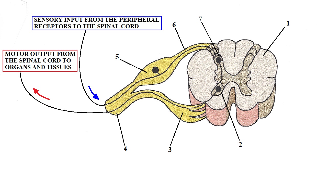
| 1 – Segment of the spinal cord |
| 2 – Anterior horn (origin of motor response) |
| 3 – Ventral root (motor commands going out) |
| 4 – Spinal nerve |
| 5 – Dorsal root ganglion (DRG) |
| 6 – Dorsal root (sensory information going in) |
| 7 – Posterior horn (sensory ‘computer’). This is also the location of the Gates which modify pain perception at the level of the spinal cord, as was shown by Professors R. Melzack and P.D. Wall (1965) |
Peripheral receptors, including nociceptors (i.e., receptors which detect harmful stimuli), located in the soft tissues and inner organs, when activated, deliver information in the form of the ascending sensory flow along the peripheral nerves and later spinal nerves to the segment of the spinal cord they are reporting to. These ascending sensory fibers split from their motor counterparts (which send motor response back to tissues and organs) just before entering the spinal cord, forming so-called dorsal and ventral roots. The dorsal root carries only a sensory component to the spinal cord while ventral roots carry motor commands from the spinal cord to the areas where peripheral receptors were initially activated.
Each spinal and peripheral nerve consists of many neurons which carry information back and forth between soft tissues and inner organs and the central nervous system. Each neuron consists of three main components: dendrites, soma and axon. Fig. 3 illustrates the structure of a myelinated neuron.
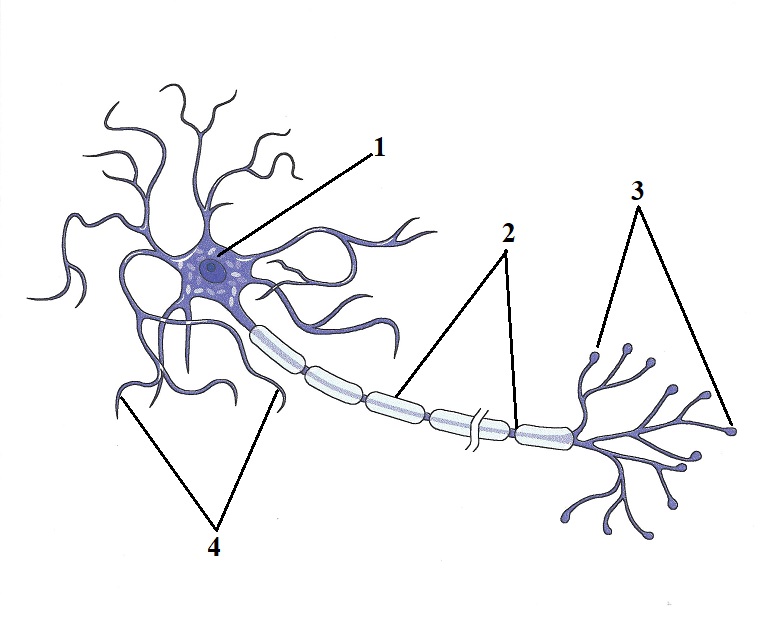
| 1 – soma or body of the neuron located in the dorsal root ganglion (DRG) |
| 2 – axon (together axons form peripheral or cranial nerves) |
| 3 – peripheral receptors in the soft tissue or inner organ |
| 4 – dendrites |
| Soma – body of the neuron |
| Dendrites – provide communication between the bodies of neighboring neurons |
| Axon – long (sometimes several feet) projection of the neuron’s body. Combined axons form cranial or peripheral nerves, which, like communication highways, connect the CNS (brain and spinal cord) with organs and tissues. |
| Peripheral receptors – initial component of the neuron which detects any changes in the inner or outer environment |
As soon as information from the receptors in the soft tissue and organs via the sensory part of the spinal nerve enters the spinal cord, it meets a sensory computer which analyzes the data and alters this data before sending it further up to the brain. This initial modification happens in the gates which are formed by special cells called substantia gelatinosa and they are a critical part of the Gate Control Theory of Pain proposed by Professors Melzack and Wall in 1965. The presence of these gates kills ‘new’ pain science, since it shows that other parts of the nervous system, besides the brain, modify sensory stimulation including nociception and thus alter pain perception before it reaches the brain.
Now let’s come back to Fig. 1 and Fig. 2 where you can see an enlargement of the dorsal root called the dorsal root ganglion (DRG). In this enlargement, all bodies of the neurons (see Fig. 3) which work with this particular segment of the spinal cord are located. It is ironic, but when I studied anatomy in medical school, I always thought that such large anatomical structures as DRGs must play a bigger role than just a repository for bodies of neurons.
In April of 2017, the Journal of Clinical Investigation published the results of a five-year study (Du et al., 2017) conducted by an international group of scientists from the USA, the United Kingdom, China, Japan, and Malaysia. This study is considered a breakthrough in modern pain science and it still reverberates through the world medical community.
As we discussed above, the bodies of neurons are tightly packed together in the DRGs and, as shown in Fig. 3, have short projections called dendrites. For years, it was known that dendrites inside DRGs, besides holding bodies of neurons together, have receptors which work with the same neurotransmitters which control the nociception (i.e., formation of pain perception) at the level of the brain. However, no one before the authors of this study asked the very simple question: Why do neurons in DRGs located outside of the CNS carry the SAME receptors and neurotransmitters which are used by the brain to form pain perception (i.e., nociception)? Thus, the initial intention of this study was to find the answer to this question.
The authors conducted a series of brilliant experiments which examined functions of DRGs. Here is a short description of one of them. A microcannula was inserted into the DRG of experimental animals (rats), and it delivered a small dose of the neurotransmitter GABA inside the DRG. GABA is a major inhibitory neurotransmitter within the CNS which suppresses the pain analyzing system and it also decreases tone in the skeletal muscles. As we mentioned above, it was known before that dendrites of the neuron bodies located in the DRG carry GABA receptors.
Thus, each DRG works as a small brain (this term was coined by Prof. N. Gamper, one of the study’s lead researchers), which alters initial sensory information, especially nociception, while letting into the CNS only the information which is needed to be delivered to the brain. In such a case, the brain becomes more of a receiver than an actual producer of pain perception! In other words, the brain receives already modified and altered nociception and it transfers it to our consciousness. Thus, the brain plays a lesser role in the formation of pain perception than we thought it did before this study was published.
Of course, according to the Neuromatrix Theory of Pain, the brain can also independently generate pain perception – for example, in cases of phantom or psychosomatic pains. However, even in this case, the constant replication of pain perception by the brain still relies on the initial activation of nociceptors and further modulation of pain perception in Gate I (DRG) and Gate II (posterior horn of the spinal cord) BEFORE these stimuli even reached the brain.
The results of this study triggered earthquakes among pain scientists and physicians around the world since it opened completely new horizons in the future clinical application of its findings. A therapy that targets nociception at the level of DRGs, without involving the brain itself, may become the only way to stop the opioid epidemic, which kills 50,000 people each year in the USA alone.
All these people continue to die because of the dominance of the false notion that to control pain we need to target the brain itself, since this is where pain perception is modulated and formed. Technically speaking, these are the same false ideas which continue to be spread within the massage community about the formation and treatment of pain. This is why this breakthrough study has such importance for everyone who is trying to address various somatic and visceral pains.
We followed reactions of therapists to this recent study and we noticed that some are incorrectly reading its results. Some therapists believed that this new gate introduced by Du et al. (2017) denied the presence of gates described by Professors Melzack and Wall, which are located inside the spinal cord in the posterior horns (see Fig. 2). However, this is a completely incorrect reading, since the authors of this study specifically mentioned that their work didn’t cancel or diminish the role of the initial gates introduced by Professors Melzack and Wall in 1965:
“Thus, our study proposes an additional [bold by JMS] mechanism to the broadly accepted Gate Control Theory of Pain, which postulated that integration of ascending peripheral and descending central pathways in the superficial dorsal horn can “gate” nociception.”
Du et al. (2017)
The only difference is that Du et al. (2017) suggested labeling DRG as Gate I and substantia gelatinosa as Gate II. In Fig. 4 we used the illustration from the original article where Gate I and Gate II are in red font.
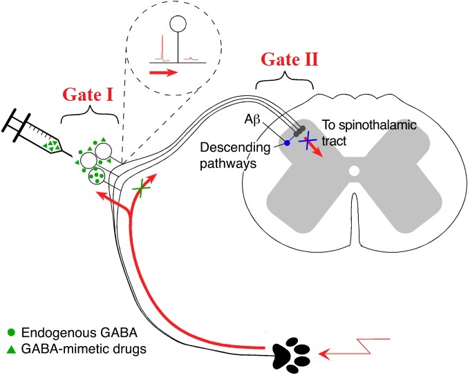
IMPACT OF STUDY ON ‘NEW’ PAIN CONCEPT AND MASSAGE THERAPY
This study is an exceptional piece of science which puts everything in the correct perspective. As we now know, the formation of pain perception is not the prerogative of the brain only. The spinal cord (Gate II) alters it and even such structures as DRGs, which don’t even belong to the CNS, are greatly altered and modify pain perception.
Thus, this study offers airtight proof that pain perception is a very complex event and many components including the brain itself play an equally important role in pain formation.
Unfortunately, a group of therapists still follows the false notion that the brain is the only place where pain perception is formed and modified, and their views still circulate and mislead other therapists in the field.
We’ve waited until the end of this article to explain our views on Prof. Moseley’s work and on the work of his followers in massage therapy. We don’t want readers to get the sense that we consider them completely wrong. The ideas of Prof. Melzack’s Neuromatrix Theory which Prof. Moseley spreads are absolutely correct. The major difference is that Prof. Melzack used the Neuromatrix Theory to explain the formation of pain in specific cases, while Prof. Moseley and his followers in MT incorrectly generalized the Neuromatrix Theory to all cases of somatic pain.
Secondly, followers of Prof. Moseley’s work in the massage therapy profession introduced a new treatment option called Dermoneuromodulation. This treatment option is completely correct but what is incorrect is that it was promoted to therapists as the ultimate clinical solution to all cases of somatic pain.
We reviewed the technical arsenal of Dermoneuromodulation and with 100% assurance we can testify that working with skin and stretching peripheral nerves are indeed very effective clinical tools. Our students around the country are using these techniques on a daily basis. However, they are only components of somatic rehabilitation, since the other three equally important types of soft tissues with their own peripheral receptors (connective tissue, skeletal muscles and periosteum) MUST be evaluated and addressed as well.
As a final note – techniques advocated by Dermoneuromodulation have been known and used in somatic rehabilitation for more than 100 years. Actually, they were the first stepping stones in the development of the modern Medical Massage concept. Techniques exactly similar to Dermoneuromodulation’s were successfully used within the areas of cutaneous reflex zones called Head’s Zones as early as 1903 (Professor A.E. Sherbak). We strongly believe that re-naming already existing therapies and fragmenting the bigger picture of the integrative approach to somatic rehabilitation jeopardizes future development of the clinical wing of the massage therapy profession and its eventual incorporation into modern medicine, where it was successfully used in the past.
We consider the study we reviewed in this article of such importance that we ask our readers to share this article or the original study with as many therapists as possible to stop confusion while practicing and promoting pain science based on data rather than personal opinions.
REFERENCES
- Du X, Hao H, Yang Y, et al. Local GABAergic signaling within sensory ganglia controls peripheral nociceptive transmission. J Clin Invest. 2017;127(5):1741-1756. doi:10.1172/JCI86812
- Melzack R. Phantom limbs and the concept of a neuromatrix. Trends Neurosci. 1990;13(3):88-92. doi:10.1016/0166-2236(90)90179-e
- Melzack R, Wall PD. Pain mechanisms: a new theory. Science. 1965;150(3699):971-979. doi:10.1126/science.150.3699.971
- Mohan R, Jensen KB, Petkova VI, et al. No pain relief with the rubber hand illusion. PLoS One. 2012;7(12):e52400. doi:10.1371/journal.pone.0052400
- Sherbak AE. Further experimental studies of physiological effects of mechanical oscillations. Physiology Review. 1903;9:641-649;10:722-733.
Category: Medical Massage
Tags: JMS 2017 Issue #3
