
In the previous issue of JMS, in the ‘News From The Clinic’ section, we published the clinical case “Medical Massage vs. Pneumonia’s Residuals”:
MEDICAL MASSAGE vs. PNEUMONIA’S RESIDUALS
This case was sent to SOMI by our former student Curt Lezanic, LMT, CMMP, who has developed a very successful Medical Massage practice in San Antonio, Texas. Besides helping patients with various somatic abnormalities, recently Curt started to branch out his practice by helping patients with different chronic visceral diseases who were referred to his clinic by local physicians.
Of course, Medical Massage can’t cure chronic visceral pathologies, but it can greatly help physicians treat and manage the intensity of symptoms and the frequency of flare-ups. Medical Massage can do that by eliminating the somatic components of the chronic visceral disorders which developed secondarily as a reflex reaction of the soft tissues to the abnormal function of the affected inner organ.
While a majority of the therapists greatly enjoyed and learned from Curt’s publication and his clinical expertise, a significant number of therapists dismissed the publication with comments like “Fantasy” and even personally attacked its author with comments like “Fallacy,” “Misinformation,” etc. These critics didn’t produce any supportive material and based their comments on ‘just because I said so.’ The material Curt shared with therapists was pure clinical observation and, of course, didn’t require any references. That was the main foundation for the unjustified attacks.
These attacks illustrated a very unfortunate reality that some therapists don’t have even the slightest idea how far they can go in helping patients when they know what to do, how to do it, and understand the importance of the place Medical Massage occupies in modern medicine. Therapists who continue to have such erroneous views not only undermine the clinical value of massage therapy but also dismiss Osteopathic Medicine in general, since Medical Massage is an integral part of it.
Modern medicine since 1947 (Prof. Korr, 1947) has clearly established the scientific and clinical fact that inner organs and soft tissues, which share innervation within the same segments of the spinal cord, are in mutually productive reflex relations, or, in other words, they are constantly talking with each other. Medical Massage engages these reflexes when it alters the functions of the affected inner organ via soft tissue work. It is astonishing to observe that some therapists deny the existence of the entire branch of medicine which is supposed to be the ‘bread and butter’ of their profession!
Here are a couple of quotes from scientific publications where, in contrast to some therapists, physicians expressed completely different opinions about the clinical prospects of MT application for chronic visceral abnormalities:
From: Rehabilitation Medicine: Principles & Practice, 1999
“Massage stimulates cutaneous receptors and spindle receptors in the skeletal muscles. These produce impulses that reach the spinal cord which once there conceivably can produce myriad effects…. allowing for such a system, massage could have distant visceral effects”
Prof. J.J. Rechtien, DO, PhD, Prof. M. Andary, MD, T.G. Holmes, MD, Prof. J.M. Wieting, DO
From: Physical Medicine & Rehabilitation, 2005
“All somatic and visceral tissues can be negatively affected if they are innervated by the nerves from a spinal segment that receives afferent information from an area of somatic dysfunction. If this is true, then it follows that alleviation of somatic dysfunction via massage results in optimized function of the somatic and visceral systems that are innervated by the same spinal segment”
Prof. J.W. Atchison, DO, S.T. Stoll, PhD, W.G. Gilliar, DO
I would like to finish this introductory part with quotes from two great scientists, physicians and authorities in the clinical application of massage therapy, Dr. J. Weiss, MD from the USA and Professor Aksenova, MD from Russia:
“The reflex value of massage is probably the most important of all. From this point of view we may retain a conventional estimate of the merits of massage.”
Dr. J. Weiss, MD (1952), USA
“The treatment of any chronic disorder of the human body has to include the appropriate method of massage therapy.”
Prof. A.M. Aksenova, MD (1998), Russia
Therefore, we decided to publish a follow-up post which illustrates the successful impact of MM therapy in even more complex clinical cases than Curt’s and provide the requested reference material.
In our clinic in Phoenix, we regularly work with patients who suffer from various chronic visceral abnormalities, and we observe results similar to those reported by Curt in his publication. To support Curt’s clinical case, we have chosen a patient from our clinic who suffered from Chronic Emphysema.
MEDICAL MASSAGE vs. CHRONIC EMPHYSEMA
CLINICAL HISTORY
The 82-year-old male had been successfully treated in our clinic for different somatic problems (Chronic Lower Back Pain and Rotator Cuff Pain) as well as neurological syndromes (Sciatica and Benign Paroxysmal Positioning Vertigo). All these abnormalities had been eliminated, and he was seen only for supporting sessions, since the patient is a dedicated golfer.
Several months ago, it was obvious that the patient had started to breathe more heavily than he usually did, and we suggested that he not waste time and go to see his pulmonologist ASAP.
The clinical history of this patient is very complex, especially regarding his pulmonary function. In 1991, he was diagnosed with left-sided breast cancer and went through a total mastectomy with lymph node removal, long chemotherapy and radiation therapy. As a result of radiation, the soft tissues on his left chest developed moderate fibrosis, which compromised his respiratory function, and every 6 months his pulmonary function was examined by a pulmonologist.
For years, he was regularly checked for possible cancer activity, and with proper medications he became cancer-free. However, in 2015, his CEA started to climb, and a newly developed cancer spot was detected in his left lung. Cancer therapy started immediately, and besides adjusting the medication, he was treated with gamma-knife.
Gamma-knife allows oncologists to deliver radiation precisely to the affected part of the lung. However, to do that correctly, the markers must first be inserted into the lung around the newly discovered spot to guide the radiologist. While he was getting therapy, his left lung collapsed, creating a condition called pneumothorax. After lung restoration, his breathing never fully recovered and became further compromised. To keep his airways patent, he was prescribed an inhaler, which assisted him with breathing.
As we suggested, the patient went to his pulmonologist, and after running the necessary tests, the doctor registered a significant drop in pulmonary function and diagnosed him with Chronic Emphysema. On average, his blood oxygenation was 93-94% at rest.
These numbers indicate a significant decrease in pulmonary function, and the pulmonologist suggested switching to a more potent inhaler with steroids to keep the patient’s airways open. However, the patient’s oncologist wasn’t happy with such a plan, since prolonged and regular usage of steroid inhalers would suppress the patient’s immune system, whose important job at this point was to control the cancer. Since our clinic had helped the patient with a variety of somatic and neurological problems, he asked if anything could be done to help him improve his pulmonary function.
EVALUATION
Breathing Pattern
Visual observation of the patient’s breathing illustrated that its normal pattern was profoundly disturbed. Normally, during quiet respiration, most of the gas exchange happens in the lower and middle portions of the lungs. We actively engage the upper parts of the lungs only when extra oxygen is needed, for example, during physical exercise.
Also, during quiet breathing, inhalation is an active process in which we use our respiratory muscles to expand the thoracic cage, and, in combination with a descending diaphragm, the level of oxygen in the blood is maintained. By contrast, exhalation during quiet breathing is a passive process, since the thoracic cage simply collapses when we release tension in the respiratory muscles needed for chest expansion during inhalation.
The video below illustrates the breathing pattern of our patient during forced inhalations. Please notice that during each inhalation, the patient elevates his shoulders rather than expanding his middle and lower chest.
Also, when he walks up even one flight of stairs, he greatly increases his inhalation efforts and also starts to actively exhale air as if he were doing intense exercise.
Measurement of his chest expansion, seen in the video below and conducted independently by one of our physicians, showed minimal chest expansion during each deep inhalation.
Chest and Soft Tissues
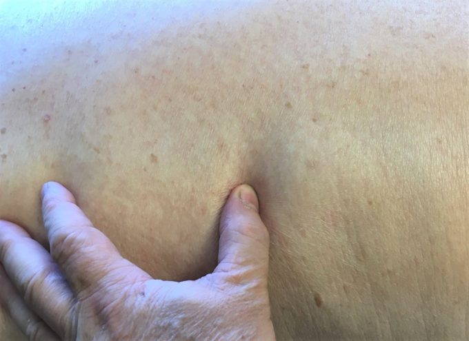
Examination of the patient’s intercostal spaces indicated that during deep inhalation there was very limited rib expansion. Fig. 1 illustrates the placement of the thumb into the 6th intercostal space at the peak of inhalation. As you can see, it is very difficult to place the thumb between two neighboring ribs at the peak of inhalation because the ribs are not driven apart.
Reflex Zones
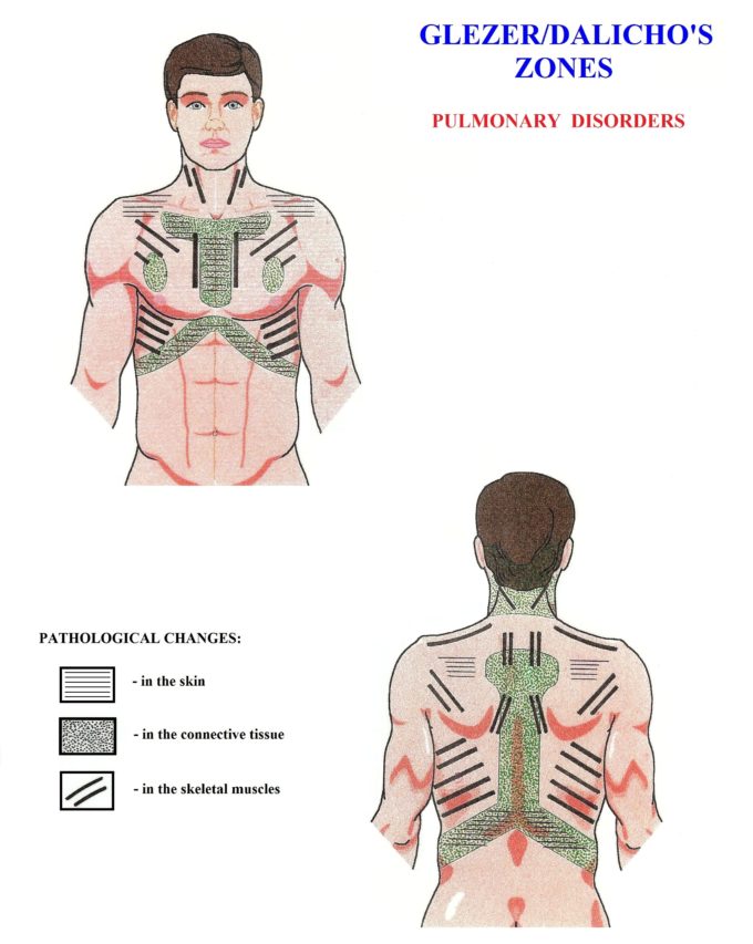
Evaluation of the soft tissues indicated the following pattern of reflex abnormalities according to the Gläser/Dalicho Zones (see Fig. 2).
1. Cutaneous Reflex Zones (CTRZ)
The Sensory Test indicated the presence of CTRZ in the left parasternal area and above the left scapula (areas with black horizontal lines in Fig. 2).
2. Connective Tissue Zones (CTZs)
Tension in the first level of CTZs (in the dermis of the skin) was detected using the first part of Kibler’s Technique (Kibler, 1953) along the parasternal area and along the lower part of the rib cage on the left.
Tension in the third level of CTZs was detected in the middle back between T4 and T12 using the Opposite Shift Technique. It reflected increased tension in the deep fascia, which separates the trapezius from the rhomboids and paravertebrals. These areas are indicated in Fig. 2 by green dots.
3. Reflex Zones in the Skeletal Muscles
Multiple locations of active TPs were recorded in the anterior scalene muscle, pectoralis major and minor, upper and lower trapezius muscle, rhomboids and intercostals.
4. Reflex Zones in the Periosteum
Active reflex zones were recorded along the occipital ridge, the medial edge of the left scapula and the lateral surfaces of the spinous processes, especially of the T5-T9 thoracic vertebrae; along the edges of the same ribs; and finally along the entire left lateral side of the breastbone. In all these areas, even slight application of pressure triggered acute pain.
TREATMENT
The patient exhibited a very complex clinical picture, which included profound local and reflex changes in the function of the entire thoracic cage. The mobility of the chest must be restored to help the respiratory muscles work more efficiently, while secondary reflex zones must be eliminated to help reduce bronchoconstriction.
In 2013, JMS published a two-part article, ‘Medical Massage For The Treatment Of Patients With Pneumonia’, which was contributed to our publication by Russian physician and scientist Dr. T.V. Mitichkina. This article was excerpted from Dr. Mitichkina’s textbook published in 2012. This textbook was written for pulmonologists to teach doctors how to prescribe Medical Massage Therapy and control its correct execution by massage therapists. Currently, this textbook is widely used in the Russian Federation as the main scientific and clinical source in continuing education classes for pulmonologists.
If you would like to read about the theoretical aspects of how Medical Massage Therapy is able to help patients with chronic visceral abnormalities, please read the first part of Dr. Mitichkina’s article:
MEDICAL MASSAGE IN CASES OF PNEUMONIA. PART I
To go over the Medical Massage protocol in cases of pneumonia and other chronic pulmonary disorders, please read Part II of the article:
MEDICAL MASSAGE PROTOCOL FOR THE TREATMENT OF PATIENTS WITH PNEUMONIA. PART II
We used the Medical Massage protocol presented in Part II of Dr. Mitichkina’s article as a framework for the treatment of our patient, with some additions needed for its application in cases of Chronic Emphysema: wave kneading of the abdominal muscles, reinforcement of abdominal breathing, relaxation of the non-respiratory muscles of the torso, decompression of the fascia and percussion along the intercostal muscles within the spaces and along the costal edges of two neighboring ribs.
RESULTS
As a result of conducting Medical Massage therapy within a 3-week span of 2-3 weekly Medical Massage sessions, all reflex zones were eliminated one by one. We started with the skin and fascia and finally got rid of hypertonic abnormalities (active trigger points and hypertonuses) in the respiratory muscles and secondary changes in the periosteum of the thoracic cage and neck.
The video below illustrates the restoration of the normal breathing pattern after the course of MM therapy. Now the patient is able to expand the lower parts of the chest and restore blood oxygenation instead of elevating his shoulders, which contributes less to the overall oxygenation process.
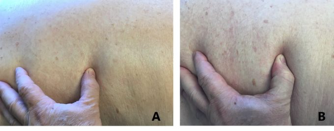
Fig. 3 below illustrates the rib mobility before and after the course of Medical Massage Therapy. You may observe how much deeper the thumb’s placement is between two neighboring ribs at the peak of inhalation. The deeper submergence of the thumb between two ribs seen in Fig. 3b indicates that the lower and middle ribs are now actively moving during chest expansion, even while the patient inhales quietly.
The video below illustrates the measurement of chest expansion conducted by an independent family physician after the course of Medical Massage therapy. Overall chest expansion increased by one inch as a result of the restoration of rib mobility and soft tissue elasticity. This is a huge improvement for this patient, since it will dramatically decrease intrapleural pressure, assist in the descent of the diaphragm and increase gas exchange in the middle and lower parts of the lungs, where the majority of oxygenation is supposed to happen.
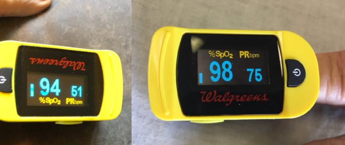
Finally, Fig. 4 illustrates the patient’s blood oxygenation at rest measured in our clinic before Medical Massage therapy started and at the end of it. 98% is normal for the resting state and age of our patient. Please remember that what separates the numbers in the two pictures is a course of Medical Massage Therapy!
LESSONS
- Therapists who regularly work on clients and patients are frequently the first who may observe even small changes in their walking, breathing, color of the soft tissues, etc. That’s why it is so important to pay attention to all fluctuations, even small ones, in the patient’s appearance or behavior.
- In the majority of somatic cases, Medical Massage Therapy by itself can be the ultimate solution to pain and dysfunction. However, in cases of chronic visceral abnormalities, Medical Massage is just another tool (in addition to medications, injections, diet, etc.) that physicians must use to help patients.
- Every chronic visceral disorder carries a somatic component, and there are no other health practitioners in medicine except MM therapists who have the tools and time to detect and decisively eliminate reflex zones in the soft tissues which formed secondarily as a CNS reaction to the inner organ dysfunctions.
REFERENCES
- Aksenova AM, Romanova MM. The effect of reflex muscle massage on the body regulatory processes of peptic ulcer patients with concomitant diseases. Vopr Kurortol Fizioter Lech Fiz Kult. 1998;(6):24-26.
- Atchison JW, Stoll ST, Gilliar WG. Manipulation, traction and massage. In: Braddon RL, ed. Physical Medicine & Rehabilitation. W.B. Saunders Co; 2005.
- Gläser O, Dalicho WA. Segmentmassage. Leipzig; 1955.
- Kibler M. Segment-Therapie bei Gelenkerkrankungen. 2nd ed. Hippokrates-Verlag; 1953.
- Korr IM. The neural basis of the osteopathic lesion. J Am Osteopath Assoc. 1947;47(4):191-198.
- Mitichkina TV. Rehabilitation of Patients with Pneumonia. Novokuznetsk Medical University; 2012.
- Rechtien JJ, Andary M, Holmes TG, Wieting JM. Rehabilitation Medicine: Principles & Practice. Lippincott-Raven; 1999.
- Weiss J. Effect of massage on muscle temperature and radiosodium clearance. Arch Phys Med Rehabil. 1952;33(7):399-405.
Category: Blog
Tags: JMS 2020 Issue #1
