
Arizona
Dr. J. Travell and Dr. S.H. Rinzler (1952) were among the first scientists who pointed out tension and spasm in the masticatory muscles as a major contributing factor to TMJ dysfunction. Despite this, other theories have also been proposed, but the muscular origin of the TMJ is considered one of the leading causes of this joint dysfunction.
According to Travel and Simmons (1983), “The trigger points (in the masticatory muscles cause increased tension and shortening of local muscle fibers, but in the absence of motor unit activity.”
Let’s decode this sentence. As a result of chronic tension, the resting muscle tone of the masticatory muscles increases, while the threshold of the muscle spindle receptors’ activation decreases, and the combination of these events triggers a condition of hyperirritability in the masticatory muscles (Kotani et al., 1980).
Now we need to briefly discuss the concepts of resting muscle tone and the condition of hyperirritability of muscle spindle receptors. The resting muscle tone is the residual tension that each muscle carries during a state of complete rest and relaxation. One of the first signs of future muscle spasm and tension is a slow rise in resting muscle tone, which the patient may not be aware of. When resting muscle tone rises, it increases peripheral vascular resistance in the affected muscle, resulting in a consequential decrease in arterial blood supply and venous and lymphatic drainage. Thus, the increase in resting muscle tone is the first clinical step in the future development of acute muscle spasm and the formation of active trigger points.
The muscle spindle receptors control the degree of muscle tension, including the resting muscle tone. As a result of various factors (see below) affecting TMJ function, the masticatory muscles begin to work under greater pressure, and they gradually reset to a new, lower threshold of activation. Such changes in the threshold of any peripheral receptors, including muscle spindle receptors, are referred to as the condition of hyperirritability. This is a main cause of the increase in resting muscle tone.
Thus, the hyperirritability of muscle spindle receptors and an increase in resting muscle tone significantly contribute to the development of tension in the TMJ, even during resting hours. As a result, articular disk wears and tears first, and Osteoarthritis of TMJ develops later.
There are many factors that contribute to the tension buildup in TMJ: chewing gum and eating hard food, a toothache on the opposite side, an incorrectly placed bridge, bruxism, hereditary factors, etc.
One of the frequent contributing factors of TMJ dysfunction we would like to mention separately is bruxism. Bruxism is especially prevalent among women, and it is characterized by the clinching and grinding of teeth at night. Excessive compression of the disk in combination with grinding movements of the lower jaw, sometimes during the entire night, can smash and, in many cases, dislocate the articular disk, causing severe TMJ dysfunction (Nagamatsu-Sakaguchi, et al. 2008). Degeneration of the TMJ triggers additional spasms in the masticatory muscles, and as a result, a vicious cycle of TMJ dysfunction is formed, which affects the patient’s everyday life.
The temporalis muscle, especially its posterior portion, is primarily activated during bruxism. Also, people may have bruxism periodically during the day as a body response to chronic stress.
CLINICAL SYMPTOMS
Patients with TMJ dysfunction complain of dull pain anterior to the ear and on the same side of the face. The pain becomes sharp when the mouth is wide open and during chewing, especially hard food. Pain also increases if the patient sleeps on the affected side because of the pressure on the TMJ against the pillow.
If the articular disk is under pressure, clicking sounds appear with every mandibular movement, and patients may report periodic ‘locking’ of the joint.
Frequently pain from the affected joint radiates to the temporal and occipital areas, and it may trigger chronic headaches which are usually located in the temporal area (Ciancaglini et al. 2001; Ballegaard et al., 2008). ALL patients with temporal headache must be examined for TMJ dysfunction, even if he or she doesn’t complain about any uncomfortable sensations in the area of TMJ.
Some patients complain about ear pain, dizziness, and even tinnitus (Rubinstein et al. 1990). These side effects of TMJ dysfunction were initially described by Dr. Costen (1934), and they constitute the so-called Costen’s Syndrome.
EVALUATION TESTS
Visual Test (VT)
The first simple test is VT. Position yourself on a patient’s side so that your eyes are level with their mouth. Now ask the patient to clinch teeth while keeping lips open. What you see is the patient bite, which is the way the upper and lower teeth are positioned in respect to each other. You may observe it from the lateral and frontal views.
The bite is very individual and changes during a lifetime as a result of lost teeth, bridges, or crowns. The bite of the patient with TMJ is always pathologically changed. It can change in two major ways: anterior-posterior and lateral.
1. Anterior-posterior changes of the bite
If the examiner views the position of the upper and lower teeth from the side, it is noticeable that in some patients, the upper and lower teeth are positioned exactly against each other (insial bite). In some patients, the lower teeth protrude slightly forward in relation to the upper teeth (maxillary protrusion), while in others, the lower teeth are slightly behind the upper teeth (mandibular protrusion). See Fig. 1, which presents three types of bites.
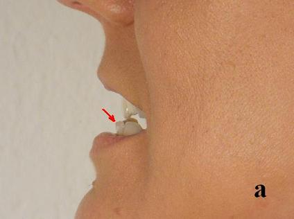
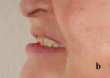

Fig. 1. Variants of the normal bite
a – mandibular protrusion bite
b – maxillar protrusion bite
c – insial bite
In many cases, there are no previous records of the bite before the patient started to suffer from TMJ dysfunction. This is why the lateral view of the bite is less informative regarding the current treatment protocol, but it can be a very important subject during and at the end of treatment.
Ask the patient for permission to take a photo of the bite from the lateral view (you don’t need to capture the entire face) before the treatment begins, during the treatment, and after the treatment ends. In many cases, comparing these pictures will reveal positive changes in the bite from the lateral view, which reflect normalization of the anterior-posterior relationship between the upper and lower jaws.
These restorative changes, documented by pictures, can also be very helpful for the dentist if a night guard is needed. They also show the patient how much the therapy has changed to restore the normal bite.
2. Lateral changes of the bite
The lateral changes of the bite are very important, because they are a definite sign of TMJ dysfunction. Ask the patient to slightly clinch their upper and lower teeth, and observe if there are any differences in the position of the upper and lower teeth on each side of the mouth. In patients with TMJ dysfunction, the spasm in the masticatory muscles pulls the lower jaw superiorly on the same side, altering the bite in a way that creates a gap between the upper and lower teeth on the opposite, unaffected side. Very frequently, patients noticed this crooked position of the lower jaw on their own and mentioned it even before the examination started.
This simple test helps confirm the diagnosis of TMJ and can be used during treatment to monitor its effectiveness. As a result of the therapy, the lateral bite deviations must be completely eliminated. Fig. 2 illustrates the anterior view of the pathological and normal bite of the patient with TMJ dysfunction.
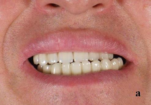
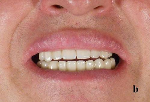
Fig. 2. Anterior view of the pathological (a) and restored normal bite (b) of the patient with TMJ dysfunction
Three Knuckles Test (TKT)
The simplest and most effective way to examine the TMJ and determine if there is any tension in the masticatory muscles is the Three Knuckles Test (Travell and Simons, 1983). This test must be conducted just before any palpatory evaluation of the TMJ and masticatory muscles. Normally, we are supposed to fit three knuckles of the 2nd-4th fingers compressed together into a wide open mouth. Ask the patient to open their mouth and try to fit their three compressed knuckles between the upper and lower teeth. If pain or stiffness prevents the patient from fitting three knuckles into the open mouth, they have spasm and shortening of the masticatory muscles, with the following pathological changes in the TMJ. Fig. 3 illustrates the results of TKT on a patient with severe TMJ dysfunction before the first session and at the end of the treatment course.
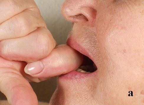
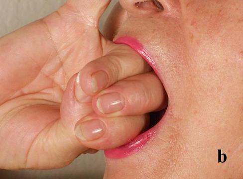
Fig. 3. Results of TKT used on a patient with severe TMJ dysfunction before the first session (a) and at the end of the treatment course (b)
Lateral Movement Test (LaMT)
LaMT is also an observation test that allows for the detection of the degree of tension in the lateral pterygoid muscles, which are responsible for the lateral or grinding movements in the TMJ.
Ask the patient’s permission to mark a spot in the middle of the chin and hold the ruler under the chin with a similar mark. The mark on the ruler must be precisely under the mark on the chin. Fig. 4 illustrates the application of the LaMT in the case of tension in the left lateral pterygoid muscle.
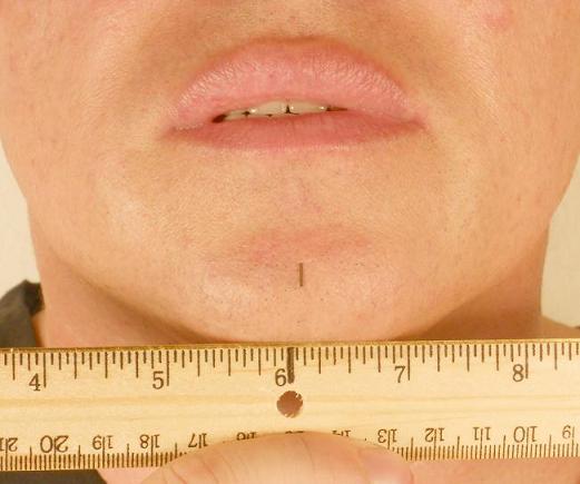
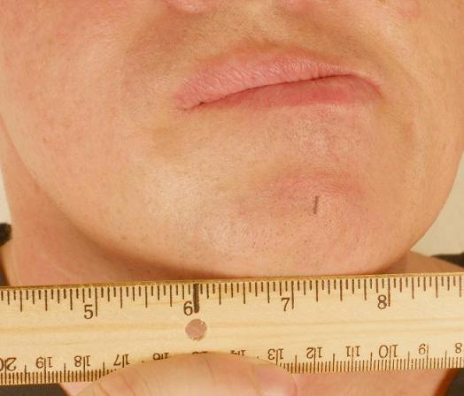

Fig. 4. LaMT in the case of tension in the left pterygoid muscle
Fig. 4a illustrates the initial position of the lower jaw before application of the test. Now ask the patient to move the lower jaw laterally first in the direction of the affected joint until they feel pain (see Fig. 4b). Notice the distance the marked point on the chin travels during the lateral movement. For the next part of the test, ask the patient to move the lower jaw laterally in the direction opposite to the affected joint. Usually, this movement is more painful and restricted if tension in the lateral pterygoid muscle is present (see Fig. 4c). Notice and record the difference between lateral movement to each side.
Please notice that during lateral movement to the left, the marked point on the chin moved more than an inch along the ruler, while during lateral movement to the right, the patient was able to move the lower jaw only slightly because he felt pain in the left TMJ.
Palpation Test (PT)
PT is the first point of contact with the affected TMJ, and it provides the practitioner with a wealth of important information. First, ensure that the patient relaxes the masticatory muscles and that the mouth is slightly open by itself. Now palpate both temporal areas, TMJ areas, as well as along both lower jaws. Apply moderate pressure and try to feel if there are any abnormalities in the tension and structure on the affected side compared to the normal side.
The practitioner needs to examine and compare abnormalities in each of the three areas separately: temporal, TMJ, and along the lower jaw. The video below presents the application of the PT in each area.
Dynamic Test for Leateral Pterygoid Muscle (DT)
The DT enables us to examine the position of the condyloid process within the TMJ and also assess tension in the lateral pterygoid muscle (LPM). The video below illustrates the application of DT. Pay attention to the position of the fingertips in the external auditory passages.
Place the tips of the index or small fingers in both external auditory passages (Part 1 of the video) so that the padded parts of both fingertips are pressed against the anterior walls of the passages. In Part 2 of the video, you see that the practitioner’s index fingers are pushing upward on the anterior wall of the ears. Now ask your patient to slowly open and close their mouth, concentrating on the sensation in your fingertips (Part 3 of the video). You will feel that the bony resistance (i.e., condyloid process) of your fingertips pressed against moves forward as the patient continues to open their mouth. If you compare sensations from both sides, you may notice that on the affected side, this movement of the condyloid process forward, you will feel as a decrease of bony resistance, and it appears earlier. This is a sign of the increased tension in the LPM.
There is another important tip for the correct execution of DT. Essentially, all patients with TMJ dysfunction experience a clicking sensation in the joint when the mouth opens. The moment of click will significantly impair your ability to examine the LPM using this test. To eliminate this distracting factor, ask the patient to slide the tip of the tongue all the way backward along the roof of the mouth before opening their mouth and keep it in this position while DT is applied. Pay attention to the position of the tongue during the application of DT (see Part 3 of the video).
Trigger Point Test (TPT)
A palpatory examination will reveal several trigger points in the masticatory muscles.
1. Temporalis muscle
The trigger points in the temporalis muscle are located above the zygomatic arch in the temporal area. The video below shows the examination of the active trigger points in the temporalis muscle. The dashed line in the video indicates the lower edge of the zygomatic arch, and the white dots indicate the location of trigger points.
During the examination, ensure that the patient relaxes all masticatory muscles and doesn’t clench their teeth. The best way to control this matter is to observe if the patient maintains a small gap between upper and lower teeth (see video of masseter muscle examination).
Two trigger points in the temporalis muscle, located just above the ear, are especially active in cases of Costen’s Syndrome or are directly associated with temporal headaches resulting from TMJ.
2. Masseter muscle
Trigger points in the masseter muscle are located on the lateral side of the face below the zygomatic arch (dashed line in the video). The video below shows the examination of the active trigger points in the masseter muscle.
During the examination, ensure that the patient relaxes all masticatory muscles and doesn’t clench their teeth. The best way to control this matter is to observe if the patient maintains a small gap between the lower and upper teeth during the examination.
3. Lateral Pterygoid Muscle (LPM)
The LPM should be examined below the zygomatic arch and in front of the TMJ. The video below shows the examination of the active trigger point in the LPM. The dashed line in the video indicates the lower edge of the zygomatic arch. Pay attention to the position of the thumb in the video.
4. Digastric muscle
The digastric muscle should be examined just below the chin as shown in the video below. The white arrow in the video indicates the direction of the pressure.
Evaluation of the Posterior Neck
Evaluation of the patient with TMJ dysfunction should be combined with an assessment of the posterior cervical muscles. There is a considerable number of publications that link TMJ dysfunction with chronic tension and spasm of the posterior cervical muscles (Nicolakis, 2000; Sonnesen et al., 2001; Wiesinger et al., 2007, etc.). It is also worth remembering that one in three people who are exposed to whiplash trauma is at risk of developing delayed TMJ symptoms that may require clinical management (Sale, 2007), and 53 % of patients with Fibromyalgia have TMJ pain of different intensity (Balasubramaniam et al., 2007).
The clinical correlation between TMJ dysfunction and tension in posterior cervical muscles was noticed long ago. Halbert’s (1958) statement may explain this phenomenon: “…there is a close anatomo-functional relationship between the masticatory system and the cervical region and scapular centric, and the postural alteration of the head leads to a disadvantage to muscular biomechanics.”
Unfortunately, the obvious clinical correlation between TMJ dysfunction and chronic tension and pain in the posterior neck created an incorrect assumption that adjustments and manipulations of the cervical vertebrae would help patients with TMJ. There are countless clinics where TMJ is treated using a series of quick cervical adjustments, despite the fact that all research data confirm its muscular origin. As it was correctly pointed by Matheus, et al., (2009): “The relationship between craniocervical disorder and TMJ dysfunction may be strongly related to the muscular component rather than the articular component.”
We will not discuss the evaluation of the posterior cervical muscles here, but we would like to make it clear to all practitioners working on TMJ cases that this is a crucial component of the patient’s examination and treatment protocol.
Other Pathological Conditions that Mimic TMJ Dysfunction
This is a very important issue for patients who suffer from TMJ dysfunction. On a weekly basis, we see patients in our clinic who were misdiagnosed and consequently incorrectly treated for TMJ dysfunction, while the real problem was triggered by a completely different cause. Expensive day guards and night guards were made, medication with severe side effects was prescribed, and even surgeries were performed on patients who were incorrectly diagnosed with TMJ dysfunction. This is why we would like to discuss this important subject separately.
First of all, we would like to emphasize that clicking and even occasional locking of the jaw isn’t the ground for intensive therapy. Only if the patient experiences pain in the area of the TMJ and/or restriction of the lower jaw’s movement, the practitioner or physician should consider treatment options. In all other scenarios, stretching during the day and before meals is usually enough to avoid hard food.
It is a very common mistake when clicking in the TMJ during mouth opening, in combination with headache or tinnitus, which immediately leads to the diagnosis of TMJ dysfunction. Yes, every patient with a temporal headache should be examined for TMJ dysfunction. However, the temporal headache, even in combination with joint clicking, is irrelevant if there is no pain and restriction of mouth opening (TKTest) or lateral movement (LaMT). If the TMJ examination we discussed above didn’t confirm the preliminary diagnosis of TMJ dysfunction, other causes for the headache should be considered first.
So, what pathological conditions can mimic TMJ dysfunction and send even an experienced health practitioner on the wrong path?
1. Greater Occipital Nerve Neuralgia (GONN) and/or Minor Occipital Nerve Neuralgia (MONN)
These pathological conditions, especially MONN are very frequent causes of wrongly diagnosed TMJ dysfunction. The minor occipital nerve innervates the temporal area and outer ear. Even the slightest irritation of the minor occipital nerve on the lateral part of the occipital ridge can trigger a temporal headache, and if the patient experiences clicking in the TMJ, an incorrect diagnosis of TMJ dysfunction is frequently made.
Greater occipital nerve innervates the scalp on the top of the head and is less frequently associated with temporal headache. However, in severe cases of GONN, the tension in the cranial aponeurosis and scalp spreads to the temporal area, potentially triggering a a temporal headache secondarily. The tension in the posterior cervical muscles is a direct cause of GONN and MONN.
The major differences of temporal headache as a result of MONN are:
1. In case of temporal headache as a result MONN the pain frequently originates on the back of the head and radiates to the temporal area.
2. Frequently, the patients have other sensory abnormalities in the temporal area: tingling, burning pain, and local numbness.
3. There is no increase in the intensity of headache during or after chewing.
4. Three Knuckles Test and Lateral Movement Tests are negative.
2. Temporal Arteritis (TA)
TA is inflammation, narrowing, and loss of elasticity in the temporal arteries. TA may also mimic TMJ dysfunction because it causes temporal headache as well as restriction of movements in TMJ.
The major differences of TA are:
1. TA affects patients after 55.
2. TA accompanied by mild fever and fatigue.
3. Temporal headache is constant and it may get worse during chewing.
4. TA accompanied by stiffness and pain in the neck, shoulders, back, and legs.
5. TA is also accompanied by visual abnormalities.
6. Patients with TA exhibit weakness of masticatory muscles and may exhibit pain in the area of the TMJ. They chew their food slowly and have difficulty with hard-to-chew foods.
3. Trigeminal Nerve Neuralgia (TNT)
Patients with TNT may also mimic TMJ dysfunction because this pathological condition may also trigger temporal headache.
The trigeminal nerve is a cranial nerve, and although it is primarily a sensory nerve, it also provides motor branches to the masticatory muscles. One of the first side effects of TNT is spasm in the masticatory muscles and TMJ dysfunction.
The major differences of temporal headache as a result of TNN are:
1. Initially, pain originated on the face, and it still mainly affects the face.
2. The pain is much more intense, and it combines with burning or itching.
3. Pain may increase its intensity during chewing, but the major difference is its constant character. Pain bothers patients 24 hours and very frequently dramatically increases its intensity at night.
4. The Three Knuckles Test and Lateral Movement Tests can be positive. However, restrictions of mouth opening or lateral movements of the lower jaw are often a result of intense facial pain rather than pain in the TMJ area.
Join the Science of Massage Institute for the Medical Massage educational program based on science and its clinical application. We educate and train therapists to become massage clinicians: http://www.scienceofmassage.com
Balasubramaniam R, de Leeuw R, Zhu H, Nickerson RB, Okeson JP, Carlson CR. Prevalence of temporomandibular disorders in fibromyalgia and failed back syndrome patients: a blinded prospective comparison study. Oral Surg Oral Med Oral Pathol Oral Radiol Endod, 2007 Aug;104(2):204-16.
Ballegaard V, Thede-Schmidt-Hansen P, Svensson P, Jensen R. Are headache and temporomandibular disorders related? A blinded study. Cephalalgia, 2008 Aug;28(8):832-41.
Ciancaglini R, Radaelli G. The relationship between headache and symptoms of temporomandibular disorder in the general population. J Dent, 2001; 29:93-8.
Costen J.B. A syndrome of ear and sinus symptoms depend upon distributed function of the temporomandibular joint. Ann. Otol, 1934, 43:1.
Halbert R. Electromyographic study of the head position. J Can Dent Assoc, 1958;24(1):11-23.
Kotani H., Kawazoe Y., Hamada T. Quantitive Electromyographic Diagnosis of Myofascial pain-dysfunction syndrome. J. Prosthet Dent, 1980, 43 450-456.
Matheus RA, Ramos-Perez FM, Menezes AV, Ambrosano GM, Haiter-Neto F, Boscolo FN, de Almeida SM. The relationship between temporomandibular dysfunction and head and cervical posture. J Appl Oral Sci, 2009 May-Jun;17(3):204-8.
Nagamatsu-Sakaguchi C, Minakuchi H, Clark GT, Kuboki T. Relationship between the frequency of sleep bruxism and the prevalence of signs and symptoms of temporomandibular disorders in an adolescent population. Int J Prosthodont, 2008 Jul-Aug;21(4):292-8.
Nicolakis P, Nicolakis M, Piehslinger E, Ebenbichler G, Vachuda M, Kirtley C. Relationship between craniomandibular disorders and poor posture. Cranio, 2000;18(2):106-12.
Rubinstein B, Axelsson A, Carlsson GE. Prevalence of signs and symptoms of craniomandibular disorders in tinnitus patients. J Craniomandib Disord, Summer; 4(3):186-92. Review.
Sale H, Isberg A. Delayed temporomandibular joint pain and dysfunction induced by whiplash trauma: a controlled prospective study. J Am Dent Assoc, 2007 Aug;138(8):1084-91.
Sonnesen L, Bakke M, Solow B. Temporomandibular disorders in relation to craniofacial dimensions, head posture and bite force in children selected for orthodontic treatment. Eur J Orthod, 2001;23(2):179-92.
Travell J., Rinzler S.H. The myofascial genesis of pain. Postgrad Med, 1952, 11:425-434.
Travell J. Simons D.G. Myofascial pain and dysfunction. The trigger point manual Vol. I. Williams&Wilkins, Baltimore, 1983.
Wiesinger B, Malker H, Englund E, Wanman A. Back pain in relation to musculoskeletal disorders in the jaw-face: a matched case-control study. Pain, 2007 Oct;131(3):311-9.
Dr. A. A. BIPAR, D.D.S., M.S.
Dr. Bipar has an unusual background for a physician. He graduated with a degree in engineering from Louisiana State University in 1981 and worked as an engineer for ExxonMobil. In 1989, he enrolled in the University of Texas Dental School and graduated in 1993 with a Doctor of Dental Surgery degree. He later obtained degrees in Doctor of Periodontics and Implant Surgery, as well as Plastic and Reconstructive Surgery.
Dr. Bipar’s unique combination of medical and engineering backgrounds helped him to become one of the world’s most recognized dentists and oral reconstructive surgeons. In 2009, the American Research Council recognized him as among the top ten dental surgeons in the United States.
Dr. Bipar is a member of the American Dental Association and the International Academy of Periodontics. He teaches at the Arizona School of Dentistry and actively lectures worldwide on behalf of the International Academy of Periodontics.
Dr. Bipar utilizes various modalities for his patients, including medical massage, as part of an integrative treatment protocol for temporomandibular dysfunction, neuralgia of cranial and peripheral nerves, and other conditions.
He lives in North Scottsdale, Arizona, with his wife and two children. His hobbies are sports, cars, and travel.
For Dr. Ross Turchaninov’s bio, please click here: Medical Massage Courses & Certification | Science of Massage Institute » Editorial Board
Category: Medical Massage
