
We consider both parts of this article precious professional pieces. First of all, the article illustrates the author’s profound understanding of the science behind massage therapy and her great clinical skills, while it also shows her deep compassion and dedication to patients who desperately need help.
This last aspect brings us to the second important feature of the article. While reading articles and various publications on Oncology Massage, it is obvious to us that they present an important, but only partial, picture of a much more complex clinical issue. Yes, full-body massage sessions are an important tool to reduce the stress that accompanies each cancer patient and ravages their souls from the inside. However, it is only one side of Oncology Massage, and another, even more important aspect of Oncology Massage is completely absent from all articles and publications we’ve read so far in American Oncology Massage literature. Vanessa Sifontes fills this unfortunate gap, and her articles demonstrate how far therapists may and must go to help cancer patients.
So, if you are practicing Oncology Massage, please consider branching out further into Oncology Massage Rehabilitation (OMR), because there is a very limited number of therapists who do it correctly, and cancer patients are in great need of your services. While reading this article, you may witness firsthand how much therapists can help patients who are going through cancer therapy. Oncologists compete for Vanessa’s treatments because they see how much her therapy helps cancer patients. Please consider adding OMR to your basic Oncology Massage!
ONCOLOGY MASSAGE REHABILITATION. PART II: TREATMENT STRATEGY
In Part I of this article (see ONCOLOGY MASSAGE REHABILITATION. PART I: NEWEST SCIENTIFIC DATA, COMMONLY USED CANCER TREATMENTS AND THEIR SIDE EFFECTS), we discussed the general principles of Oncology Massage Rehabilitation (OMR) for patients as well as the most common side effects of modern oncology treatment.
In Part II, I would like to discuss the treatment strategy and how manual therapy and massage may diminish the side effects of cancer therapy and help soft tissue rehabilitation. To better illustrate the concept of rehabilitation, I will use cases of successful somatic rehabilitation from our clinic.
FASCIA REHABILITATION
It would be impossible to discuss manual rehabilitation of cancer patients without a clear understanding of fascia’s role. Fascia, as defined by Schleip et al. (2012), is a “fibrous network, a single continuity within the body from the undersurface of the skin, all the way down to the nucleus of the cell.”
It is imperative to remember that fascia is also a sensory organ, composed of six times as many sensory nerves as the retina, which is thought to be the richest sensory organ in our body (Grunwald, 2017). Addressing cancer patients with OMR leads to the restoration of fascia structure and function, which are greatly affected by cancer and oncotherapy.
During OMR sessions, the therapist stimulates mechanoreceptors located in the fascia, restores normal bioelectricity within soft tissues, reduces tension in the extracellular matrix (ECM), regulates the production of cytokines (substances that play a major role in local inflammation of tissues and organs), increases ATP (the energy source for muscle contraction and relaxation) production by the mitochondria, and contributes to the reconstruction of the entire fascia by stimulating normal collagen production. Clinically, OMR improves neuromuscular efficiency, decreases muscular hypertonicity, improves ROM, restores soft tissue drainage, and decreases pain and discomfort.
Cancer starts at the cellular level, and in order to successfully rehabilitate cancer patients and address the side effects of its treatment, we have to work at the same level. This is why addressing the fascia and muscles with myofascial work, as a component of OMR, is a critically important first line of defense against soft tissue fibrosis due to radiation, chemotherapy, or inactivity, which many cancer patients suffer from during long periods of oncotherapy.
As we discussed in Part I, the rigidity of the ECM is one of the key factors that affect the cancer microenvironment in the affected tissues and contributes to tumor growth, invasion, and malignancy (Butcher et al., 2009). There are no other clinical tools to decrease the rigidity of the ECM besides manual work, which allows the restoration of tissue elasticity and maintenance of the ECM’s ‘fluidity.’ This is why OMR is such a critical component of cancer therapy.
Another aspect is the loss of muscle activity during oncotherapy due to fatigue triggered by chemotherapy and radiation. Unused skeletal muscles quickly become atrophic, and this triggers fascia scarification due to excessive connective tissue proliferation, which further limits muscle contractions and diminishes the range of motion. Utilizing all of the tools in the therapist’s toolbox greatly facilitates functional gains.
Let me define the common steps of fascia rehabilitation for Head and Neck Cancer (H&NC) patients. Let’s look at which muscles, and the fascia that covers them, are in the radiation (XRT) field, and how their damage triggers soft tissue constrictions and weakness. Fig. 1 illustrates a head and neck radiation field dose map.
The red-colored area is the location of the primary cancer, and its color indicates the largest power of radiation (70Gy) delivered to the tumor. The change in color represents a lesser radiation impact (24Gy) on the soft tissues around the tumor. However, repetitive XRT, even with lesser power, has a profound impact on the function of soft tissues.
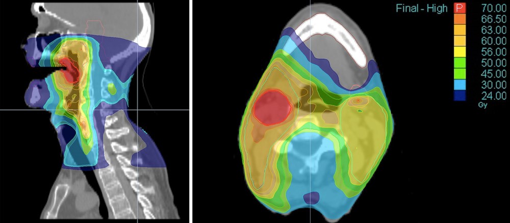
Looking at the potentially affected muscles (Fig. 1) from a functional perspective, it is obvious that they are responsible for rotation, lateral flexion, flexion, and extension of the cervical spine. XRT greatly affects cervical muscles on all levels, making them short and tense due to a dramatic increase in pressure in the extracellular matrix. This pressure, besides limiting the tissues’ elasticity and function, is one of the major contributing factors in cancer metastasizing (as we discussed in Part I). Thus, decompressing soft tissues and the extracellular matrix, besides restoring tissue function, will contribute to the stability of oncotherapy’s clinical results.
However, there is another aspect of soft tissues in the neck – they are key participants in the act of swallowing. The swallowing mechanism is one of the most intricate mechanisms in our body’s somatic functions. It relies on precise interactions between many somatic structures, and even the slightest delay or failure of even one of them greatly affects food and liquid consumption. I compare the simple act of swallowing with a perfectly performing orchestra which flawlessly performs a symphony.
When neck muscles become tight and fibrotic due to XRT, the range of motion of the patient’s larynx dramatically decreases, and the consequences could be catastrophic. The most common side effect is food and liquid aspiration into the patient’s airway and lungs, which is the major cause of pneumonia. Mix pneumonia with the immunosuppression that all patients with cancer go through, and it becomes a deadly complication.
LYMPHATIC DRAINAGE
Another aspect of the OMR is its impact on the lymphatic system. Travell and Simons identified tension in the scalenes, subscapularis, teres major, and pectoralis minor as one of the possible causes that affect lymphatic drainage from the upper body. In 1994, Kuchera and Kuchera reinforced the fact that scalenes “have shown reflexly to suppress lymphatic duct peristaltic contractions.”
Here is another example of how important OMR is for the rehabilitation of patients with H&NC. Behind the mandibular ramus on the posterior surface of the external pterygoid muscle in the pharyngobasilar fascia (Fig. 2, blue line) lies a pack of retropharyngeal lymph nodes (see Fig. 2, green line), which are later connected to the chain of deep lateral cervical lymph nodes (see Fig. 2, red line).
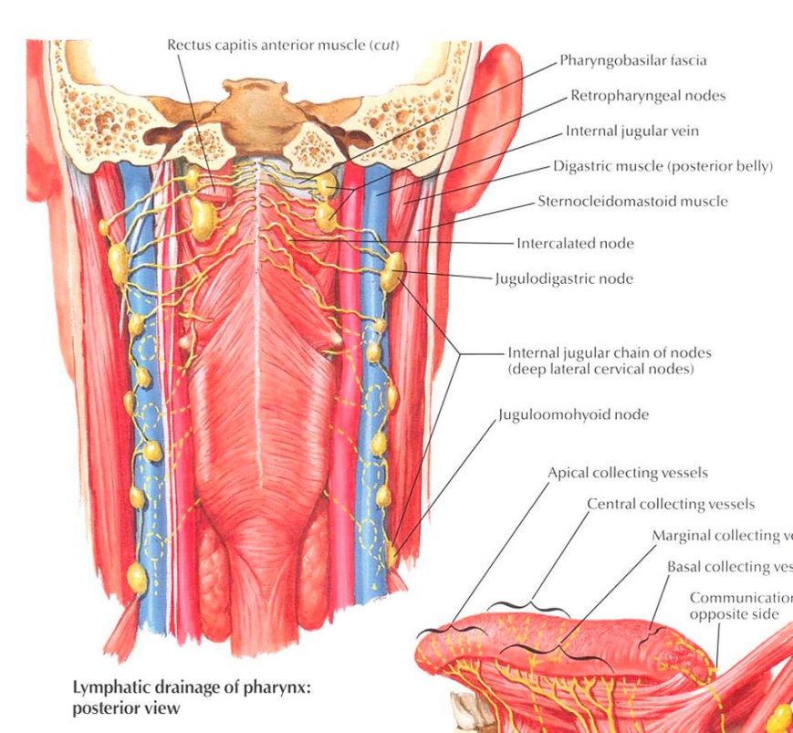
This pack of lymph nodes is a very important component of the deep system of cervical lymphatic drainage, and even in a normal anatomical scenario, it operates in a very tight spot, squeezed between the upper part of the pharynx and the arch of the atlas, while the belly of the rectus capitis muscle is positioned on top of it.
Therefore, releasing tension in these muscles and decompressing the first cervical vertebra will greatly assist in the deep lymphatic drainage, which, if it fails, is responsible for internal or deep lymphedema. Fighting and preventing deep lymphedema is critically important for the rehabilitation of H&NC patients, since there is very limited space in the neck, and any additional pressure, even in the form of extra fluid, triggers a variety of secondary complications.
Thus, therapists can restore and assist in deep lymphatic drainage by targeting soft tissues in the area behind the mandible and first cervical vertebra, and this becomes vital support for H&NC patients in dealing with the side effects of oncotherapy. Let me illustrate the clinical significance of OMR for the H&NC patients based on experiences from our clinic.
CLINICAL CASE #1
In 2018, we treated a patient who had completed treatment for Squamous Cell Carcinoma of the base of the tongue back in 2005. At the initial evaluation, the patient presented with reduced cervical ROM and painful and constant spasms of the cervical muscles throughout the entire day. The intensity of spasms and pain increased greatly at the end of each neck movement and every time he was getting in and out of bed. The patient’s initial cervical ROM was:
Cervical Rotation to the right – 39 degrees
Cervical Rotation to the left – 35 degrees
Lateral Flexion to the right – 14 degrees
Lateral Flexion to the left – 14 degrees
Flexion – 44 degrees
Extension – 13 degrees
Our therapy focused on addressing the drainage, fascia, and tension in the levator scapulae, upper trapezius, scalenes, SCM, and pectoralis minor muscles. At the end of the fifth session his ROM measurements were:
Cervical Rotation to the right – 68 degrees
Cervical Rotation to the left – 70 degrees
Lateral Flexion right – 20 degrees
Lateral Flexion left – 20 degrees
Flexion – 55 degrees
Extension – 32 degrees
The patient’s neck never went into spasm after the first session, and to this day, spasms have not returned. The patient does his daily stretching sessions religiously, and despite living in NM, he flies to CO twice a year to let us work on him as a supportive measure.
CLINICAL CASE #2
A 51-year-old female with a long (30 years) history of tobacco use was diagnosed with Squamous Cell Carcinoma of the buccal mucosa, which had spread to the submandibular salivary gland. The tumor was encapsulated with no further extension or lymph node involvement.
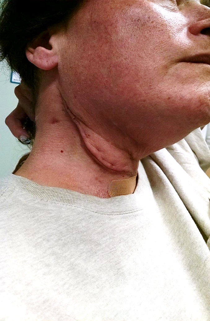
A modified radical neck dissection on the right side and a partial anterior glossectomy with an anterolateral flap were performed by a surgeon. Fig. 3 illustrates her neck at her first visit to our clinic. Severe edema above the scar all the way to the right side of her face is clearly visible. Pay attention to the red discolorations on the cheek and chin above the scar due to the severe venous stasis.
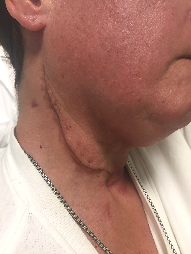
We started therapy twice a week in order to improve neck drainage and scar elasticity. Fig. 4 illustrates the patient’s neck after 10 sessions of OMR. Changes in scar density with restoration of color on the face are notable, but the patient still exhibited submandibular edema above the scar.
At approximately the same time, the patient began XRT, while we continued to work on her neck. Fig. 5 illustrates the patient’s neck after the first radiation session. Brown skin pigmentation on the cheek is a skin reaction to XRT.
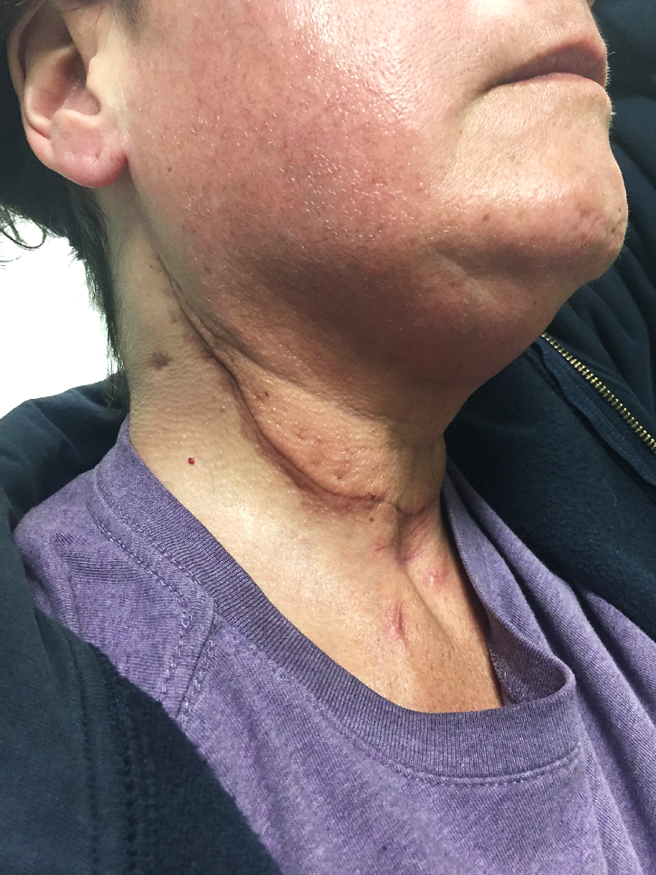
Fig. 6 illustrates the clinical picture after two weeks of radiation therapy. Her skin now exhibits severe radiation dermatitis with skin damage and scaling.
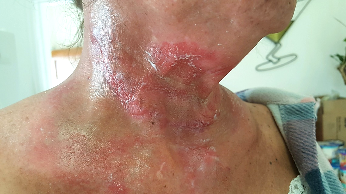
Fig. 7 illustrates the neck of our patient after three weeks of XRT. At that time, thanks to correct OMR, the symptoms of her radiation dermatitis improved, but newly developed tissue fibrosis with muscle shortening is clearly visible (indicated by the yellow circle). These changes required our immediate attention in order to prevent permanent scarification and minimize functional loss in ROM.
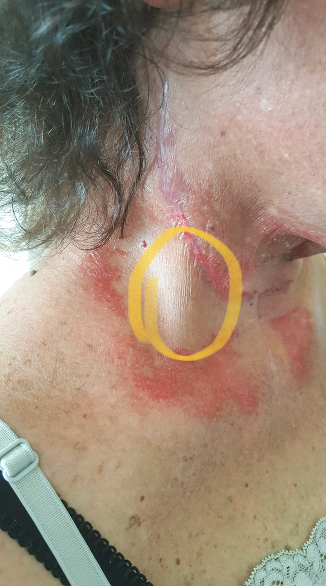
Yellow circle – newly developed soft tissue fibrosis
Fig. 8 illustrates our patient’s neck at the first and last session of somatic rehabilitation.
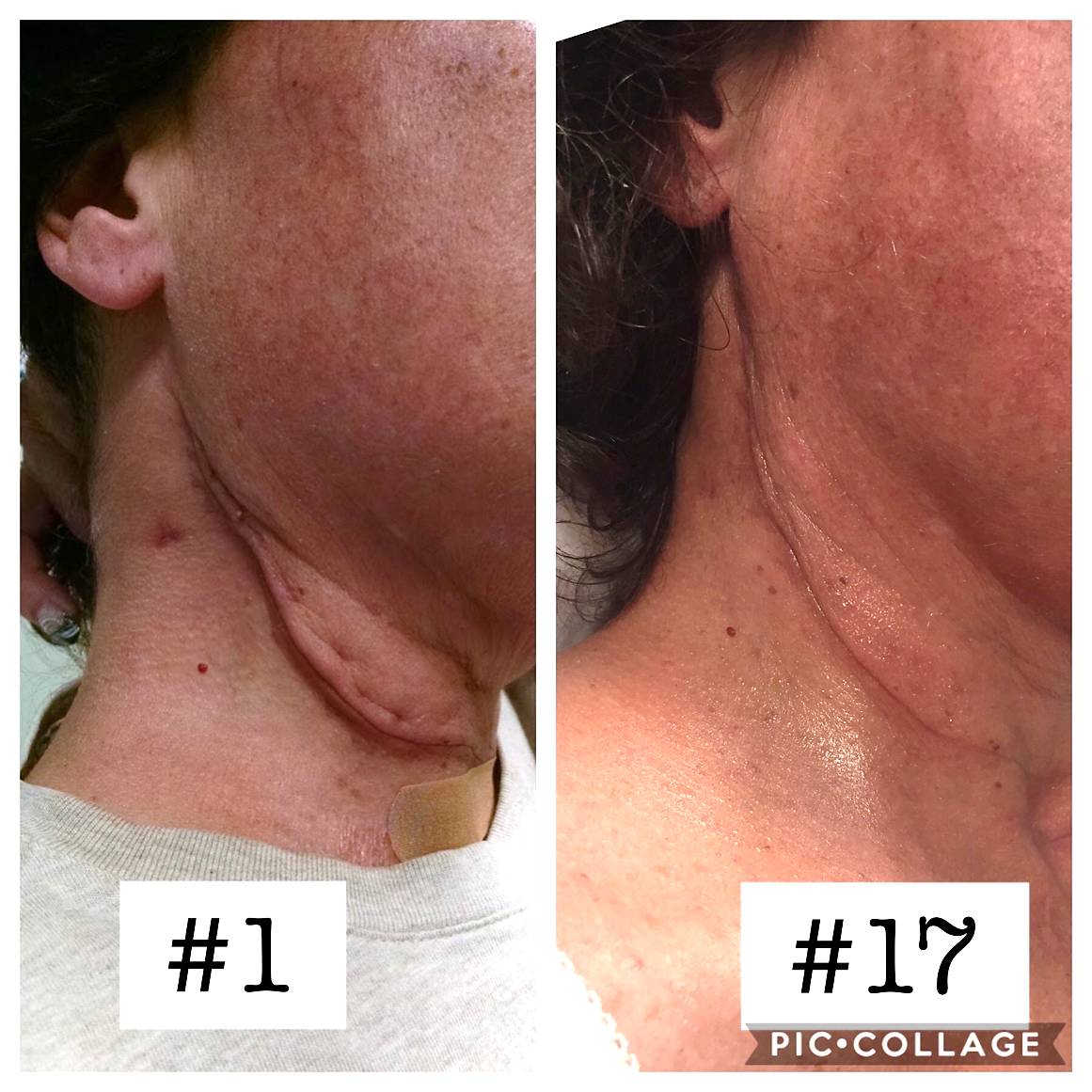
The reddish color of the skin on the face and neck after session #17 is the result of a cumulative burn by XRT, which disappears with time. Redirection of the lymph drainage almost completely eliminated the edema, and it greatly helped with the formation of a scar with low rigidity and less stiffness in the surrounding tissues. No tissue fibrosis is visible (please compare with Fig. 7).
We were able to decrease the symptoms of radiation dermatitis while successfully fighting soft tissue fibrosis. Today, her scar is barely noticeable, with full cervical ROM and normal swallowing and breathing. The patient completely recovered, returned to full work and a productive social life, and recently got promoted at her job.
CLINICAL CASE #3
A 46-year-old male was diagnosed with Stage IV Nasopharyngeal cancer in 2007. The patient’s treatment started with chemoradiation therapy (XRT with cisplatin and 5-FU), with initially good results.
In 2012, the neck cancer came back, and the patient underwent a subtotal neck resection with a full dose of neck radiation for the second time. In 2017, the patient’s cancer came back again, and this time the surgeon was forced to do a neck resection with a pectoralis major flap, since there were no soft tissues left on the neck. Surgery was followed by nasopharyngeal radiation for a third time. All these XRTs triggered severe soft tissue fibrosis, which is visible as a string of tension on the patient’s lateral neck all the way to the lower jaw. Fig. 9 illustrates the initial clinical picture.
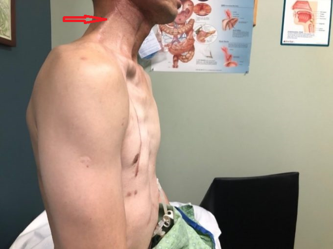
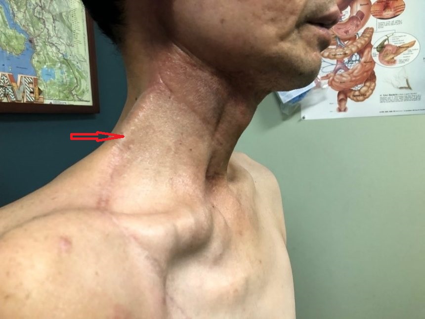
Red arrows – area of soft tissue fibrosis
Here is the patient’s Cervical ROM before and after somatic rehabilitation in our clinic:
Cervical rotation to the right:
Original – 25 degrees
After the therapy – 53 degrees.
Cervical rotation to the left:
Original – 50 degrees
After the therapy – 65 degrees.
Mouth opening:
Original – 15mm
After the therapy – 21mm.
Fig. 10 illustrates the patient at the end of somatic rehabilitation with soft tissue fibrosis on the right dramatically decreased. The string of tissue tension on the right lateral neck is less visible, and the soft tissues became soft on palpation.
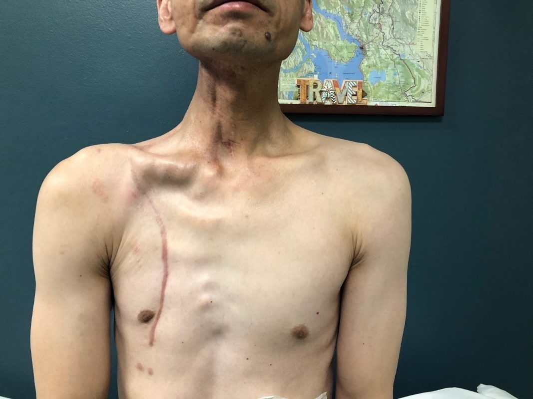
The time difference between the initial evaluation and the first improvement was 8 sessions.
Currently, the patient is on immunotherapy (Keytruda), which has been shown to have good results, especially with recurrent cancer. The patient is a FedEx delivery driver, and he has fully returned to his job.
These patients are the simplest clinical cases from our clinic, but they greatly illustrate cancer patients’ need for a correctly applied protocol of OMR (MLD + Myofascial work) as soon as cancer therapy starts. Losing precious time may produce grave complications for H&NC patients and it may dramatically affect their somatic functions.
The more experience I gain through research, work on patients, and communications with colleagues, the more frequently I find myself returning to the basic concepts of soft tissue rehabilitation: Cell. Sarcomere. Fascia. Therapist’s Hands.
REFERENCES
- Butcher DT, Alliston T, Weaver VM. A tense situation: forcing tumour progression. Nat Rev Cancer. 2009;9(2):108-22. doi:10.1038/nrc2544
- Grunwald M. Homo Hapticus. Droemer; 2017.
- Kuchera WA, Kuchera ML. Osteopathic Principles in Practice. Greyden Press; 1994.
- Schleip R, Findley TW, Chaitow L, Huijing PA, eds. Fascia: The Tensional Network of the Human Body: The Science and Clinical Applications in Manual and Movement Therapy. Churchill Livingstone Elsevier; 2012.
- Travell JG, Simons DG. Myofascial Pain and Dysfunction: The Trigger Point Manual. Williams & Wilkins; 1983.
Category: Medical Massage
Tags: JMS 2019 Issue #2
