
The Science of Massage Institute (SOMI) is proud to collaborate with massage schools that have built their curriculum on SOMI’s science-based textbooks and publications. These schools guide new generations of therapists, shaping their clinical skills and mindset from the very first day of their education and into their future professional careers.
One such school is the Institute for Massage and Bodywork Therapy in Fayetteville, NC. Its director, Brenda Howell, LMT, CMMP, a former SOMI student, carries the torch of massage science with intellectual rigor and unwavering dedication to the profession. Through her leadership, Brenda continues to light the path for new therapists and their patients.
This clinical case was submitted by Madison Mawson, LMT, who was a student at the school at the time. Despite her early stage in training, Madison applied her clinical knowledge with remarkable precision. Using her “ten magic fingers,” she successfully resolved a seven-year-old clinical case that had resisted all conventional therapies, while debilitating surgery was the only option offered.
Pay close attention to Madison’s profound understanding of pathological changes in the human body, as well as her application of Medical Massage evaluation and clinical techniques. This case is a powerful illustration of the clinical effectiveness of Medical Massage and a testament to Brenda’s teaching and SOMI’s efforts to advance the profession.
Dr. Ross Turchaninov, Editor-in-Chief
MEDICAL MASSAGE vs. THORACIC OUTLET SYNDROME or NOT?
by Madison Mawson, LMT
North Carolina,
PATIENT’s HISTORY
The patient is a 35-year-old male diagnosed with Thoracic Outlet Syndrome (TOS). He was medically discharged from the U.S. Army in 2017 due to service-related injuries. While on active duty in 2013, he sustained a concussion and a mild traumatic brain injury (mTBI) that was never treated.
In early 2018, shortly after discharge, he began experiencing numbness and tingling down his left arm when extending it at or above shoulder level. Over the past seven years, his symptoms have progressively worsened to the point of interfering with daily life.
He consulted multiple physicians and specialists in search of treatment. A nerve conduction test on his left hand was negative, and multiple cervical spine scans showed no abnormalities. At Duke University Hospital, surgery—specifically removal of the first rib—was recommended. Notably, no healthcare provider suggested medical massage as a potential treatment option.
Additional diagnoses included: Three bulging lumbar discs due to lumbar spondylosis
CURRENT COMPLAINTS
This case is unusual because, unlike many TOS cases, the patient reported no pain in the cervical spine or shoulder complex while at rest. However, he experienced lower back pain from his lumbar discs, though treatment focused on his left shoulder and arm due to the severity of dysfunction there.
At presentation, his main complaint was an inability to raise his left arm above 90 degrees. Upon reaching this point, he immediately lost strength, and the arm and hand became completely numb. Although pain was absent at rest, he consistently felt significant weakness and described “an awareness of sensation down the arm,” which he emphasized several times.
EVALUATION
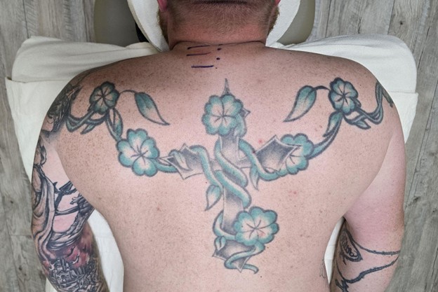
Visual evaluation:
- The patient’s right upper trapezius appeared more elevated than the left, both standing and sitting.
- Motor observation: During assessment, he repeatedly opened and closed his left hand, while the right hand remained still.
- Postural observation: In the prone position, the right scapula rested slightly higher than the left.
- Cutaneous findings: Freckles were markedly more prominent on the left side, extending from the shoulder to the lower lumbar region, which may indicate cutaneous reflex zones.
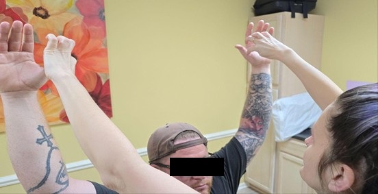
Sensory Test
During this test, I compared the feelings the patient has in each hand while in a resting position as well as with his arms fully extended above the head. With the hands in a resting position, the Sensory Test was positive in the left hand over the radial and median nerve distribution. The patient said that he could feel my touch in both hands, but felt it was more prominent in the right hand. At rest, sensation in the left hand was diminished over the radial and median nerve distribution. The patient reported being able to feel touch bilaterally, but described it as less prominent in the left hand.
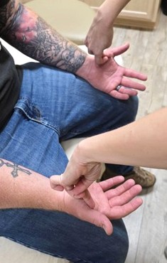
When the arms were extended overhead, the left hand went completely numb, with loss of skin sensation. These findings indicated irritation of the radial and median nerves, prompting further investigation to locate the primary trigger.
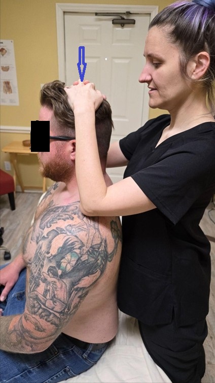
Cervical Compression Test
I applied moderate vertical compression on the top of the head (blue arrow) during the patient’s prolonged exhalation to assess possible cervical disk pathology. The results were negative, with no sensation in the arm. It was consistent with the prior cervical MRI findings.
Cervical Erector Spinae Compression Test
With this test, I examined whether the spinal cervical nerves, which contribute to the radial and median nerves, are irritated by tensed posterior cervical muscles. To help with the testing, I used a marker to show the spinous processes of C5, C6, and C7 (dots in the picture) and matching spinal nerves in the spinal groove (horizontal lines).
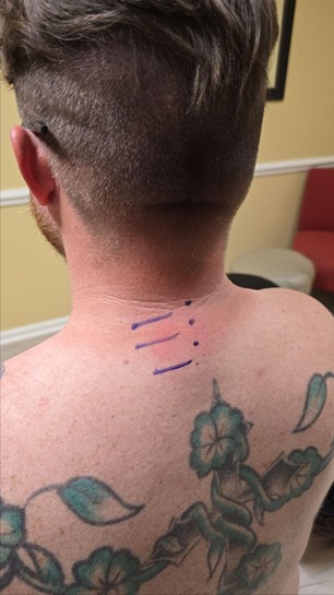
Results of testing on the left:
C5 – With moderate compression in the spinal groove, the patient feels pressure running down the left arm.
C6 – With moderate compression in the spinal groove patient felt lightheaded
C7 – With moderate compression in the spinal groove, the patient felt new sensations on the back of his left arm (triceps area) and down into his thumb
Results of this examination pointed to the spinal nerves irritation by tensed posterior cervical muscles. The tension there started to reach a critical level because the patient exhibited an autonomic reaction (lightheadedness) while I examined tension in the paravertebral. These reactions are signs of a significant imbalance within the autonomic nervous system.
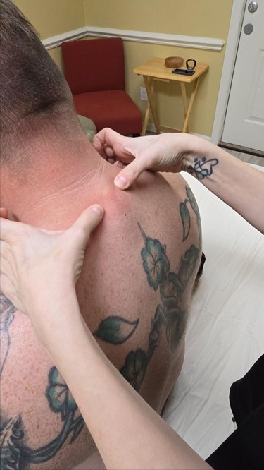
Wartenburg’s Test
Positive. Even mild pressure above and behind the clavicle reproduced uncomfortable sensations radiating into the thumb. This confirmed severe tension in the anterior scalene muscle, consistent with Anterior Scalene Syndrome and TOS.
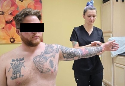
Adson’s Test
This test was immediately positive. Simply raising the left arm without head rotation produced a significant reduction in the radial pulse. When the patient rotated his head to the right, the radial pulse disappeared completely. This confirmed compression of the subclavian artery by the anterior scalene muscle, consistent with advanced Thoracic Outlet Syndrome (TOS).
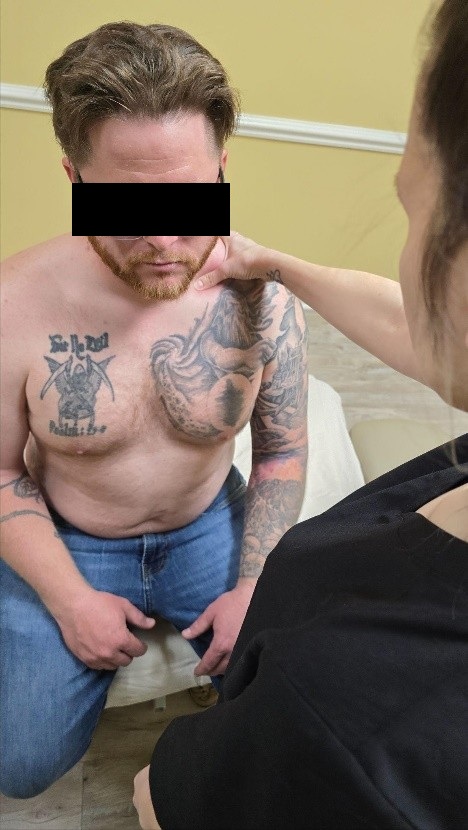
Pectoralis Minor Compression Test
Positive. Pressure over the anterior shoulder reproduced symptoms traveling down the left arm, indicating irritation of the brachial plexus by the pectoralis minor muscle.
Pronator Teres Compression Test
Positive. Compression just below the elbow crease reproduced sensations into the thumb and palm, indicating involvement of the median nerve.
SUMMARY OF FINDINGS
Although multiple tests were positive, results pointed to posterior cervical muscle tension irritating the C5–C7 spinal nerves as the primary trigger. It was the uppermost test of symptom re-creation. Thus, TOS symptoms were secondary consequences of this irritation.
The next important step in the evaluation was examination of tension distribution between the layers of the soft tissue on the posterior neck and shoulder. I used evaluation tests for each layer of soft tissues.
Cutaneous Reflex Zones
I examined the cutaneous reflex zones, keeping in mind the difference in skin pigmentation (freckles were more pronounced and abundant on the left side).
Dr. Kibler’s Test (Part I) – The fold of skin was significantly thicker on the left side than on the right. It was a sign of tension formed in the dermis due to the local interstitial edema.
Dermographism Test: After striking the skin with my fingernails on both sides of the neck and upper shoulders, red lines faded quickly on the right but persisted for over one minute on the left, reflecting increased parasympathetic activity.
Connective Tissue Zones
Dr. Kibler’s Test (Part 2): The patient exhibited restriction in the lifting fold of skin on the left side (affected) compared to the right. Part 2 of Dr. Kibler’s Test indicated the tension and scarification formed in the superficial fascia that covered the superficial skeletal muscles.
Dickle’s Test: I got the same reading as a restriction of skin mobility during the application of Dickle’s Test.
TREATMENT PLAN
The patient has been diagnosed with Thoracic Outlet Syndrome; however, during the soft tissue evaluation, the patient exhibited clear signs of irritation to the C5-C7 spinal nerves by the posterior cervical muscles at these levels. Since these nerves are major contributors to the radial and median nerves, all his symptoms, including those in his arm and hand, as well as TOS, were likely reflex reactions.
I decided to combine parts of the Medical Massage protocol for Cervicalgia and TOS. In such a case, I will decompress the superficial and deep fascia first, address the posterior cervical muscles, while relieving tension in the anterior scalene muscle. I gave the patient a homework assignment for passive stretching of the anterior scalene and posterior cervical muscles at least twice a day.
To be efficient and not waste time, I created a slideshow of the treatment steps. It helped me to follow the protocol and to control the treatment’s flow and timing. I attached the slideshow of the entire treatment plan. Here is a link to my slideshow
The original plan for the patient was to come twice a week for one month. The last two weeks, he was only able to come in once a week. I did notice we made more progress with biweekly medical massage sessions. He was seen a total of 6 times in 4 weeks. By the 4th treatment session, the patient’s tension in the posterior cervical muscles decreased significantly. I re-tested the Erectors with the Compression Test as well as the tension in CTZs. All tests were now negative, which allowed me to remove the Connective Tissue Massage from the treatment protocol and focus more on the area around the spinous processes of C5, C6, and C7, as well as the anterior scalene muscle.
RESULTS
The most significant difference was observed after the fourth medical massage session. At this point, I successfully decompressed the C5, C6, and C7 spinal nerves. Patient immediately noticed an increase in mobility within his neck and left shoulder. Decompression of the left anterior scalene muscle gradually restored proper innervation of the left arm, rendering Wartenberg’s and Adson’s tests negative.
At the end of the last session, I noticed his left scapula was even to the right when he was prone, and both trapezius muscles were even when he was standing. I consider the result of combining Medical Massage for the posterior cervical and anterior scalene muscles to be highly successful.
This impressive clinical case was written by M. Mawson, LMT, a current student of SOMI. Don’t miss the opportunity to learn Medical Massage based on clinical science and join SOMI’s Medical Massage Certification Program! Together we can change the massage industry, bringing it back to its medical roots!
ABOUT THE AUTHOR
Madison graduated from the Institute for Massage and Bodywork Therapy in Fayetteville, NC, in April of 2025 and is working her dream job massaging infants who undergo a frenectomy.
Her contact info: Madison.mawson02@gmail.com
Category: Case Studies
Tags: 2025 Issue #2
