
Medical Massage has…
- Helped thousands of patients resolve pain. Many were cases doctors had given up on.
- Prevented countless surgeries. In fact, several surgeons now refer to Dr. Turchaninov’s clinic.
- Saved athletic careers when team doctors could not resolve the issue.
- Put therapists in control of their results and significantly increased their income

I always mention to therapists who graduate from SOMI’s Medical Massage Certification Program: ‘Only now that our training has ended will you face professional ‘nightmare’ scenarios. You’ll see very complex clinical cases that nobody could have predicted and for which no Medical Massage protocol was written. You will be forced to develop your own treatment strategies based on the evaluation and clinical skills we’ve shared with you!’ This clinical case submitted to JMS by our former student, Kimberly Merryman, LMT, CMMP, who recently graduated from SOMI’s Program, is an excellent illustration of that. Yes, in the majority of somatic pain and dysfunction cases, Medical Massage is the ultimate clinical solution. However, even correctly formulated MM protocol, like any other therapies, has limitations in dealing with genetic diseases. Kimberly was presented with a rare, debilitating congenital disease called Rett Syndrome. With a correct understanding of the MM evaluation and treatment strategies, Kimberly took the challenge. She developed a Medical Massage protocol, which improved the patient’s life and relieved the burden from her family members who, for years, desperately sought help. We hope that you enjoy Kimberly’s work as we did. Dr. Ross Turchaninov, editor-in-chief Medical Massage Therapy vs. Rett Syndrome By Kimberly Merryman, LMT, CMMP Cypress, TX Patient: 30-year-old female with Rett Syndrome Medical History The patient was diagnosed as a toddler with Rett Syndrome, a rare genetic neurodevelopmental disorder that primarily afflicts girls. It is characterized by normal early development followed by regression of motor and language skills somewhere between 8 and 18 months. Life expectancy, on average, is mid-40s; death is often related to seizures, aspiration pneumonia, malnutrition, and accidents. Rett Syndrome symptoms: Cognitive impairment due to slowed brain development Delayed growth in cranial bones Seizures Slow physical development Severe gait impairment Uncontrollable hand movements Disorders of the digestive and respiratory systems This patient had all these symptoms plus severe scoliosis of 50+ degrees. As a teenager, she had corrective surgery, but over time, her spine shifted again, and scoliosis reoccurred. She had lost her ability to swallow food, so a feeding tube had been in place for several years. Due to the nature of this neurological disorder, there is uncontrollable resistance to all movements. I have yet to find articles or medical massage protocols for Rett Syndrome. Therefore, I was forced to compose my own treatment plan based on information from several articles on PT for Rett Syndrome and the knowledge I obtained with training from SOMI by Dr. Ross Turchaninov. Based on the evaluation of her tissues, I used components of various Medical Massage protocols for scar tissue management, muscle atrophy, joint dysfunction and tendinitis, lateral shift techniques to decompress superficial and deep fascia tendonitis, lymphatic drainage, etc. Clinical Presentation I started seeing this patient in October of 2023 and am working on her twice weekly. She is wheelchair-bound and non-verbal. Since she could not communicate, I had to separately evaluate every muscle, examine each joint movement, and take cues from the noises she made and reactions that the evaluation triggered. My first task was to decrease her protective muscle tension so that I could conduct an evaluation as efficiently as possible. I found that grounding her patiently using gentle touch while wearing grounding shoes seemed to stabilize her reactions in each area that I was trying to examine. The circulation in her lower extremities was very poor, and soft tissues were consistently very cold to the touch. I decided to use a heating blanket, and it helped a lot. She has restricted ROM and significant muscle atrophy in every joint. In her back, the patient had a sizeable postsurgical incision after scoliosis correction, with deep adhesions along the scar. These adhesions had fused layers of soft tissue together and prevented their normal functioning. My conclusion was that these spinal adhesions were never managed correctly and, in combination with her lack of mobility, were triggers for scoliosis to reappear years after the corrective surgery. There was no ROM in any segment of her vertebral column, and scoliosis tilted her pelvis. A Dermographism Test showed a Lasting White Reaction along her back, which indicated that sympathetic override had triggered local vasoconstriction and decreased circulation throughout the soft tissues of her back. Besides that, she had contractures formed in all joints. I could extend her hands and fingers while working on her wrist and hand joints with careful application. The patient’s hip joints had some ROM in flexion and extension, while inner and outer rotations were very restricted. Both hips are in adduction contractures. Scoliosis, which caused her pelvic and hip joints to tilt, also triggered calcifications in the hip and sacroiliac joints, creating immense pain. She exhibited severe hypertonicity in the quadriceps and hamstring muscles, which restricted ROM in the knee joints. Each patella had ascended above its normal anatomical position, and patellar instability greatly affected her knee joint functions. Unfortunately, tendinous, ligamental hyperelasticity and atrophy are common problems for patients with Rett Syndrome. The patient’s feet were fixed in plantar flexion, and periodically, muscle spasms created a lateral or valgus deviation in the ankle joints. There was no ROM in the direction of dorsiflexion. However, she did have limited ROM in medial deviation in the ankle joints. Due to the reduced ROM, muscle atrophy, and muscle hypertonicity, every joint presented with a visual deformity, and unfortunately, the patient had reached the late motor deterioration stage. Besides losing walking skills, mobility, and muscle strength, she had …
-
SOMI’s former student and exceptional therapist, Brenda Howell, LMT, CMMP, opened the Institute of Massage and Bodywork Therapy (IMBT) in Fayetteville, NC. From its inception, the school’s curriculum has been to lead future therapists into the clinical application of MT. IMBT is educating a new generation of therapists using only scientific data. In the Case of the Month section of the #1 issue of JMS (2024), we published the clinical case of an MT student, Jaci Stephens, LMT, from the same school, who worked on a patient with a complex somatic abnormality and completely restored the patient’s health while still in school! Here is a link to that publication: Medical Massage Courses & Certification | Science of Massage Institute » Table of Contents Issue #1 2024 The case you are about to read here is another example of the clinical expertise of students from IMBT. Julia Shackelford, LMT, also a student there, decisively solved (in four sessions!) a three-year-old case of Benign Paroxysmal Positioning Vertigo (BPPV) using Semont’s Protocol provided by SOMI. All other modalities failed, while this treatment completely restored the patient’s health. We would like readers to pay attention to Julia’s reasoning for justifying her treatment and how deeply students of IMBT understand the clinical application of Medical Massage. SOMI acknowledges and applauds Brenda’s efforts to educate new generations of therapists who exhibit clinical thinking and exceptional skills to help patients in complex situations. Dr. Ross Turchaninov, editor-in-chief MEDICAL MASSAGE vs. VERTIGO (BPPV) Julia Shackelford, LMT Fort Liberty, NC THE PATIENT The patient is a 71-year-old female who has been diagnosed with Benign Paroxysmal Positional Vertigo (BPPV) by her family physician. Her symptoms started one morning when she got up and immediately felt a wave of dizziness, loss of balance, and severe nausea, which she has since been experiencing for the past three years. Severe episodes of BPPV are always accompanied by nausea. Within the last year, the nausea episodes have subsided, but dizziness and loss of balance are still affecting the patient. CURRENT COMPLAINTS Episodes of BPPV are triggered by changing positions: standing up, sitting down, walking, and rotations from left to right. The patient has noticed that dizziness significantly increases in intensity when she is prone or gets up from her right side. ASSESSMENT Vertigo cannot be examined outside the body; evaluation relies on the patient’s sensations. That said, the patient’s symptoms (being prone and rotating to the right) indicate that otoliths in her vertical (head flexion/extension) and horizontal (head rotation) semicircular canals are mostly affected. TREATMENT It was very important to explain the application of Semont’s Protocol to my patient and what each movement aims to accomplish. Communication with the patient and her understanding of my treatment would be crucial components of the therapy’s success. First, I explained to her the nature of Vertigo (BPPV). Our sense of body positioning comes from two equally important sources: vision and the vestibular apparatus in the inner ears. Both sources must deliver similar data for the brain to form a perfect picture of our body positioning. Inside the inner ears are three membranous semicircular canals filled with endolymph fluid. Each time we move our head, endolymph flows through the semicircular canals. This movement of fluid bends the top of hair cells (stereocilia) located on the bottom of semicircular canals in the direction of the flow. The accumulation of debris that lands on stereocilia makes them heavier, preventing them from getting into the neutral position as soon as needed, similar to the acceleration we experience in a car or roller coaster. The delay of stereocilia getting into a neutral position does not match data from our eyes, and the consequent mismatch between the two equally important sources confuses the brain, triggering a vertigo sensation. I used Semont’s Protocol, recommended in the Medical Massage Volume I textbook and the Video Library of the Science of Massage Institute. Once the patient understood the chain of events outlined above, it helped her understand the goal of Semont’s Protocol to dislodge debris from the stereocilia and restore the proper functioning of the vestibular system. I asked her to get close to the table, close her eyes, slowly get on the table, and lay on her stomach. She needed to keep her eyes closed during the entire treatment until I let her gradually sit up. Shutting down the visual analysis helps to reset cooperation between the vestibular and vision systems more efficiently without visual interference. Semont’s Protocol starts with posterior neck and scalp therapy to loosen the posterior cervical muscles and decompress the cranial aponeurosis. The crucial area of this therapy is the small space behind the mastoid process, which is located behind the ear. That is where the minor occipital nerve, which innervates the temporal area and outer ear, emerges under the scalp. The patient’s brain triggers compensatory reactions in the form of increased muscle tone in posterior cervical muscles and restricted cervical ROM to decrease the intensity of vertigo episodes. Thus, to achieve the optimal application of Semont’s protocol, I needed the posterior cervical muscles to be completely relaxed. I started with effleurage and kneading on her posterior cervical muscles to decrease their resting muscle tone. Next, I turned the patient’s head to the right side and placed an electric massager that produced true vibration on the mastoid process. I held it for a minute and then repeated the same on the left mastoid process. I started with low-frequency vibration and increased it as soon as the …
-
Veronica Selby, LMT, CMMP, recently graduated from SOMI’s Medical Massage Certification Program. While building her theoretical and clinical skills, Veronica shifted her practice from massage therapy’s preventive, stress-reduction application to a fully operational Medical Massage Practice. Our family medicine office refers her patients, and every time she is able to deliver stable clinical results. The clinical case you are about to read is an excellent illustration of Veronica’s skills. Still, when we read this submission for the first time, we had the reasonable question: How many patients in the USA, even after successful surgeries, still suffer from pain and dysfunction because no one was able to rehabilitate their soft tissues fully? Please pay attention to how skillfully Veronica juggled various techniques and modalities she learned from SOMI and her clinical experience. While creating the Medical Massage protocol framework, Veronica brought in additional therapies, made the correct choices, and applied them at the right time. This Medical Massage concept is a perfect clinical application! Dr. Ross Turchaninov, JMS editor-in-chief MEDICAL MASSAGE VS. CONSEQUENCES OF SEVERE LEG TRAUMA Veronica Selby, LMT, CNMT, CMMP Phoenix, AZ The patient emailed me on Jan 4, 2024, asking for an evaluation. I sent her a SOMI’s intake form and set up the first appointment several days later. She had been in a car accident five months earlier and spent two months in the hospital. Her treatment was followed up by three months of physical therapy with limited progress. She was looking for an alternative clinical solution to help her with the remaining pain and to restore mobility. This woman had suffered severe fractures of her left tibia and fibula, and fragments were stabilized by surgery using metal plates and screws. Additionally, she suffered from right-side leg compartment syndrome. As a result of the accident and following surgery, a very thick layer of scar tissue had formed on both legs, triggering stiffness and pain in both ankle joints and the left knee joint at the patellar tendon of the quadriceps muscle. Thus, pain and severe restrictions in the legs and feet mobility were the driving forces for her to seek Medical Massage Therapy in our clinic. EVALUATION Severe soft tissue adhesions in both legs are visible. Her right foot is in plantar flexion, which additionally affects her mobility. Skin The patient experiences almost constant burning sensations along the lateral surface of the right leg. Mobility of the skin is severely restricted. The intensity of the burning sensation increases with even gentle palpation. Fascia and Muscles Due to the immobilization of superficial and deep fascia, mobility of the gastrocnemius, peroneal group, and tibial anterior is severely restricted. Different degrees of muscle spasm and tension are registered in the gastrocnemius, soleus, peroneus longus and brevis, tibialis anterior, and quadriceps muscles. When I tested her ROM in the left hip joint, the patient exhibited limited extension. I also noticed misalignment in the left hip when she went from sitting to standing. Compression tests ruled out possible irritation of the lumbar and sacral spinal nerves. TREATMENT My treatment plan included the layer-by-layer application of proper drainage, lateral shift techniques to increase mobility between layers of the soft tissues and slowly eliminate adhesions, scar management techniques, passive stretching, and Postisometric Muscular Relaxation (PIR) separately for the peroneal group, gastrocnemius and soleus, quadriceps, and hamstring muscles. I added cupping and hot stone massage on the legs to speed up treatment to soften adhesions. Session 1 – 1/9/2024 I started with Lymph Drainage Massage (LDM), draining the inguinal lymph nodes first and the rest of the leg. I added cupping, skin rolling, and kneading according to the patient’s comfort level to help with cutaneous reflex zones and scar tissue. Next, friction along and across fibers of leg muscles and lateral shift techniques were used. I switched to friction on the anterior, lateral, and medial surfaces of the patellar tendon on the left side and decompressed the quadriceps muscle in an inhibitory regime. I finished the session with PIR and passive stretching for all leg muscle groups. Session 2 – 1/16/2024 The patient’s mobility had increased somewhat, as had her overall discomfort, which was the expected outcome of her first session. She also mentioned that her sleep quality had significantly improved since the last session. Her primary complaint at this time was pain in her right foot upon walking for an extended period. Active dorsiflexion was painful due to the spasm and scar tissue formation along the peroneal group. I started with her lower back and hips, hamstrings, and calves. Thirty-five minutes of the session were spent on her anterior legs using LDM, active frictions, and lateral shift techniques. I then used mobile cupping with lift around the scar tissue, initially slowly increasing lift and speed. I finished working on the scar tissue by skin lifting, rolling, and using Connective Tissue Massage strokes. We ended the session by working on her leg muscles with PIR and passive stretching, concentrating specifically on the peroneal muscles. I told her that she might feel the symptoms flare up in the next couple of days, as I expected that to happen. Session 3 – 1/23/2024 Mobility continued to improve, but the intensity of the symptoms didn’t change. Symptoms around both ankle joints were still a major issue. I concentrated the entire session on the legs using the same protocol. In the end, I did some Active Release Techniques (ART) for her quadricep at the knee insertion and PIR. She noticed some burning when I applied draining effleurage strokes on her lower legs (likely from the stretched skin). Session 4 – 1/30/2024 With the same protocol, I started to work more deeply and more intensely to apply greater pressure on the entire system of her leg function. I also used hot stones to soften her scar tissue. Session 5 – 2/7/2024 The only complaint today was that the left ankle had discomfort and stiffness, but without the pain she experienced before. She …
-
Our former student, Brenda Howell, LMT, CMMP, opened the Institute for Massage and Bodywork Therapy, a massage therapy school in Fayetteville, NC. This school represents new advances in MT education with a curriculum based on massage science and its clinical applications. Although the school is relatively young, it has already generated raving reviews and support from businesses and the medical community that hire its newly graduated therapists because of their impressive technical skills and clinical abilities. To illustrate her school’s achievements, Brenda sent us two clinical cases written by her students, Jaci Stephens and Julia Shackelford. In this issue, we will publish Jaci Stephens’ contribution. We want to emphasize that Jaci was still a student in MT school at the time of submission! However, Jaci obtained exceptional theoretical knowledge and technical skills thanks to Brenda’s and other instructors’ teaching and expertise. Notice that the school requires students to collect and interpret clinical data correctly. Thus, this clinical case is a perfect example of effective MT education based on science rather than personal opinions and anecdotic experiences. Finally, Jaci illustrated how soft tissue evaluation must be done by therapists who work in the clinical setting. We are very proud that SOMI training allows therapists to help patients in very complex clinical situations and that our former students can share their expertise with new therapists. Dr. Ross Turchaninov, Editor-in-Chief MEDICAL MASSAGE vs MULTI-LAYERED SOMATIC PATHOLOGY Jaci Stephens, MT Student Medical Massage Class IMBT 2023 General Information The patient is a 43-year-old woman who has an office job. She presents with left-side chronic ear pain and headaches, which have been ongoing for over a year. The patient also complains about tension down the left side of her neck and into her shoulder. Initially, she suspected an ear infection and visited an ENT doctor, who reassured her that there was no ear infection, so no medications were needed. Instead, the physician concluded that musculoskeletal dysfunction was the cause of her symptoms and recommended that she see a somatic specialist. The physician did not perform imaging or further testing. The patient’s office work and poor ergonomic situation may contribute to her symptoms. She did not have prior neck or head trauma. Initial Complaints At the time of evaluation, the patient complained about mild tension in her left neck. However, she had taken ibuprofen roughly 5-6 hours prior, and she takes NSAIDS daily to address her pain and discomfort. She has been getting full-body relaxation massages twice a month since June and noticed some decrease in intensity. However, this relief typically only lasts 3-5 days after the massage, when her symptoms return to the previous intensity. The patient describes those symptoms as tension that starts as dull and aching, then compounds over time until it peaks as a sharp pulsating pain. On a scale of one to ten, the intensity of her pain can reach seven. At the time of evaluation, she described her symptoms as tension rather than pain. Usually, when an attack starts, it originates around the left ear and radiates to the left neck all the way to the top of her left scapula. At the peak of pain, it creates a compressive band of tension and pain around the entire head. She notices feeling ‘off-kilter’ when the intensity of ear pain increases. She doesn’t have an aura before a headache attack. The patient notices more tension, stiffness, and pain in the morning. At night, she sleeps on her left side because sleeping on the right side triggers the sensation of a ‘pull’ on her left neck and upper shoulder. She places her right leg in a hiking position and uses support pillows for additional comfort. Assessment Visual Evaluation The patient exhibits moderate kyphosis, and shoulders are rolled forward. Evaluation of active ROM in the neck shows significant restrictions. She can only rotate her head to the right a few degrees without pain. Further rotation to the right triggers pain in the left neck and ear. She must rotate her upper body to look over her right shoulder without pain. Rotation to the left is almost within normal ROM. Fig. 1 illustrates her ROM during rotation to the right. Further rotation triggers pain in the left neck and shoulder. Fig. 1. Patient’s cervical rotation to the right during the evaluation. Lateral flexion to the right (ear-to-right shoulder movement) is also limited and triggers pressure and pain in the left neck and ear. The neck’s flexion (drawing the chin to the chest) is very limited, and this movement immediately triggers sharp cervical pain, specifically in the C5-C7 area. Fig. 2 illustrates ROM in cervical flexion. Fig. 2. Patient’s cervical flexion during the evaluation Interpretation of Obtained Data Given the acute onset of pain without injury or trauma, it is most likely due to the phenomenon of hyperirritability, and this has been building for some time without the patient necessarily noticing anything of concern. While I suspect some incorrect postural habits and perhaps muscle imbalance, her clinical symptoms certainly are the compensatory reactions to hyperirritability of peripheral receptors. With this in mind, I fully expect to find reflex zones, trigger points, and hypertonus during palpatory evaluation. This also explains why she experiences a degree of relief after relaxation massages. My evaluation of the patient’s pain indicates that her dull, aching pain, which increases with active movement, is more likely an indicator of active trigger points and/or myogelosis. The sharp pain triggered by neck flexion …
-
This clinical case was contributed to JMS by a recent graduate of SOMI’s Medical Massage Certification Program, Kristi Tidwell, LMT, CMMP from Coolidge, AZ. Her experience is interesting from two perspectives. First, it illustrates the clinical value of Medical Massage as the cornerstone of somatic rehabilitation. For years, a long list of various modalities was used that failed her patient; only Medical Massage decisively eliminated HER pain and dysfunction. The second aspect of this submission is how masterfully Kristi combined components of different Medical Massage protocols, constantly adjusting the structure of each treatment session. By doing that, Kristi carefully and steadily built up an effective clinical response, restoring normal elasticity and functions of the soft tissues while resetting the patient’s sensory and motor cortex. Dr. Ross Turchaninov, Editor-in-chief MEDICAL MASSAGE vs. FRUITLESS PAIN MANAGEMENT By Kristi Tidwell, LMT, CMMP Coolidge, AZ The patient is a 62-year-old retired female. Her symptoms appeared several decades ago with right side lower back and hip tightness and discomfort. Initially, symptoms started as a tightening across the top of the right gluteal area and later transformed into pain, which spread down along her lateral thigh. Symptoms progressively worsened, and in late 2018, she started to feel severe left hip pain to the degree that she couldn’t walk to the mailbox. By then the pain had become burning, especially along both lateral thighs. Any extra physical activity, such as bicycling, increased pain and discomfort. The intensity of the bilateral hip pain progressed to such a degree that she could only walk about 800 steps and then had to stop and rest for 10-15 seconds before being able to continue walking. Since the first symptoms developed, the patient went through many different therapies, none of which provided stable clinical results: Trigger point injections along the entire length of her lower back and outer thighs, totaling 32 in 2019 Cortisone shots in the trochanteric bursa, two in 2019 and in April 2023 Spinal manipulations Spinal decompression treatment Physical therapy treatment Regular Massage Therapy Foam roller fascial release Massage gun, 3x weekly since November 2022 Radial Pressure Wave (RPW) 15 sessions, May-July 2023 Weekly in StretchLab All these treatments are standard protocols for “responsible pain management” in medical facilities. I have worked in a similar medical office for nearly two decades and observed first-hand the application and results of commonly accepted pain management. Chiropractors, physical therapists, and nurse practitioners designed treatment plans strictly on the location of the pain and other symptoms. For example, the initial diagnosis of this patient’s right hip pain and dysfunction was “elevation of the right hip.” No one tried to detect the initial trigger but instead treated her secondary symptoms. Medical Massage Therapy Before SOMI’s Medical Massage seminars and hands-on training, I worked on the patient using sessions of Therapeutic Massage with elements of Medical Massage focusing on areas of the patient’s complaints: Lumbar, Gluteal, Hamstring, and IT band. She felt some improvement but continued to suffer from the acute, bilateral burning pain in her hips and thighs. During Ask-and-Show training with Dr. Ross, I brought up this case. We developed a new evaluation and treatment strategy to identify and eliminate the initial trigger rather than chasing the pain ghost. Evaluation: Detailed evaluation of the patient’s soft tissues in the lower back and hip area provided the following data: Active connective tissue zones in the second (superficial fascia) and third (deep fascia) levels were detected in the lumbar area. However, the degree of fascial tension was far more significant on the right side, creating a visible crease that followed the last rib (see Fig. 1). Fig. 1. Fascial tension below the last rib was detected during the initial evaluation. 2. There were trigger points in the thoracic and lumbar erectors. 3. There were trigger points in the right Quadratus Lumborum Muscle. The upper TP below the 12th rib was especially active. 4. Even mild application of pressure during the Compression Test in front of the right sacroiliac joint immediately triggered referred pain to the gluteal area and thigh. Thus, the Compression Test confirmed irritation of the L5 spinal nerve between the last lumbar vertebra, sacrum, and right SI joint. 5. There were periosteal TPs along the last rib and the SI joint and sacrum 6. Examination of the patient’s thighs showed the presence of very significant fascial restrictions and muscle tension in the middle thigh, almost fist-sized on the right. It involved the vastus lateralis, biceps femoris muscle. 7. Both iliotibial bands showed significant fascial adhesions and restrictions of soft tissue mobility and elasticity, especially in the upper and middle thirds of both IT bands. The degree of tension was less in the lower third of the IT bands above the knee joints. The first session of Medical Massage: I started with the Medical Massage Protocol for the Lumbalgia concentrating only on lumbodorsal fascia and lumbar erectors. I separately worked on the skin within affected dermatomes, then on the superficial fascia, and finally on the lumbar erectors (insertions and muscle belly). I used superficial friction, skin rolling, the inhibitory regime of MT, engaged H-Reflex, Trigger Point Therapy, and finally Postisometric Muscle Relaxation. By the end of the first session, the patient demonstrated an immediate decrease in pain intensity in the right lumbosacral region, and a Compression Test to examine the L5 spinal nerve in the area of the right SI joint was negative! I gave her detailed instructions as homework to decrease tension in the Lumbar Erector and QL muscle. I also encouraged her to share my recommendations with her Stretchlab therapist …
-
This clinical case was submitted to the Journal of Massage Science by Cheri Conklen, LMT, from Phoenix, Arizona. Cheri is a current SOMI student, and her clinical expertise and technical abilities are growing from seminar to seminar. Cheri’s submission is an excellent illustration of clinical reasoning and the efficiency of Medical Massage therapy. It took Cheri three sessions using the skills and knowledge she’s received from SOMI training to decisively help her patient with a complex combination of somatic abnormalities. She correctly identified the initial trigger and addressed it with Medical Massage protocols while eliminating secondary syndromes that had formed in the patient’s body. Besides evaluation skills, Cheri exhibited the ability to combine steps from different Medical Massage protocols to develop an ideal treatment strategy for her patient. Dr. Ross Turchaninov, Editor in Chief THREE MEDICAL MASSAGE SESSIONS vs. COMPLEX COMBINATION OF SOMATIC DYSFUNCTIONS by Cheri Conklen, LMT Phoenix, AZ The patient, 21 years old, works as a secretary. She has suffered from severe headaches, peripheral vision loss, and nausea for several weeks. She also complains about the significant restriction of ROM in the right shoulder, which locks periodically. Recently pain in the right shoulder increased, and she’s had difficulties getting dressed. She visited different medical facilities, but the treatment didn’t significantly relieve her symptoms. EVALUATION The patient has evident discomfort due to severe headaches. Further visual observation revealed significant elevation, medial rotation of the right shoulder, and restrictions in the cervical ROM. Applying the Cervical Compression Test ruled out acute pathology of the cervical disks. The Sensory Test was negative in all cervical dermatomes and ruled out the irritation of the peripheral nerves on her neck and upper extremity. The Wartenberg’s Test was negative, ruling out brachial plexus irritation on the anterior neck. Palpation indicated severe muscle spasms in the posterior cervical and upper shoulder muscles, with the epicenter of pain and tension in her paravertebral muscles on the level of C5-C6. Active trigger points were registered in the vertical portion of the trapezius, splenius capitis, and levator scapulae muscles on the right. Examination of the ROM in the right shoulder joint revealed active abduction only to the level of 90 degrees and very painful medial rotation. All portions of the deltoid and supraspinatus muscles were sensitive during the palpation. My clinical reasoning is based on the evaluation results and the nature of this patient’s job. No disk or anterior scalene muscle pathologies triggered her symptoms. Since her job entails so much computer time, she initially developed an acute spasm in her posterior cervical muscles on the C5-C6 level. As a result, her greater occipital nerve was compressed in the suboccipital space, triggering a Tension Headache. Its intensity slowly worsened until her tension headache became a severe Cluster Headache with a visual deficit and autonomic reactions in the form of nausea. As a reflex reaction to the acute spasm in the posterior cervical muscles and the presence of the Cluster Headache, she secondarily developed tension in the deltoid and supraspinatus muscles after initial clinical symptoms were fully active. The probability of that was increased by the innervation pattern of the deltoid muscle, which is supported by the axillary nerve originating from C5-C6 of her cervical spine. Finally, the supraspinatus muscle was innervated by the suprascapular nerve, which originates from C5-C6. FIRST SESSION Based on my theory and SOMI’s Medical Massage training, it is evident that control of the patient’s headache was the main priority. Using SOMI’s Medical Massage protocol for Chronic Headaches, I concentrated on the C5-C6 level and performed detailed work in the suboccipital space, addressing obliques capitis inferior and superior muscles. Next, I used Scalpotherapy to decompress the cranial aponeurosis. I finished the headache part of the session by working on the patient’s face and using Eye Therapy to eliminate her Cluster Headache. At this point, the patient stated that the headache was gone. After I deactivated trigger points in the vertical portion of the trapezius muscles as well as slenius capitis and addressed the levator scapulae at its insertions to the transverse processes of C5-C6, the patient reported a decrease of discomfort in the right shoulder. Despite Wartenberg’s Test being negative, I drained the anterior scalene muscle and passively stretched her anterior neck. SECOND SESSION The patient reported no headache before we started the second session after two days, and her peripheral vision was fully restored. I used the same treatment regime again but now concentrated on the right shoulder using steps from SOMI’s MMM protocol for Rotator Cuff Syndrome. I released protective tension in the trapezius muscle, which gave me access to the supraspinatus muscle. I finished my work on the posterior shoulder by addressing her deltoid, teres, and latissimus dorsi muscles. Before passive stretching, I used heated stones to enhance therapy additionally. After turning the patient on her back, I released tension in the middle and anterior portions of the deltoid muscle. I finished the decompression of the shoulder joint by working on her pectoral major and minor muscles. Elevation of the right shoulder significantly declined after passive stretching. THIRD SESSION After a third session combining Medical Massage protocols, all clinical symptoms were gone, and functions were restored. Today the patient is free of headaches, her peripheral vision is fine, and her right shoulder is in the proper position with the full range of motion. About Author Cheri Conklen, LMT studied Massage Therapy at the Sedona School of Massage. She continued her massage education for Lomi Lomi on the island of Maui in Hawaii. Her most recent studies have been in Medical Massage with the Science of Massage Institute located in …
-
A very frequent complication of surgery is direct trauma of the cutaneous nerves or their compression from resulting scar tissue. Both unfortunate scenarios may trigger a condition called hyperesthesia. Hyperesthesia is associated with an abnormal increase in sensitivity to touch and pressure. Such stimuli will trigger electric shock type of sensations in the affected areas. Some patients may also complain about numbness, tingling, and burning sensations. These extremely uncomfortable symptoms may last for months, years, and sometimes for the rest of a patient’s life. Tianna Beebe, LMT from Appleton, Wisconsin, and a current student of SOMI encountered this complicated pathological situation at the beginning of 2023. The patient had suffered from postsurgical nerve trauma for months before Tianna brought her to almost complete recovery, using a combination of Medical Massage and the application of Essential Oils. Notice how masterfully she developed and managed a treatment plan, from lymphatic drainage and scar tissue stretching to slowly desensitizing the affected nerves and resetting the patient’s sensory cortex. Also, please pay attention to how meticulously she recorded her patient progress. This case is an excellent illustration of an important fundamental concept of Medical Massage: building clinical response. Dr. Ross Turchaninov, Editor in Chief MEDICAL MASSAGE WITH AROMATHERAPY vs POSTSURGICAL NERVE TRAUMA By Tianna Beebe, LMT Appleton, Wisconsin A 64-year-old woman presented with significant numbness and pain in her right thigh and right lower abdominal quadrant following complex surgery performed in June 2022. Very frequently throughout the day, she experienced electric shock-type pain in the areas of both incisions. Here is a list of her surgical procedures: Right retroperitoneal dissection Right common and external iliac artery endarterectomy with the removal of foreign body (stent) Percutaneous cannulation of the right common femoral artery Right external iliac artery bovine pericardial patch angioplasty Right common femoral and superficial femoral endarterectomy Right superficial femoral artery bovine pericardial angioplasty Surgeries were performed with two incisions; one in the lower right quadrant of her abdomen and another in the right upper anterior thigh. Beginning ten days after surgery, she had weekly 90-minute massage sessions for three weeks, then every two weeks for the next five months, to address tension and stress, with no intentional focus on the loss of sensation in her legs and abdomen. No significant clinical progress was made in the affected areas. In January 2023, we began targeted treatment to address her symptoms with a combination of Medical Massage and Aromatherapy using Peppermint essential oil (EO) three times per week with 30-minute sessions. 1st Session: I started the session by marking the borders of the affected areas, using a Sensory Test (gentle scratch with my fingernail) to determine the distribution of numbness or sensory deficit that she felt along the affected dermatomes. Results are presented in Fig. 1. Fig. 1. Borders of areas with sensory deficit a – Borders of numbness in the lower abdomen b – Borders of numbness in the thigh. In this picture, my patient’s index finger points to the upper point of the sensory deficit on the upper thigh I started lymphatic drainage from the foot up to the knee for two minutes, then from the knee to the top of the thigh for two minutes, avoiding direct contact with compromised areas. Before working directly on the affected regions, I mixed three drops of Peppermint EO with a small amount (dime size) of lotion. I started to use draining effleurage strokes with light pressure along the affected areas and correlated the speed and intensity of my strokes with the patient’s sensations. I added light cross-fiber friction along the adductors for six minutes, moving from the medial to lateral aspects of the thigh, followed by effleurage. The patient reported a warm sensation from the Peppermint application. I shifted my attention to the scar on her upper thigh, applying cross-fiber friction for four minutes. She immediately reported the electric shock-type of pain when even mild pressure was applied directly to the medial side of the scar. She also reported a sensation of tingling down to her toes. The pain stopped as soon as I eased the pressure of my touch. I started to work in the lower right quadrant of her abdomen, applying the same mixture of lotion and EO in a clockwise direction for one minute. I also used draining effleurage from the median line of the abdomen to the anterior superior iliac spine (ASIS) across the affected area for seven minutes. Finally, I concluded the session with cross-fiber friction to the scar in the lower abdomen for two minutes. The patient again reported electric shock sensations that wrapped around her waist. 2nd Session: There were no changes in the Sensory Test results compared to the previous session. However, the patient reported that her right leg and abdomen had felt different since the last session, specifically less intense electric pain in her right leg. I followed the same treatment protocol. 3rd Session: The patient reported a flare-up the previous night, experiencing several burning sensations and electrical shocks down her right leg to her big toe. I started with the same protocol. When I applied even mild pressure on the medial edge of her rectus femoris muscle (the spot indicated in Fig. 2), she felt intense, ‘exploding’ electric shock radiating pain down along the medial leg all along the way to her toes. Fig. 2. Area in the rectus femoris muscle where mild pressure irritates branches of the femoral nerve During this session, I added work in the patient’s lower back, using simultaneous counter-pressure with my left hand on the spinal erectors near L5 and the sacrum while my right-hand applied pressure around the ASIS. She reported fewer tingling sensations during the cross-fiber friction on the scar located on her thigh. 4th Session: The patient reported that her right leg was feeling much better: overall, more sensation and less intense electric-type pains. She did notice a new discomfort in her right groin and sciatica-like pain down the back of her right leg; however, she …
-
This Case of the Month was submitted to JMS by Richard Abisia, LMT, CMMP – the latest graduate of SOMI’s Medical Massage Certification Program. When you read Richard’s case, please pay attention to his evaluation’s accuracy and careful patient’s guidance to correct homework. However, Richard’s ability to masterfully formulate and constantly adjust treatment strategy is even more impressive. It allowed him to move from session to session slowly but steadily building stable clinical response and decisively helping patients in very complex situations. Dr. Ross Turchaninov, Editor in Chief MEDICAL MASSAGE vs MIXED SOMATIC SYNDROMES by Ricard Abisia, LMT, CMMP Phoenix, AZ A forty-two-year-old male came to our clinic with bilateral chronic right middle back pain, numbness, and tingling in the 1st-3rd fingers on the right hand. Also, he had bilateral chronic lower back pain more prominent on the right and the pain level was consistent around 5 to 6 (on a scale of 1-10 with 10 being severe). The patient cannot walk distances over 100 feet with quickly escalating pain. The Cervical ROM and ROM in the shoulder joints are greatly reduced, and the patient cannot reach behind their back. The patient cannot sleep if his arms are flat on the bed and he must have both arms on a pillow for support to relieve shoulder and middle-back pain which is significantly worse in the morning. PATIENT’S HISTORY The patient is obese and works as a food chief inspector. The patient recalls a previous injury to their middle/lower back approximately 15 years ago when they were hit from behind by a baseball bat. This recent pain started several months ago, and the patient was first treated by a chiropractor without clinical success. Considering the presence of neurological symptoms in the form of Radial Nerve Neuralgia, the patient was treated with Radiofrequency Nerve Ablation followed by plasma injections. The goal of the treatment was to block the inflammation in the spinal nerves and relieve the patient’s chronic pain while helping the affected nerve to regenerate slowly. According to medical sources, the ablation is effective within 3 to 15 months. The patient improved significantly after six months however the pain soon returned. Repeat ablation was suggested but due to insurance conflicts, there was no further treatment. EVALUATION I conducted a layer-by-layer soft tissue evaluation, and it revealed the following: Fascia: Applying Kibler’s Technique confirmed the presence of active Connective Tissue Reflex Zones in the first level (in the skin) and the second level (in the superficial fascia) bilaterally from C2 to T12. Applying the Opposite Shift Technique indicated the presence of tension and adhesions formed in the third level of Connective Tissue Zones (in the deep fascia) within the same distribution. Skin: During an evaluation of the cutaneous reflex zones using a Sensory Test (slow bilateral striking of the skin), the patient reported less sensation on the right side along the paravertebral line. Skeletal Muscles: Examining the Reflex Zones in the Skeletal Muscles confirmed the presence of active trigger points in the mid-thoracic iliocostalis as well as trapezius and levator scapulae muscles bi-laterally. Evaluation Of Peripheral Nerves: A negative Spine Compression Test ruled out acute nerve compression by the degenerated disk. However, stressing the C5-C8 spinal nerves with the Nerve Compression Test paravertebrally triggered local and radiating pain patterns. Wartenburg’s Test used to evaluate the presence of brachial plexus irritation from a tensed anterior scalene muscle was positive bilaterally. Finally, a Compression Test by the upper Quadratus Lumborum fibers under the last rib was positive on the right indicating possible irritation of the upper lumbar spinal nerves there. TREATMENT 1st Session I saw my initial goal in blocking the pain analyzing system locally to decrease the intensity of chronic pain and give the sensory part of the patient’s brain time to rest and reset itself. I started with drainage of the area performing effleurage starting from T12 to the axilla on the right (most affected). My next step was to decompress superficial fascia and use it as a tool to balance the autonomic nervous system. To do that, I used Connective Tissue Massage on the entire posterior thoracic region within the patient’s comfort zone, avoiding triggering autonomic reactions. I drained the tissues again. The next step was applying the “Big Fold” Technique to decompress deep fascia and relax paravertebral muscles. I followed with effleurage again and concentrated on the posterior cervical spine, especially C5-C8 levels decompressing paravertebral tissues there. Finally, I applied different kneading techniques on each part of the trapezius muscles and finished with effleurage. Homework: I taught the patient to stretch the posterior cervical muscles during long exhalations following morning showers and to use this stretching routine at least three times daily. 2nd Session The patient reported that he could sleep with less discomfort and keep his arms on the bed without pillow support. Local pain in the right mid-back intensified from soft tissue release during the previous session, which is an expected reaction. Testing of superficial and deep fascia indicated that they were no longer unrestricted, and the patient could capitalize on the fascia decompression results—the Nerve Compression Test on levels C5-C8 became negative. During the evaluation, the patient exhibited a positive Wartenburg’s Test, which indicated an irritation of the brachial plexus by the anterior scalene muscle. Since this irritation was the likely cause of the middle back …
-
This clinical case was submitted to JMS by our former student Ben Keyes, LMT, from Florida. Ben works on various somatic problems, and his main focus is sports trauma and rehabilitation. Ben is an exceptionally well-educated therapist who fully grasps the concept of Medical Massage. Stable clinical Medical Massage results come from two equally important components: the correct identification of the initial trigger and an optimal blend of different treatment modalities within one session of Medical Massage. When you read Ben’s clinical case, please pay attention to his clinical thinking, which allowed him to collect the necessary data, correctly interpret it, prioritize treatment options and build up a clinical response from session to session. This clinical case from SOMI’s student is an excellent example of the treatments we reference in our Periosteal Massage articles published in this and previous issues of JMS. Dr. Ross Turchaninov, Editor in Chief MEDICAL MASSAGE VS ACUTE ‘TENNIS ELBOW’ by Ben Keyes, LMT, Winter Park, FL Patient History: Patient, I successfully helped with lumbar pain before coming into the clinic complaining of pain she developed while playing tennis. She is 36 years old and has been playing tennis and practicing with a coach 3-4 times weekly for more than six months. First Appointment: Monday Complaints: The patient was experiencing pain on the lateral surface of the right elbow joint, which started as mild discomfort approximately five weeks prior. The intensity of symptoms gradually increased, leading to sharper and more frequent pain lasting long after each workout or tennis game. She did not have pain throughout her arm but noticed discomfort in her right shoulder. There was no correlation between shoulder and elbow pain, and the patient was not taking any prescribed medications. The patient is sure that she has Tennis Elbow (TE). Evaluation: A Motor Test revealed that the grip of her right hand was noticeably weaker. The skin over the dorsal right hand felt cooler to touch than on her left. A Sensory Test indicated a sensory deficit along the C-6 Dermatome. This meant that her C-6 Sclerotome–innervation to the Lateral Epicondyle of the humerus–was most likely affected. An active and passive cervical ROM test demonstrated a restriction in lateral flexion and discomfort during passive cervical extension. These sensations are not present during active extension. I performed Cervical traction and compression tests while the patient was seated, with no change in symptom intensity. A Wartenberg’s Test was negative on the left but positive on the right, sending an ache down to her right shoulder and stopping short above the elbow. A Trigger Point Test for the right ASM was negative, and all other tissue seemed normal to the touch. An elbow and wrist Motor Test showed restrictions in wrist extension and forearm supination. When I eccentrically resisted wrist flexion, it created pain at the Lateral Epicondyle. I applied a Compression Test on the periosteum of the Lateral Epicondyle, and it immediately triggered a similar pain to what the patient felt during a workout or game. Thus my theory of an impacted C6 sclerotome was correct. She exhibited a positive Trigger Point Test for the Extensor Digitorum Muscle. Conclusion: It was evident that the patient indeed had TE. Nevertheless, an evaluation pointed to mild radial nerve irritation by the anterior scalene muscle on her anterior/lateral neck as an initial trigger to the inflamed periosteum of the lateral epicondyle. My first goal was to decompress the brachial plexus irritated between the anterior and middle scalene muscles. I started with Medical Massage Protocol for Anterior Scalene Muscle Syndrome (ASMS), which I learned during the Science of Massage Institute’s training (Medical Massage Courses & Certification | Science of Massage Institute). At the end of the first session, Wartenberg’s Test was negative on both sides. A Motor Test showed that both hands’ ability to squeeze was symmetrically restored. The Sensory deficit on the right hand and forearm was gone, and the skin’s temperature in both the hands and forearms was restored. Finally, Cervical ROM was restored with no more tightness during passive and active extension. The patient had an upcoming tennis tournament in five days and hoped for a quick single-session solution to her pain and dysfunction. She was disappointed that I did not work on her elbow–her primary concern. I set realistic expectations of the clinical results she could expect, considering she had already suffered from TA for five weeks before treatment. Second Appointment: Wednesday Two days after the first appointment, I reevaluated her arm’s functions via a Motor Test and also re-examined her skin temperature and sensory deficit. All tests appeared to be normal. However, Wartenberg’s Test was still positive but less acute on the right, and her active and passive ROMs were slightly deficient in left lateral flexion. She reported feeling the same level of pain while serving the ball. I again applied the ASMS protocol followed by a Wartenberg Test, which turned negative following treatment. Since she was seated and securely draped by a sheet, I examined her Deltoid Muscle through Resistance and Trigger Point Tests–I detected no abnormalities. And yet, applying a Compression Test for the pectoralis minor muscle sent new sensations down to her Right Lateral Epicondyle, which was where she felt elbow pain. Tension in the pectoralis minor muscle, similar to ASM, may irritate the brachial plexus. I, therefore, decided to address the pectoralis minor muscle with frictions, kneading, and active concentric and eccentric contractions of the Pec Minor muscle. At the session’s end, the patient reported a warm sensation …
-
This clinical case was presented to the Science Of Massage Institute by Jennifer Chason, who is the newest graduate of our Medical Massage Certification Program. Jennifer’s ability to conduct detailed clinical evaluation and her excellent clinical observation skills stand out in this case. While training the therapists in Medical Massage, we emphasize two important points: Always know the source of soft tissues’ innervation the therapist works with Continually read and correctly interpret signs and signals the body exhibits during the evaluation and treatment session. Jennifer did precisely that, allowing her to eliminate the patient’s eight-year history of chronic suffering. Let’s pause for a second: on one side, eight years of pain, endless medical testing, different procedures including surgery and various medications, and on the other side, ten sessions of Medical Massage Therapy! Thank you, Jennifer, for all your efforts to master Medical Massage therapy, and now it pays off beautifully! Editor in Chief, Dr. Ross Turchaninov MEDICAL MASSAGE VS CHRONIC MIDDLE BACK PAIN WITH INTERCOSTAL NERVE NEURALGIA by Jennifer Chason, MS LMBT, CMMP Spartanburg, NC The patient is a 55 year old male who works as an IT manager. COMPLAINTS The patient experienced pain wrapping around the anterior ribs and “knife-like” pain under the edge of the rib cage on the left. He experienced shortness of breath (SOB) when at its worst, and the pain sometimes spreads to the shoulder. The pain level often gets to 7-8/10 but is not that bad every day. The SOB is not tied to exertion. His pain begins within moments of getting up in the morning but will stop within minutes if he lies down. Work (walking on incline or stairs, standing to cook, etc.) exacerbates it, and driving or even riding in a car for 10 minutes will cause a quick flare-up. CLINICAL HISTORY The patient works long hours as an IT manager, but his job requires varied tasks. His problem originated eight years ago while he was driving. He felt a sudden sharp pain in his left abdomen that got progressively worse. He has given up driving the car and has difficulty driving his truck more than short trips. Moving around at work seems to help a bit sometimes. He reported no pain in coughing, sneezing, or deep breathing. The chronic pain he experienced dramatically affected the quality of his life for eight years! During these eight years, the patient had extensive medical testing: X-ray, CT, MRI, colonoscopy, breathing study, laparoscopic abdominal surgery, which ruled out abdominal and thoracic disorders which may trigger his symptoms. Also, he had spinal fusion on the level L4-L5 to eliminate radiating leg pain due to intervertebral disk degeneration. Six years ago, he also had cervical fusion on the C5-C6 and C6-C7 levels due to pain/tingling/numbness in the left arm. All these surgeries didn’t change his left trunk pain. Finally, to control his lateral trunk pain six months before our first meeting, an electric nerve stimulator was implanted on the level T5. However, the patient didn’t notice the difference. The patient had numerous physical therapy sessions without any improvement. Also, he tried massage therapy and saw that it brought a couple of hours of relief but the pain returned with the same intensity. INITIAL EVALUATION Chronic muscle spasm in the left side is visible, so skin with superficial fascia is drawn into a crease on the left side of the body below the armpit. Fig. 1 illustrates visual observation. Fig. 1. Posterior view of patient’s back General skin thickness in this area was so intense that it was impossible to use Dickle’s technique to examine the mobility of the fascia. The patient didn’t exhibit abnormal dermographism reactions or pain along the spine. The soft tissues in the patient’s left neck looked and felt tight and restricted, but Wartenburg’s Test was negative. Compression Test for pectoralis minor muscle, posterior cervical muscles were also negative. All these tests ruled out the irritation of the brachial plexus on the anterior neck and shoulder. Finally, examination of the cervical spine with a vertical compression test didn’t indicate spinal nerves compression or irritation. Active ROM in both shoulders was normal and pain-free. Erector spinae muscles were not particularly tender but the left latissimus dorsi and especially serratus anterior muscles harbored active trigger points that elicited a Jump Sign with even gentle palpation. That explained the deep crease on the left side of the patient’s body below the armpit (see Fig. 1). Palpatory examination of the diaphragm along its insertion indicated a very painful anterior ribcage. The patient’s rib pain corresponded with T7-T8 dermatomes and sometimes may radiate to the level of T10. MEDICAL MASSAGE PROTOCOL As a first step, I decided to focus on the neck/shoulder muscles since just being upright and under normal gravity initiated his trunk and abdominal pain. As evaluation showed, the lower cervical and upper thoracic myotomes are involved with latissimus dorsi and serratus anterior muscles mostly affected. Also, my evaluation indicated that the patient had symptoms of T7 intercostal nerve neuralgia with possible diaphragm spasm developed as a secondary reaction. I decided that my treatment strategy would reduce tension in the cervical and middle back paravertebral and postural muscles of the neck and upper back. I planned to add Medical Massage Protocol for Intercostal Nerve Neuralgia later. Session 1 The patient’s pain level was moderate (6/10), and it radiated to the anterior ribcage and left shoulder. My first goal for the session was to control the pain and tension in the cervical and middle back muscles, including scalene muscles. Although Wartenburg’s test was negative, the tension in the anterior neck was visible. I started with the inhibitory regime of massage therapy for the scalene …
-
This clinical case sent to JMS by one of our former students, Allen Stanley, LMT, CMMP, CMLDT strongly illustrates the value of Medical Massage Concept. Most of the Medical Massage (MM) sources incorrectly represent MM as an independent clinical modality. However, modern medicine sees MM as flexible and inclusive of different techniques and modalities previously tested in the clinical practice. Thus, a Medical Massage practitioner is a therapist who has enough clinical knowledge and expertise to read the patient’s body and to create individually designed treatment protocols using different treatment options offered by MM. Allen exhibited a deep understanding of MM because he could correctly prioritize and use MM techniques to step-by-step address post-surgical edema, restricted mobility of soft tissues, and the scarification of grafted skin. His efforts greatly paid off by returning pain-free functionality to his patient. Dr. Ross Turchaninov, Editor in Chief MANUAL REHABILITATION OF BURN PATIENTS by Allen Stanley, LMT, CMMP, CMLDT The patient is a 42-year old female who works as the owner and operator of a trucking business. In early August of 2021, the patient suffered second and third-degree burns from spilled boiling water on her lower legs and feet. The burn trauma was so severe that she was transported to a burn center in Gainesville, Florida, where she had multiple surgeries and skin grafting mostly on the right leg and both feet. Before the accident, I worked on this patient to decrease tension and multiple adhesions in her fascia and other connective tissue structures developed after several C-sections. Thus, the fascia which covered her lower extremities was already affected due to deep scars and adhesions formed on her lower abdomen. EVALUATION Clinical Interview I saw patient for the first time at the beginning of October when her doctor gave permission for further rehabilitation of soft tissues. The patient could hardly walk into the clinic due to the pain and tension in the upper thighs, legs and feet. In her thighs the significant tension was in the areas of Rectus femoris, Vastus Lateralis, and Vastus Intermedius. In these areas the skin was harvested for grafting. However, the most painful areas were the lower legs and feet, where grafting was done. Aside from severe pain, tension, and numbness, the patient couldn’t tolerate anything touching her feet, even bedsheets. This indicated that the patient suffered from a severe case of causalgia (extreme skin hypersensitivity due to hyperirritability of peripheral receptors) as an indicator of reflex zones formed in the skin. Visual Evaluation A visual evaluation of the patient’s skin indicated insufficient venous drainage on both feet and legs but with right affected the most. The soft tissues exhibited glossy skin reflecting light due to the peripheral edema, skin pigmentation and peeling. Signs of peripheral edema were also registered along the posterior aspect of L5 dermatome and the anterior aspect of L2-L3 dermatomes. On anterior and medial surfaces of both thighs areas of skin harvesting indicted with red, swollen areas (see Fig. 1). Fig. 1. Skin at the inner thigh where the skin was harvested Fig. 2 demonstrate the clinical picture at the evaluation session Fig 2. The initial clinical picture on both feet and legs Palpatory Evaluation A Sensory Test (decrease of skin sensitivity) was positive along the affected dermatomes (L2-L3 and L5). A Compression Test indicated pitted edema on both feet and legs up to the knee-level. A palpatory examination of the skin harvesting areas in the thigh and grafted skin in both feet and legs confirmed significant skin tightness and decrease of the skin’s elasticity. It was impossible to examine the skin’s mobility since skin couldn’t be moved under any pressure applied laterally to shift it. Forming and lifting a fold of skin could only be done above the unaffected tissues. Peripheral edema and skin tightness in the feet can be seen in Fig. 2. Another piece of the evaluation puzzle was the slightly edematous soft tissues in the middle and lower back. Mild edema spread to the posterior thighs and was confirmed by positive Sensory Tests. Therefore, the evaluation confirmed severe adhesions formed in the areas of skin’s harvesting and skin grafting which, in combination with the initial trauma, greatly affected drainage. It created a vicious cycle as the fluid retention triggered the formation of new adhesions and the hardening of preexisting ones that contributed to further drainage failure. The peripheral edema compressed the cutaneous nerves triggering sensory abnormalities in the form of tingling and numbness. Finally, fluid retention in the traumatized tissues overwhelmed the lymphatic system forming secondary milder edema in the upper parts of the body up to the middle back. TREATMENT The main goals of my therapy were to: 1. Control the edema and restore proper drainage from all compromised areas. 2. Restore elasticity and proper function of the grafted tissues. 3. Normalize skin’s innervation. The patient couldn’t lay on her stomach, so I started her face up in a supine position. I started with MLD on the head and cervical lymph nodes to open up the lymphatic system and worked my way down to her thoracic areas, draining upper watersheds along the anterior-axillo-anastomosis (AAA) and the axillo-inguinal anastomosis (AI). I used Vodder’s Technique combined with proper breathing, starting with a normal rate and slowly increasing it times ten. It is an excellent tool to assist lymph movement and enhance MLD. I incorporated work on her legs and feet during the following sessions using MLD. I added small half-circle strokes with …
-
This clinical case was submitted by our current student Don Lozon, LMT from Salt Lake City, Utah. We would like readers to pay attention to how perfectly Don examined the patient’s soft tissues using all necessary tests and evaluation techniques and detected all pathological changes layer by layer. It gave him all the needed information to formulate an individually designed treatment strategy that worked perfectly for the patient despite Don having very limited time to help this patient. This case is a perfect example of why we at SOMI concentrate so much on therapists’ evaluation skills. Overall clinical success lies in the practitioner’s abilities to ‘read’ tissues, to understand and correctly interpret the nature of symptoms. All these skills Don exhibited perfectly, and we are very proud to work with like-minded therapists to guide them towards hard clinical skills. Dr. Ross Turchaninov, Editor in Chief MEDICAL MASSAGE VS SEVERE WHIPLASH AND LIMITED TIME Don Lozon, LMT, Salt Lake City, UT A 31-years old male patient was referred from the Chiropractic Office for Medical Massage for Whiplash after a car accident which occurred a month prior. The patient was treated by DC for three weeks two times per week. Unfortunately, I had only four days to work on him before he moved out of state. CLINICAL INTERVIEW The patient’s car was hit on the passenger side in a T-Bone accident while he was in the driver’s seat. Immediately after the car accident the patient felt bi-lateral neck pain. The pain quickly migrated to the back of the head triggering severe headache. Symptoms were pronounced on the right side. At the time of evaluation, pain and cervical muscle stiffness were especially acute in the mornings and acute occipital headache with pulsating pain appeared daily in the late afternoon. The patient also felt pain on the right anterior shoulder. ASSESSMENT Visual assessment shows local soft tissue inflammation in the suboccipital area with skin redness below the hairline which explained the presence of pulsating pain in the upper neck. Tissues in the suboccipital area were very sensitive to the touch. Cervical ROM was significantly restricted in all directions: flexion, extension and rotation. Repetitive cervical movements triggered cervical and occipital pain so the patient tried to keep his head still. 1. Examination of Cutaneous Reflex Zones (i.e., dermatomes): Sensory Test indicated presence of sensory alarm points for C4, T2, T6 and T7 dermatomes on the right. 2. Examination Of Fascia and Connective Tissue Zones (CTZs) Palpation and testing revealed fascial adhesions formed within the fascia as a reflex reaction to the original trauma. The main foci of tension were located in the right upper back at 1st level (dermis of the skin) and 2nd levels (superficial fascia) along right upper trapezius muscle; 3rd (deep fascia) level of CTZs in middle back along lower part of left the trapezius muscle. 3. Examination of Reflex Zones in the Skeletal Muscles (i.e., Myotomes): Further palpatory examination showed that C2-C4, C4 myotomes bilaterally were mostly affected. Active trigger points were detected in both trapezius muscles (right is worse) in all three divisions. Trigger Point Test in the lower left trapezius muscle activated tension in the middle and upper parts of the same muscle. Examination of the suboccipital area revealed the presence of a significant muscle spasm. Even mild pressure during palpatory examination of the suboccipital area triggered withdrawal and worsening of the headache. Splenius capitis and oblique capitis superior muscles were mostly affected. Also, right palpation detected significant tension in the right sternocleidomastoid (SCM) muscle just below the mastoid process. 4. Functional Muscle Tests ‘Shrug’ test for the trapezius muscle positive on the left side. Trapezius resistance Test #1 positive bilateral. Trapezius Resistance Test #2 positive with pain referral down shoulder. 5. Examination of Reflex Zones in the Periosteum (i.e., Sclerotomes): The patient exhibited active periosteal trigger points in the right clavicle (C4), C4 lateral side of spinous processes and along the right sternum on the level of T3 6. Evaluation of the Greater Occipital Nerve Initial presence of acute headache and its worsening during the palpation of the suboccipital area pointed to the possible irritation of greater occipital nerve. Indeed, even the slightest application of the Compression Test on and just above the occipital ridge immediately triggered pain radiation to the top of the head and occipital headache. Both Gernstein’s Point on the top of the head were active which indicated scarification and shortening of cranial aponeurosis. Thus, it was clear that the greater occipital nerve was compressed by soft tissues at its emergence under the scalp. TREATMENT My Objectives: To decrease pain and eliminate hyperirritability of nociceptors (i.e., pain receptors). Eliminate symptoms of reflex zones in all tissues within affected dermatomes, CTZs, myotomes and sclerotomes. Decompress the greater occipital nerve. Restore function I followed the Medical Massage protocol for Cervicalgia from the Video Library of the Science Of Massage Institute. As I learned during SOMI’s Medical Massage seminars, the key component of successful rehabilitation of soft tissues is their decompression on a layer-by-layer basis. Therefore, I started with preparation of soft tissues using massage therapy in the inhibitory regime. Next, I addressed the cutaneous reflex zones within the affected dermatomes using skin kneading and local stretching; to eliminate tension in fascia, I used Connective Tissue Massage; to eliminate local vasoconstriction in the trigger points I used Trigger Point Therapy; to reset muscle spindle receptors in the affected skeletal muscles I used Postisometric Muscular Relaxation; and finally I addressed periosteal trigger points with Sherbak’s friction and periosteum at the …
-
This clinical case was sent to JMS by our current student Luis Carreras, LMT, from Mobile, Alabama. His previous training and teaching experiences allowed Luis to quickly absorb SOMI’s materials and immediately use them in his clinical work. The importance of this case in the correct vision of somatic rehabilitation Luis used so well. He correctly identified triggers of somatic abnormalities, including visceral pathologies, and created a treatment plan unique to the patient. By pure coincidence, this case is a perfect clinical illustration to our Part 3 article about reflex zones formation published in the current issue of JMS: Luis’s submission is a great example of what we envisioned therapists do after SOMI’s Medical Massage training: the ability to absorb clinical material but use suggested data creatively since no two patients have precisely the same clinical pictures. Correct identification of variants in the clinical picture combined with free clinical thinking is the foundation for a successful treatment strategy. This clinical case is an excellent illustration of that! Dr. Ross Turchaninov, Editor in Chief MEDICAL MASSAGE VS CHRONIC VISCERA-SOMATIC DISORDERS by Luis Fernando Carreras Perez A fifty-one-year-old male patient came to our clinic with complaints about severe lower back pain (8-10 levels), radiating to both posterior legs with each step. Recently it became so bad that he started to use the walker. He cannot drive anymore, and his sister drove him to the appointment. The patient has an overly complicated previous medical history. He had two kidney transplants in 2002 and a second in 2014, gallbladder removal in 2003, and open-heart surgery in 2012. Also, the patient had severe Diabetes, which triggered Gastroparesis (i.e., partial paralysis of the stomach). As a result of his stomach’s malfunction, he developed significant muscle atrophy. He lost 105 lbs in less than a year. Evaluation The patient can’t keep himself straight, and right-side Scoliosis is visible. Considering his medical history’s complexity, I expected a clinical combination of local and reflex changes in the soft tissues. Evaluation of reflex changes in the soft tissues I based on clinical presentations of viscera-somatic and viscera-motor reflexes according to Glezer/Dalicho Zones. The intensity of cutaneous reflex zones was evident from the first look on the patient’s back. He had rush along T5-T9 dermatomes which matches with stomach innervation by T4-T9 segments of the spinal cord. The patient also exhibited an Excessive Red Dermographism reaction in Glezer/Dalicho zones matching stomach and gallbladder dysfunction. Excessive Red Dermographism reaction within Glezer/Dalicho zones associated with stomach abnormalities is a direct indicator of reflex zones built up in the soft tissues, especially the skin and fascia. It results from misbalance within the autonomic nervous system, which controls functions of the stomach and soft tissues that share the same level of innervation. The patient also exhibited tension in the connective tissue structures in the skin and superficial fascia (i.e., active Connective Tissue Zones of the first and second level). Examination of the skeletal muscles indicated active trigger points, hypertonus, and myogelosises in paravertebral muscles, especially on the level T4-L2, left Quadratus lumborum, trapezius, both rhomboids, and levator scapulae muscles. The patient also exhibited positive Wartenburg’s Test, indicating and tension in the right anterior scalene muscle. The periosteal reflex zones were detected in the spinous processes of thoracic vertebrae and an occipital ridge. Medical Massage Therapy Pain in the soft tissue (local hyperalgesia) was very intense, and I decided to start with the inhibitory regime of massage therapy to suppress the overactive pain analyzing system. I used repetitive effleurage techniques in the direction of lymph drainage combined with light electric vibration and hot stones application. It gradually reset nociceptors (i.e., receptors of pain analyzing system) in the affected soft tissues and slowly restored the patient’s pain tolerance threshold. Restoration of pain threshold allowed me to apply very slow frictions gradually and later skin rolling. I used skin rolling within Glezer/Dalicho Zones in the directions of Connective Tissue Massage. I finished the first 30 minutes with a combination of electric vibration and hot stones on the back. These preparations allowed me to desensitize the peripheral sensory system and significantly decrease his brain’s overload. Now the patient’s body allowed me to apply Medical Massage techniques in the full protocol. I started with decompression of the paravertebral muscles, gradually eliminating active trigger points and hypertonus there. I used classic steps of MM: kneading in the inhibitory regime + fixed electric vibration + ischemic compression + Postisometric Muscular Relaxation and passive stretching. The same protocol applied to the left Quadratus lumborum, trapezius, and right levator scapulae muscles. At the end of the session, I addressed the patient’s Scoliosis with appropriate stretching and balancing techniques. After the first session, the pain intensity dropped decrease from 10 to 2! After three following sessions, the pain level was stable on the level 1-2. During the next set of sessions, I started to mix in Medical Massage protocol components to decompress the sciatic nerve since peripheral neurological symptoms still bothered the patient. After eight Medical Massage therapy sessions, the patient was able to start physical therapy to recover lost muscle mass and strength. I used three more sessions while he was in PT to help him with muscle soreness after the exercises. Currently, the patient finished his PT and finally can walk without any support. The patient got back his appetite and regain 30lbs! The patient’s gastroenterologist registered great improvement in his Gastroparesis …
-
This Case of the Month was contributed to JMS by SOMI’s former student, Carolynn Anderson, CMMP, LMT from Bristol, TN. For years since Carolyn finished SOMI’s Medical Massage Certification program she kept in touch with us and it was very gratifying to observe how the complexity of her questions and patients she was working with changed with time. Years of SOMI’s work was paid back in our former students who can independently solve very complex clinical cases on a daily basis. Dr. Ross Turchaninov Editor in Chief MEDICAL MASSAGE VS MULTILARED LOWER BACK TENSION By Carolyn Anderson, CMMP, LMT Bristol, VA Male client, 43 years old. Counselor to high school teens, married with two children. MAIN COMPLAINTS Low back pain across the belt line anywhere from 4 to 6 on the pain scale, especially when sitting and driving. Also, he reported aching pain and numbness down the lateral aspect of the right leg to the knee from a 4-6. In three weeks, the client was supposed to go to Colorado for ten days of a 40-mile back country hike trip with nine high school students. The trip also included summiting 2 of 14,000’ mountains in 4 days. Because of the pain he was really concerned about the plane ride, driving the van, and let alone backpacking 40 miles up two mountains. PATIENT HISTORY AND CLINICAL EXAMINATION Client has been athletic from elementary school. Basketball was his sport of choice. While making a play as a college freshman he was trapped at the basket and fell on his sacrum, resulting in severe low back pain. At 26 he was riding a moped at 30 mph, lost control and ran into a telephone pole. The ignition key laid his knee open requiring surgery. I mention these two incidences because these traumatic situations may have contributed to the overall picture. He said that he has always had tight muscles, especially lower back, and hamstring muscles. He had received several steroid injections which brought some relief. Also, client liked to run until last October, 2019 when he felt a sharp pain in his low back and he could barely return to the house. An MRI, done at that time confirmed an L4-L5 disc rupture. Since then he has had two-disc surgeries one week apart caused by an incomplete surgical procedure the first time. After the operations he did PT for four weeks, has had chiropractic adjustments, and started to practice yoga daily. Sensory Test confirmed sensory deficit in the common peroneal nerve distribution. He did not have night pain and did feel rested in the morning. That indicated pressure builds up within L4-L5-S1 segments during the day and its unloading itself during the night. Also, the client mentioned that movement decreased the pain intensity. That was the sign of his symptoms coming mostly from the soft tissue component of the spine rather than operated disks. Thus, during the day, active engagement of the ‘muscle pump’ and restoration of muscle oxygenation brought him relief from the lower back pain. These were first encouraging observations. There was no sensation of spreading pain associated with Fibromyalgia and he didn’t have autonomic reactions associated with his lower back pain (i.e., headache, nausea, sweating, etc.) as well as abnormalities in Dermographism Test. Their lack indicated absence of significant disbalance within functions of the autonomic nervous system. With mild to moderate pressure along the erector spinae muscles, they exhibited abundant active trigger points all way up to the middle back. Examination of skin elasticity and mobility indicated significant buildup of tension in the lumbodorsal fascia. Examination of the quadratus lumborum muscles on both sides indicated the presence of the active trigger points which were more pronounced on the right. The Compression Test applied to the upper and lower QL muscle on the right triggered increase in tingling and numbness on the lateral leg. The Compression Test applied on the level of L4-L5 in front of right sacroiliac joint triggered mild pain radiation to the right leg. The client showed moderate restriction with lumbar flexion. Thus, evaluation of the client’s soft tissues in the lumbar area indicated significant restriction of the tissues’ elasticity and mobility which is more likely in combination with scar tissue formation after two spinal surgeries contributed to the moderate irritation of the L4-L5 spinal nerve and that was the cause of his lower back and right side sciatica pain. TREATMENT Right from the beginning my client and I formed a very good therapist/client bond. I gained his trust by explaining what I was going to do and as I proceeded. He was reassured that at this point it was a mostly soft tissue problem and we have the chance to decompress his affected vertebral segments. Client was relieved to know that his back tightness/pain more likely won’t result in another surgery in the future. My plan was to keep him on a maintenance program at least once a month if Medical Massage is going to solve his issues. Gaining the client’s trust can’t be understated, as Dr. Ross has emphasized to us in the article, “Clinical Application of the Gate Control and Neuromatrix Theory of the Pain” published in Journal of Massage Science. Science of Massage Institute » HOW MASSAGE HEALS THE BODY: CLINICAL APPLICATION OF GATE CONTROL AND NEUROMATRIX THEORIES OF PAIN The healthy placebo affect is absolutely needed to gain compliance from the brain to allow resolution of the client’s issues. …
-
This clinical case is exceptionally thought through with beautifully orchestrated treatment. Gerry Ivanov, LMT from Switzerland, was able to solve an extremely difficult and seemingly unsolved clinical case by integrating various techniques and modalities of somatic rehabilitation and attacking the patient’s difficult situation from several directions. Also notice how cleverly and patiently he increased pressure on the patient’s body slowly building up clinical response. Despite that Medical Massage was the cornerstone of an entire treatment process we decided to name this clinical case as ‘Integrative Therapy’ rather than ‘Medical Massage’. Gerry greatly illustrated for us how important is correct integration of techniques and modalities if therapists would like to reach stable clinical results in a relatively short time. Dr. Ross Turchaninov, Editor in Chief INTEGRATIVE THERAPY VS SEVERE SPONDYLOSIS AND FUNCTION LOST Gerry Ivanov, LMT Geneva, Switzerland The patient is a 61-year old male who owns a home-based business. For almost three years now he has had acute pain in the lower back which greatly affects his range of motions and everyday activities. During these years the patient was unsuccessfully treated with medications and physical therapy and in several hospitals and clinics in Moscow and Switzerland. EVALUATION Complaints During First Visit: Intense bi-lateral pain in the lower back, general weakness in both legs especially the right, numbness in the left hip and anterior-lateral thigh. Sleep pattern is greatly affected, and the patient is physically and emotionally exhausted from pain and lack of night sleep. Clinical Evaluation: The patient exhibited a complex clinical picture which indicated severe irritation of the L1-L5 lumbar spinal nerves and lumbar plexus. Measurement of his legs indicated that right lower extremity 1.6 (half of inch) is shorter due to the pelvis tilt. Examination of the cutaneous (i.e., skin) reflex zones indicated the presence of sensory deficit (paresthesia or ‘pins and needles’) along the left iliotibial band, leg and all way down to the top of the foot. It indicated irritation of the sciatic and common peroneal nerves on the left side. Dr. Solomon’s compression points to test for possible sciatic nerve irritation were positive on both lower extremities. Examination of the reflex zones in the skeletal muscles indicated the presence of the active trigger points in quadratus lumborum muscle on the both sides, gluteus maximus and piriformis muscles. Further testing indicated the presence of significant muscle weakness in: iliopsoas, gastrocnemius, extensor hallucis longus and tibialis anterior muscles. The patient’s biceps femoris was so hypercontracted that it was simply impossible to perform Lasegue’s Test to examine the degree of sciatic nerve inflammation. Tension is biceps femoris greatly contributed to inflammation and adhesions formed along the sciatic nerve. Thus, the patient exhibited motor deficit within sciatic (common peroneal and tibial nerve distribution) as well as the femoral nerve. In the MRI report the radiologist described advanced spondylosis with disc herniation on the levels L3-L4; disc protrusions L2-L3, L4-L5 and L5-S1; a synovial cyst of the left transverse joint on the level L3-L4 and multiple Schmorl’s hernias at levels T12-L2. Degeneration of itervertabral disks caused deformation of the dural sac and compression of the cauda equina (or bundle of L1-L5 spinal nerves). Considering the complexity of the clinical picture and lack of positive dynamic with conservative therapy the only solution offered to the patient was spinal surgery. TREATMENT A radiologist’s report as well as our evaluation confirmed a very difficult and complex clinical case. The patient was unsuccessfully treated in various clinics and hospitals, but his treatment history indicated that all therapies were done with very limited sets of clinical tools. Thus, it was obvious that the integration of methods and modalities of somatic rehabilitation was missing. I decided to give the patient a last chance with conservative therapy and to customize an integrative treatment protocol. My idea was to carefully combine clinical modalities and slowly increase the complexity of the therapy, in the affected area, while the patient was going through the treatment process. Here is the treatment protocol I designed for the patient: 1. Medical Massage in an inhibitory regime starting from lower extremities. The goal is to release the tension and rebalancing actions of large muscle groups. Strokes were directed laterally and proximally. Fig. 1. Work with gluteal muscles 2. Acupuncture – S5-CS2 on periost depth, bi-lateral, and also in Vastus Latetralis, Iliotibial Tract, Gluteus Maximus, Gluteus Medius, Piriformis, Quadratus Lumborum. Fig. 2. Acupuncture in the lower back 3. Trigger Point Therapy massage in Quadratus Lumborum, Piriformis, Gluteus Maximus, Gluteus Medius, Biceps Femoris, Semitendinosus and Adductor muscles. 4. Myofascial Release in the thoracic and lumbar areas. Fig. 3. Myofascial work on the level L4-S1 5. Radial Shockwave 10 and 15 Hz, 2.5 – 4 bar administered on Biceps Femoris, Semitendinosus, Adductors group, Gluteal group, and Sacrospinalis. Fig. 4. Application of Radial Shockwave 6. Sacroiliac and Hip joints stretching. Fig. 5. Stretching of Sacroiliac Joint 7. Stationary and Mobile Cupping paravertebrally along the lumbar and thoracic spine and both legs. 8. Electric Vibration along thoracic and lumbar spine to additionally control the pain-analyzing system. 9. Low level Laser Therapy administered on all branches of L3 – S1 spinal nerves. 10. Pressure release in the lower back using PIR (Postisometric Muscular Relaxation) and tractions in the lower back and lower extremities. Fig. 6. PIR for piriformis (a) and hamstring (b) muscles. Perl’s Traction (c) 12. At the end I applied Bee venom cream in all areas of pain. I also suggested taking immunotherapy support formulas: INFLAM, …
-
This clinical case was sent to the Journal of Massage Science by SOMI’s current student Brenda Howell, LMBT, MS. We think that this submission contains a lot of great information therapists can learn from. However, three things caught our attention: 1. An extremely detailed patient’s examination Brenda conducted at the beginning, starting with visual evaluation. This is exactly what SOMI trains our students to do – always see the bigger picture and conduct soft tissue examination layer by layer. 2. A deep understanding of mechanisms behind the pathological changes Brenda registered. It is not enough to detect abnormalities in the soft tissues or body arrangement and try to fix them. Clear understanding of mechanisms behind their formation is the cornerstone of future clinical success. It gives the therapist the golden opportunity to constantly adjust treatment strategy. 3. Brenda very carefully and thoughtfully designed her treatment strategy for a very complex case with a variety of original and later developed clinical symptoms. It allowed her to correctly prioritize necessary therapies and slowly and carefully peeled off layers of somatic abnormalities one by one. It is obvious that even at this point in her training Brenda exhibits full theoretical and clinical readiness to successfully practice Medical Massage therapy and help patients in the most desperate situation. Dr. Ross Turchaninov, Editor in Chief MEDICAL MASSAGE vs BODY TRAUMA By Brenda Howell, LMBT, MS North Carolina Patient History Accident at work occurred on July 12, 2011. While working, the patient reports that she was trying to push open a heavy steel door while at the same time (unknown to her) another person was pulling the door open. The second person was surprised to see her and let go of the door. The force of the door swung back towards the patient and hit left side of her body. The force of impact was so powerful that it knocked her across the room and into the opposite wall about 10-12 feet away. The patient was in excruciating pain. She underwent SAD with distal clavicle excision early 2012 and left ulnar nerve transposition in 2013. Since then the patient has had multiple rounds of PT that are both land and aquatic based. Current Complaints Patient experiences constant pain throughout her body that averages on the pain scale 8-9. She had so many areas of complaints that we decided to concentrate on her most pressing pain areas. During the Initial evaluation we focused on the pain and numbness that radiates from her cervical spine to the shoulders and mid back. We also focused on her severe headaches and blurred vision. The patient describes the pain in the neck and middle back as fluctuating between sharp, aching, burning and pulsating. She specifically pointed to the following areas: between the C2 and C7 vertebrae, top of her head and supraclavicular fossae bilaterally. The patient also pointed to the pattern of pain radiation which refers from the area of C7 up her neck, over the top of the head and behind her eyes, forming a bilateral Cluster Headache. The pain is often accompanied by nausea and the sensation of heat which wrapped around the head and face. These sensations become very prominent when the headaches flare up. She feels the sensation of constant pull in the front left of her chest when she even slightly turns her head or when she lifted her arms. She feels that her chest squeezed which affects her normal breathing. The patient reported that her sleep pattern is deeply affected. She has difficulties falling asleep and she is unable to sleep through the night and as a result, she does not feel rested when waking in the morning and that is another contributing factor to the intensity of her symptoms. Even mild physical activities significantly increase pain intensity. The patient also reported numbness and tingling sensations paravertebrally between C2 and C7. Numbness has spread bilaterally to her shoulders, and all way down to her wrists and hands. She also mentioned tingling sensations along her rib cage under her breasts and down both legs and to her feet. Assessment Visual Evaluation The patient walked in heavily relying on her cane for support. She had difficulty sitting and standing and she held herself in a very guarded manner. Both shoulders are rotated anteriorly and elevated. Her head was constantly held slightly to the right and a kyphotic curve in the upper thoracic is very prominent. The range of motion in her cervical spine was extremely limited. She could only move her head to the left and right a few degrees without causing extreme pain. Fig. 1 illustrates our patient’s initial cervical ROM. Fig. 1. Patient’s initial cervical ROM The intensity of pain (patient pointed to the both supraclavicular fossae) increased dramatically with even slight movement of the head in flexion/extension. Technically, she can’t look both up and down. Visual signs of peripheral edema were present in the patient’s arms, hands (especially on the left side) and legs. Interpretation of obtained data: As a reaction to the pain she suffered for so long her brain developed a very complex system of compensatory reactions in the form of dramatic postural changes and restriction in the ROM. The presence of the peripheral edema she didn’t have before indicates significant delay in the venous and lymph return, which is due to inactivity and in case of upper extremities due to the pressure on the subclavian veins in supraclavicular fossae Sensory Test: During the application of the Sensory Test along the paravertebral lines the patient reported much more sensation on the right side compared to the left. Interpretation of obtained data: Sensory Test conducted paravertebrally indicated the presence of cutaneous reflex zones on the left since the patient reported many more sensations on the right side …
-
Nancy McNamara, CMMP, LMT is one of newest graduates form our Medical Massage Certification Program. For more than 2 years we’ve worked with Nancy and we gradually built her professional expertise and clinical thinking. The results are exceptional and therapists may observe them firsthand while reading Nancy’s submission to JMS’s Case of the Month Contest. Please pay attention to the depth of Nancy’s evaluation skills and how carefully but with clinical precision she was able to peel off layers of soft tissue dysfunctions and years of pain and suffering. Seeing such profound impact our former students are able to make in patients’ lives makes us proud of the effectiveness of SOMI’s program. Dr. Ross Turchaninov, Editor in Chief MEDICAL MASSAGE VS 30 YEARS OF PAIN AND MISERY by Nancy McNamara, CMMP, LMT A gentleman in his mid-60s (Mr. J) came to my office on 7/01//19 with Right Side Lower Back Pain (Lumbalgia). Periodically he also experienced pain radiation to the dorsal surface of both feet along the 3rd and 4th phalanges and bi-lateral pain and numbness in his feet. All his life he worked in a sitting position as a CPA. His pain and numbness in his feet, which is mostly centered around both greater toes, has a 30 year-old history after he had a Laminectomy to relieve pressure of bulging discs L4 and L5 while he was in his 30s. Later on, the patient had two more surgeries to remove debris from the original back surgery and a third surgery to clean up scar tissue. His recent symptoms were diagnosed by a neurologist as not being associated with impingement of the spinal nerve on the level of the spine, but rather from peripheral nerve compression by muscles. The patient was prescribed Zoloft as a muscle relaxant. He complained that medication did nothing to resolve his pain and cramping but made him “feel sick and psychotic.” Originally his symptoms began in the right leg and foot and after 10 years of on and off “moved to the left.” He clearly remembers that symptoms on the right foot manifest themselves approximately three years after his laminectomy for L4 and L5. Over the years the patient was treated by number of physicians and physical therapists but didn’t get any stable clinical results. For the last 8 months the patient had been confined to his recliner and could not even walk around a grocery store without severe cramping in both legs, pain and sensory deficit in his feet. EVALUATION Complaints His current complaints are: pain on the dorsal surface of his feet more prominent on the right; bi-lateral lower back pain especially prominent on the right; right gluteal pain (the patient said ‘right here in a circle” as he pointed to the area over the right posterior superior iliac spine and gluteal area). Evaluation of the Soft Tissues in Lumbar Area First of all, I wanted to rule out acute compression of the spinal nerves in the last lumbar segments. A Vertical Compression Test on spinous processes of L4 and L5 didn’t trigger any new sensory abnormalities (pain, burning, tingling, numbness) down to the leg and foot. a. Fascia My next step was to examine fascial tension in his lower back. Both parts of Kibler’s Technique pointed to severe tension accumulation in the first level of Connective Tissue Zones (dermis of the skin) and the second level of connective tissue zones (superficial fascia which cover lumbar erectors) on the level L4-L5. I was unable to even barely pinch fold skin in these areas. Examination of the third level of connective tissue zone (i.e., deep fascia) using Lateral Shift Technique indicated the presence of great tension in the deep fascia which separates lumbar erectors from the quadratus lumborum muscle. In other words, the patient developed heavy scarification of deep fascia with severe adhesions formed between two muscle layers which replaced the normal network of fibrotic bridges with high elasticity. b. Skin The Sensory Test to examine the presence of cutaneous reflex zones didn’t show significant differences between right and left sides, but this finding was uninformative since the patient had already exhibited profound sensory abnormalities and simply couldn’t differentiate intensity of his sensation during the test application. What was obvious is the Dermographism Test. It pointed to severe parasympathetic tone predominance in the lumbar area which was an additional indicator of long presence of chronic pain which actively disturbed balance within autonomic the nervous system. c. Skeletal muscles Next, I detected the presence of active trigger points and great tension in lumbar erectors and quadratus lumborum muscles. d. Periosteum Examination of the periosteum indicated very active periostal trigger points along the right iliac crest, especially at the insertion of erectors and QL muscles. However, these local findings in the patient’s lower back can be also the results of spinal surgery done more than 30 years ago. Examination of the Soft Tissues in Lower Extremities Examination of the gluteal area indicated the presence of active trigger points in gluteal muscles. An especially active trigger point was detected in the right piriformis muscle. Also the patient exhibited so significant tension is hamstrings and posterior leg muscles that Compression Test as well as Tinnel’s Test for both soleus canal (for tibial nerve) and tarsal canal (for common peroneal nerve) were very positive. Thus, it was almost impossible to determine if either the Tibial nerve or Peroneal nerve are compromised or both. So …
-
This Case of the Month is contributed to JMS by Steve Smith, LMT, CMMP from Loveland, CO who recently graduated from SOMI’s Medical Massage Certification program. Generally speaking, we suggest to our students not to rush and take classes slowly while building clinical expertise. Steve took each offered seminar within a year and it was fascinating to see how quickly he progressed. From general questions Steve had initially, while trying to master the Medical Massage concept, he evolved in Medical Massage practitioner with fully developed clinical reasoning, evaluation abilities and excellent technical skills. Besides giving students a solid clinical expertise, SOMI’s goal is to trigger clinical thinking in combination with professional curiosity. While we provided the protocol for Parkinson’s Disease in the Medical Massage Vol I. textbook, we didn’t give Steve hands-on training in this therapy. He used the treatment framework from textbook as well as a case study of Parkinson’s Disease therapy submitted to JMS by one of our former students, Juan Luis Ordaz Sabag, DVM, CMMP, LMT from Mexico. Steve successfully applied this inofrmation to his first patient with this debilitating disease with very good results. With regular application of MEDICAL MASSAGE PROTOCOL, Steve will be able to significantly slow further development of Parkinson’s Disease of course in combination with medications and exercise. Dr. Ross Turchaninov, Editor in Chief MEDICAL MASSAGE VS PARKINSON’S DISEASE by Steve Smith, LMT, CMMP, Littleton CO I first became aware of the MEDICAL MASSAGE PROTOCOL for Parkinson’s Disease with the case study submitted by Juan Luis Ordaz Sabag, DVM, CMMP, LMT from Torreon, Mexico, and published by the Journal Of Massage Science: https://www.scienceofmassage.com/2019/03/medical-massage-vs-parkinsons-disease/ I recently had the opportunity to begin working with a 78 year-old gentleman who was diagnosed four months previously with Parkinson’s Disease. Six months prior, he began to have symptoms including a slight tremor of his right hand, and his left leg. Upon his first visit, I noticed him walking slowly and deliberately with a shuffle gait. His neurologist who initially diagnosed the Parkinson’s Disease started him on medication to reduce the tremors. EVALUATION My patient reported no pain symptoms. Past medical history included bilateral hip replacements nine years ago. As a result, he had chronic tightness around his hip joints and used assistive devices for dress shoes and socks. It was difficult to determine how much of the shuffling was due to the hip replacements or Parkinson’s. Upon physical examination, he presented with extreme rigidity in the muscles of all extremities including the upper and lower back. He reported no pain during evaluation. Also, the connective tissues zones of the first and second level were prominent in his right arm. He did not have any sensory abnormalities in the extremities. TREATMENT After the evaluation, I discussed the protocol with the patient. Four consecutive Medical Massage sessions were conducted using the Parkinson’s Disease protocol followed by a four-day break, and another four daily sessions. The Parkinson’s Disease protocol I performed is found in “Medical Massage, Vol. I, by Ross Turchaninov MD, (pages 374-376). I consulted with Dr. Ross about the details of this case before and during the treatment. Step 1: Work on middle and upper back: Effleurage of neck and shoulders, then relaxation of the paravertebral areas of the middle back. Sherback’s friction especially around C7 Repetitive circular friction and fixed, permanent electric vibration in areas where cutaneous branches emerge under the skin. Step 2: Vertebral-Basilar Arteries Increase and maintain blood supply to the brain with work on upper neck and skull. Permanent manual vibration 15-20 seconds, release, repeat 2-3 times. Location is just below the mastoid process in front of the lateral edge of the paravertebral muscles. Step 3: Repeat step 1 on lower back Concentrate on paravertebral areas of L1-S1 Step 4: Work on posterior affected extremities First with lower then upper extremities Begin with effleurage in direction of venous and lymph drainage, switch to effleurage with unequally distributed pressure Step 5: Apply kneading techniques Begin with more distal extremities and moving proximally. Protocol #1 Step 6: Work on areas of insertions Longitudinal then cross-fiber friction, then 5-10 intermittent compressions on tendinous parts of affected muscles to inhibit H-reflex. Step 7: Felp’s technique On each segment of lower and upper extremities 5-10 seconds of passive maximal flexion, then extension with gently shaking extremities Step 8: Repeat kneading Begin with more distal extremities and moving proximally. Protocol #1 Step 9: Gentle PIR or passive stretching PIR is preferred to affected extremities Step 10: Turn supine and repeat steps 4-9 Anterior surface of lower then upper extremities RESULTS During the treatments, I noticed a gradual decrease in the overall muscle rigidity and soft tissue adhesions in the areas of connective tissue zones. The patient was given homework of gentle movement in the swimming pool for not more than 30 minutes. He reported that the pool at his complex was out of commission for the foreseeable future. Once in operation he would comply. Also, I noticed a marked decrease in muscle rigidity in his upper and lower extremities and he reported that he started to enjoy the sessions. Two weeks after the last treatment, my patient saw his Neurologist for the regular check up. She was amazed how much his symptoms reduced. The tremor in his right hand was nearly gone and she wanted to see him again in five months, with no increase in his medication. I decided with the patient that I should see him every two weeks in order to keep the peripheral symptoms of his Parkinson’s Disease at bay. We agreed to do another set of intensive sessions in the fall as recommended by Dr. Ross. Steve is a Licensed massage therapist in the State of Colorado. He has always had a passion for helping people achieve health in order to lead active lives. He was the owner of a CrossFit gym for 10 years. During this time he found …
-
This Case of the Month is contributed to JMS by Mike Devo, LMT from Chicago who recently graduated from SOMI’s Medical Massage Certification program. From the very first seminar Mike took, it was obvious he had great promise to be an excellent Medical Massage practitioner. He was very thoughtful, paid attention to details, which are so important in MM practice, and took his time to move forward while immediately testing on his clients the clinical information he learned from SOMI. This clinical case is a great illustration of Mike’s professional success, his ability to solve complex cases and help patients who sometimes have lost the hope to recover. Dr. Ross Turchaninov, Editor in Chief MEDICAL MASSAGE vs SEVERE SOFT TISSUE CONTUSION by Mike Devo, LMT, CMMP Recently, one of my regular clients had a bad ladder fall in her garage and she injured her head, arms and right thigh and buttocks. She went to the local clinic initially who sent her to the hospital for X-rays because of the blow to the head. Luckily, they were negative. While she sustained some minor cuts and trauma on her hand the most significant and painful injury was the large contusion she sustained on her right thigh and buttocks. The area was very painful to the touch for the first few days and it was over a week before the pain eased up somewhat and she came in for treatment. I took a picture before our first treatment (see Fig. 1 below). She said it was actually worse before. Fig. 1. Initial clinical picture I asked her if she did not mind, I was going to take my time and use some new techniques on her I learned in SOMI’s Medical Massage Seminar. As luck would have it, I just took Dr. Ross Turchaninov’s class on soft tissue trauma and MEDICAL MASSAGE PROTOCOL to deal with it a few months back. Now, I could put that new training into practice. I do a lot of lymphatic drainage in my daily practice. However, I would have shied away from working on a hematoma of this magnitude in the past. She trusts my work and agreed to whatever needs to be done to help the healing process. EVALUATION I explained to my client that the light-colored area in the middle of the hematoma is where the brunt of the trauma occurred – the epicenter of the impact if you will. This caused the explosion of ruptured blood vessels and bleeding into the surrounding soft tissues to occur giving the tissues the darker color. Thus, the main area of the impact was on the right buttock with secondarily areas formed down the right thigh. These areas suffered secondarily due to blood flow from injured capillaries directed down by the force of gravity and continuing contractions of the injuries muscles before her coagulation system was able to stop bleeding. All soft tissues below the area of initial impact became saturated with coagulated blood. Palpation was very painful and soft tissues in the area of hematoma were very hard, almost like a rock and very warm to touch. Starting with palpation from normal colored tissues I slowly moved closer to the area of impact, detecting the demarcation line which is the border between normal tissues and the first sensation of firmness in the soft tissues due to blood saturation. Detecting the demarcation line gave me a sense of how to direct my treatment strokes to help soft tissues recover faster. If a hematoma of this size isn’t treated properly, it may organize and forever change the elasticity of the soft tissues. Organization of hematoma means the deposit of collagen fibers inside of the tissues saturated with blood and formation of a rigid network of scar tissue. TREATMENT Even though we began treatment past the initial period (24-48) hours, I started the session with some abdominal lymph drainage pumping techniques. I thought it would still have a positive effect before draining the right leg. I performed LD on the left unaffected leg first. I followed that up with LD strokes directed to the right Inguinal lymph nodes on the right side, and after that worked with gentle strokes around the contusion. Switching the client to the prone position, I did some general TM and Swedish on the left unaffected leg first, including Effleurage With Unequally Distributed Pressure. Moving to the right leg I started work on the main contusion on the right buttocks. Starting from the proximal periphery of the contusion on buttocks all the way to the upper thigh directing LDM strokes to the right inguinal nodes (See Fig. 2 ‘1’). In the more distal areas, I directed the LDM strokes against lymph flow first and later around the contusion, but not through it (see Fig. 2 ‘2’). As Dr. Ross explained there is too much damage to the lymphatic vessels’ walls and now, they are unable to drain the remnants of damaged cells, inflammatory debris and hematoma itself. That is why it is more efficient to create a detour of drainage around the contusion even if it goes against the main direction of drainage. This was a totally new concept to me. I think it greatly enhanced the recovery time in this case. Fig. 2. The direction of strokes I used to drain the hematoma By our second session, the firmness of the contusion had decreased by fifty percent. The tissue in the area of hematoma became more mobile as well – not hard as a rock during our initial session. I performed the same steps as the first session. This visit I added work on the injured site. Starting with effleurage strokes above the injury, adding strokes on the injury site and then adding strokes below the site, all the while increasing pressure above and below, but always keeping the pressure on the injury site below the pain threshold. I would check with my client frequently about the pain level. Moving to the …
-
This interesting clinical case submitted to JMS by one of our students Laura (Bennetti) Righter, CMMP, LMT, who recently graduated SOMI’s Medical Massage Certification program. At the very beginning of training we warn our students that they should be ready to face more and more complex cases as soon as they start to deliver stable clinical results. This case is great illustration of that. However, there is another very important aspect of this case. We also insist that therapists should never blindly follow thoughts and treatment strategies of other health practitioners and it doesn’t matter how qualified they are. While getting needed information from everyone the only basis for real time clinical decision is what therapist found during the soft tissues evaluation and how to read these data correctly. Laura greatly illustrated such vision and it allowed her to properly identify initial trigger and solve the complex clinical issue. Dr. Ross Turchaninov, Editor in Chief MEDICAL MASSAGE VS TRIGEMINAL NERVE NEURALGIA by Laura M. (Bennetti) Righter, CMMP, LMT Michelle, 50, came to me with a Temporomandibular Joint Dysfunction (TMJ) diagnosis established by her family physician and orthodontist. The initial onset of the symptoms of pain in her jaw began in May 2018. She stated that she was under a lot of stress, applying for a new work position and assumed that her symptoms could be stress related. After initial onset, the pain had been so severe that she went to the local emergency room on June 7, 2018. She was given an X-ray, CT scan, MRI, blood tests, EKG, EEG as there was a concern for a brain tumor due to the intensity of the pain she was experiencing. All testing came back normal, she was discharged and told to follow up with family physician. Her family physician diagnosed her with TMJ dysfunction and advised her to purchase an over the counter night guard. She had mentioned to her PCP that after researching her symptoms she found that her symptoms almost exactly matched the Trigeminal Nerve Neuralgia (TNN), but he quickly dismissed her thoughts telling her that it is very rare and unlikely. She was taking over the counter Aleve for pain, but after the night guard it was not helping anymore. The night guard usage caused her symptoms to worsen significantly. With little answers she sought out alternative options and found the Wellness Center where I work. She had been receiving cranial and myofascial treatments from another colleague in the office which gave her temporary relief and helped her manage pain but did not eliminate her symptoms completely. Her visits with my colleague over the summer had dwindled due to scheduling and because she was not experiencing severe pain anymore. On her own, she had made an appointment with an orthodontist who told her that he was unsure if it was TMJ or Trigeminal Nerve Neuralgia and gave her a prescription for PT. On October 4, 2018 she woke up with severe pain and decided she needed to call off work. She called our office to see if she could get in with my colleague who happened to be unavailable that day and so she was booked with me. Upon initial visit she was in severe pain and I decided to conduct evaluation mostly relying on verbal description of her symptoms. Thus, my main focus during first session was to relieve tension and allow her to adjust to my future treatments. In my first session I used myofascial release in the neck and shoulders combined with scalpotherapy. While working on her I explained basic ideas of Medical Massage I learned from SOMI. Michelle also reiterated that based on her research she thinks that she suffers from the Trigeminal Nerve Neuralgia rather than anything else. After the session when the intensity of her symptoms subsided, I did the Eyeball Palpation Test to determine if indeed the TNN was causing her severe pain. I found that the first part of the Eyeball Palpation Test was positive on the right side, as her eyeball stuck out of the orbit significantly more than the left. The second part of Eyeball Palpation Test was positive, as the patient reported acute pain in the right eyeball and behind the eye as soon as I applied mild repetitive vertical compressions on both eyeballs with my index fingers. There was also more resistance to the applied vertical pressure on the right side compared to the left and her right eyeball didn’t sink into the orbit the same way as left did. I explained to the patient what this testing means and that I would strongly suggest coming in for full medical massage clinical evaluation following application of the TNN Protocol. She had already made an initial appointment with physical therapy prior to her first appointment with me. She decided that my questioning, as well as the palpation tests I performed, well surpassed everything the emergency room physician, her family doctor, orthodontist and the physical therapist did. She decided to postpone PT treatment and go ahead with the clinical intake, proceeding with medical massage. MEDICAL MASSAGE EVALUATION During the clinical intake the patient described that she experienced lower jaw pain with extra sensitivity on the right side; ear pain mostly on the right, but sometimes felt on the left, but with lesser intensity. She reported having pain in two teeth IN THE MIDDLE on both sides. She also stated that besides having lower jaw pain, it also hurt to swallow, reporting her pain to be a 5 on a pain scale of 0 to 10. Sensory Test: Striking the skin of the forehead with my both index fingernails revealed a sharper sensation in the ophthalmic division of the trigeminal nerve on the right side. However, when I tested the maxillary (below the orbit) and mandibular (along lower jaw) divisions of the trigeminal nerve, the patient reported electric shock-like sensations on the …
-
Theresa Brumble, LMT is close to graduating from our SOMI Medical Massage Program. She recently opened her own clinic where she actively practices MM as part of an integrative approach to somatic rehabilitation. During the time we worked together Theresa grew tremendously as a therapist and also as a clinician. This case she submitted to JMS is a great illustration of Theresa’s ability to gather clinical data and correctly interpret it to develop an individual treatment strategy. Dr. Ross Turchaninov MEDICAL MASSAGE VS CHRONIC HEADACHE By Theresa M. Brumble, RT(R)(CT), LMT, LLLT This clinical case was very important to me. My patient is a longtime friend for more than 30 years. She has suffered from tension headaches for most of her adult life. A little more than 10 years ago she was treated by a local neurologist who prescribed Topomax, a nerve pain reducer used to prevent migraines. She also used Relpax to treat migraines. She is very health conscious and did not want to take prescription drugs any longer. She has been on these 2 drugs for more than 10 years. She learned of my recent training with the Science of Massage Institute (SOMI) while I was treating her knee with a combination of Medical Massage and deep tissue laser. I had just received SOMI’s training for Chronic Headache due to Occipital Nerve Neuralgia and she wanted to give it a try after having suffered for so long. Initial Symptoms And Evaluation My patient works from home and sits at a desk for about eight hours a day. She takes breaks periodically, but seems to always have headaches and neck pain. Mostly the pain was concentrated in upper and mid trapezius which radiated to her left shoulder and up the back of her head. Her ROM was limited flexion and with rotation to the left. Examination of cutaneous reflex zones Initial evaluation of the skin using the Dermographism Test indicated an overactive parasympathetic division of the autonomic nervous system. Tissues over upper shoulders and neck were really warm to the touch and puffy. Examination of the connective tissue zones The Kibler’s Technique, which is a resistance to skin’s vertical lift indicated the presence of tension in superficial fascia which covered fibers of the upper and middle portions of the trapezius. The Dickle’s Technique with prominent ‘Orange Peel Sign’ confirmed the same findings on the left side along the trapezius muscle. Examination of reflex zones in the skeletal muscles The patient’s upper and middle portions of the left trapezius were more prominent and appeared almost swollen. Her left shoulder was elevated more than the right. Wartenberg’s test was positive for the tension in the Anterior Scalene muscle especially on the left. That can be a contributing factor to trapezius muscle tension. Further evaluation of muscle tension confirmed the presence of active trigger points in Trapezius, Splenius Cervicis, Multifidus, Levator Scapulae at the distal attachment and muscle belly superiorly, as well as Splenius Capitis and clavicular head of the Sternocleidomastoid. Examination of periostal reflex zones Periosteal trigger points were active along the Superior nuchal line of the Occiput, in all spinous processes of the cervical vertebrae as well as the spinous processes of T1-T-4 and T-11 & 12 where the trapezius attaches. Treatment I began her treatment on March 26, 2018 which included deep tissue laser therapy for 4-10 minutes on the entire upper back and neck posteriorly and anteriorly. The laser protocol for headaches states every other day for two weeks then evaluate. I added my newly found knowledge of Occipital Nerve Neuralgia after laser therapy. I began treating each layer of tissue with effleurage, cupping, Graston and deeper kneading to soften the tissue. I also used electric vibration to the trapezius and erector muscles to release active trigger points. Her periosteal trigger points were very painful on the first visit especially at the trapezius attachment to the occipital ridge. I won’t describe MM protocol for Chronic Headache since it can be reviewed in previous JMS publications. Here are links: MEDICAL MASSAGE PROTOCOL: https://www.scienceofmassage.com/2013/01/the-role-of-medical-massage-in-the-clinical-management-of-chronic-headache-part-iv-medical-massage-protocol/ Treatment of secondary symptoms: https://www.scienceofmassage.com/2013/04/the-role-of-medical-massage-in-the-clinical-management-of-chronic-headache-part-v-treatment-of-secondary-symptoms/ After two visits, her headaches were reduced in intensity and frequency. By visits five and six, she was happy to report she had zero headaches. However, she was still taking Topomax. After two weeks, we determined that we would extend her treatments to one time per week. She was very faithful with her homework stretching routine. We also adjusted her work environment to be ergonomically correct and made a few other adjustments. After two weeks of once-weekly treatments, she only complained of one headache. Her periosteal trigger points were no longer active and the tension in the trapezius reduced substantially. At this point we switched to one treatment every two weeks. After the second visit, she had zero headaches! She went to her neurologist with the information of our treatment and requested to come off of her Topomax. The doctor started reducing her dose. We extended treatment again to 6 weeks due to scheduling. In that time, she was totally off Topomax and reduced her Relpax substantially, which was only as needed and occasionally replaced with Tylenol. Outcome She now sees me for maintenance visits every six weeks. She is off her Topomax and uses Tylenol for intermittent headaches. There is no original tension in fascia as well as reflex zones in the skeletal muscles (especially trapezius) and periosteum. Both divisions of her autonomic nervous system are now balanced. She may have an occasional headache now and then, but they are usually from sinus issues. I was so very grateful to be able to use my knowledge to help my dear friend! Thank you so much. I have the utmost respect for Dr. Ross Turchaninov for bringing this knowledge to the United States and sharing it with us. We are all now better equipped to help our patients out of pain! I’m so grateful to have had the opportunity to learn and continue to learn from him! Theresa Brumble, …
-
This exceptional clinical case was contributed to JMS by our student Juan Luis Ordaz Sabag, DVM, CMMP, LMT from Mexico who recently graduated our Medical Massage Certification Program. Juan Luis has a long and productive relationship with SOMI and once was featured as Person of the Month in issue #3, 2015. He lives and successfully practices Medical Massage in Terreon, Mexico. Being a Doctor of Veterinary gave Juan Luis a better and quicker understanding of the important role Medical Massage plays in patients’ treatment. He meticulously documents in pictures and notes every one of his clinical cases and the progress of its treatment. It makes his contributions to JMS priceless because readers can learn a lot from real clinical photos and author’s notes. While reading Juan Luis’s case, notice how creative he became with his clinical decisions. This is what we expect from our students who eventually become Massage Clinicians. It is a mistake to blindly follow the treatment protocol offered by any educational source. It puts the therapist’s mind in a box mentality which has nothing to do with clinical reality. In medicine, including MT, nothing is written in stone! In the textbook or during the class you presented only with average scenarios while the clinical reality is much more complex. The efficient therapist arranges techniques and modalities not based on recommendations from educational sources, but only on the detailed evaluation of clinical symptoms. This case is a great example of the creativity we expect from our students when we finally free their minds from the box mentality. Dr. Ross Turchaninov MEDICAL MASSAGE VS MULTILAYERED PATHOLOGY By Juan Luis Ordaz Sabag, DVM, CMMP, LMT Torreon, Mexico In May a 36 year-old male visited our clinic with complaints about pain in the middle back, upper right shoulder with ‘pins and needles’ sensations in his right arm and face. He also noticed that recently he developed uncontrollable muscle jerking. He’s had these sensations on and off for the last 10 years, but in the last 12 months they have really intensified. During past years he was treated by a family physician and physical therapists and consulted by several orthopedists and neurologists. The patient was diagnosed with intervertebral disk herniations on the levels C3-C4 and C5-C6. He also has mild lumbar scoliosis to the right. After years of different therapies, he was recommended cervical surgery, which he would like to avoid. EVALUATION Detailed questioning identified the following pain pattern: patient feels acute pain in the posterior neck, especially the suboccipital area, right upper shoulder, middle back, anterior neck bi-laterally, both parasternal. Aching pain radiates along his entire right upper extremity. Burning sensation in the middle back between the scapula and vertebral column and on the hand along the ulnar nerve distribution. All of that indicated significant sensory deficit within distribution of many spinal nerves. Multiple active trigger points were detected in the muscles of the right neck, shoulder and upper extremity (anterior scalene, lower and upper trapezius, rhomboids supraspinatus, pectorals). Active Connective Tissue Zone in the skin and superficial fascia were detected in the middle back and in the posterior shoulder with thickening of skin, stretch marks and pigmentation which suggested the presence of Cutaneous and Connective Tissue Zones in the first (skin) and second (superficial fascia) layers. The significant restriction of the trapezius muscle’s mobility indicated the presence of the Connective Tissue Zones in the third level (deep fascia) in the middle back. Periostal trigger points were active in the spinous processes of the lower thoracic vertebrae between T9 and L1. Wartenburg’s Test which indicated tension in anterior scalene muscles was positive bi-laterally, but with extreme withdrawal on the right. It pointed to the irritation of both brachial plexus with its compression on the right. Motor Test on the right showed hand weakness in Ulnar and Radial Nerve Distributions. The X-ray indicated presence of mild right-side scoliosis. Here are visual observations of my patient before treatment: Depression of right shoulder Weakness of the right shoulder Swollenness Of The Soft Issues Compared To The Left Visible Protrusion Of Spinous Processes Of Lower Thoracic Vertebrae Bi-Lateral Stretch Marks In The Lower Thoracic Level With Skin Pigmentation Over It TREATMENT It was obvious that over the years the patient developed chronic, multilayered pathology and it must be approached as a mix of reflex and local abnormalities. To address all issues and not leave even one stone unturned I decided to combine treatment protocols suggested by the Science of Massage Institute and based their mix on the data I obtained during the patient’s evaluation. I started first with the application of Anterior Scalene Muscle Protocol to free brachial plexus from irritation and after that add Trapezius Muscle Syndrome Protocol. Thus, first of all I targeted the reflex component of my patient’s clinical picture. Following two treatments the patient reported less tension in the neck, upper and middle back. The uncomfortable sensation in the upper extremity were still present. The patient also exhibited great sensitivity along the 5-10 intercostal spaces on the right side and to address that I decided to add elements of Rhomboideus Muscle Syndrome protocol. As a result of application of the combined treatment I noticed significant decrease in the fascial tension and restoration of skin elasticity in the middle back and the patient reported that he no longer feels tingling and numbness in the right side of the face. After two more sessions Wartenberg’s Test became negative which means that ASM freed brachial plexus from direct irritation. The patient had mild to moderate sensory abnormalities only on the forearm and hand with full restoration of sensations on the back, shoulder and arm. Considering the weakness of posterior shoulder muscles, I added elements of MEDICAL MASSAGE PROTOCOL for Rotator Cuff Syndrome. After adjusted treatment I noticed improvements in the skin’s elasticity and complete disappearance of fascial tension in the right posterior shoulder which was a very prominent symptom from the very beginning. Also, the function of his posterior …
-
CASE OF THE MONTH Sarah Waldhorn, LMT is SOMI’s student and she is currently progressing through our Medical Massage Certification program. From the first class she took with SOMI it was obvious that Sarah was going to become an excellent Medical Massage practitioner. She absorbed theory and clinical skills like a sponge and all of that paid off since she is now able to solve more and more complicated clinical cases. Sarah’s contribution to JMS is a great illustration of her clinical reasoning and practical skills. Dr. Ross Turchaninov MEDICAL MASSAGE VS CHRONIC BILATERAL LOWER BACK TENSION by Sarah Waldhorn, LMT Jake is a 43 year-old self-employed electrician. He first came to me at the end of 2017 at the chiropractic office where I have hours once a week, complaining of chronic low back pain that somehow grew to an all-pervasive intensity. He decided to come in when he realized he was holding his low back and complaining of pain “all the time.” He was very skeptical that anything could help. I asked him to point to where the pain was. His hand scanned his sacroiliac joint and the ilium as well as smack in the middle of the right glute/piriformis. Ok, I said, let me see if I can help. He came to me without having seen any previous massage therapists, and with a severe distrust in chiropractic care, as he did go to a chiropractor and he came out of that much worse for the wear. During initial evaluation it was obvious that his entire back was very tense, but his low back was nearly impenetrable during the palpation. He exhibited severe hypertonicity in his low lumbar erectors and Quadratus Lumborum (QL) on both sides. I spent the first session just focused on effleurage, kneading in inhibitory regime and frictions on the lateral side of his lumbar erectors, QL, gluteal group and piriformis. I also addressed all attachments to the periosteum of sacrum, ilium and sacroiliac joints. Since his soft tissues exhibited very low mobility I worked specifically around the sacrum and ilium by ‘pulling’ as much as I could all soft tissue from bone. My goal was to decrease possible adhesions which formed between layers of the soft tissue especially at the insertion into the periosteum. After just one session he said that he felt great and told me I “saved his life.” (!) I started seeing him privately and sporadically and always focused on his low back and piriformis, as it would tighten up. It was always on his right side. Occasionally he would have pain on the right lateral calf along distribution of common peroneal nerve. This is where he exhibited tension in the peroneal muscles. I always addressed this area as well. RECENT CLINICAL PICTURE Lately, he came with severe pain on his left lateral calf as well along the lateral edge of the left foot, at the insertions of peroneus longus and brevis muscles in the middle of his foot. His right lateral calf at the same areas also pained him, but not nearly as much as his left. I asked how his back and gluteal area on the left felt. “Fine,” he shrugged, “no more than usual tightness. It’s just in my left calf and foot.” He then explained he had gone on vacation with his wife and slept in a strange bed, but that was it. I suspected that the strange bed had triggered tension in the left QL, around the SI joint and also in the piriformis muscle. I was thinking that that initial tension in the lower lumbar back entrapped the sciatic nerve or to be more precise, the common peroneal part of it and that triggered intense pain on the left calf and foot. The fact that he felt nothing in his low back didn’t deter my opinion; on the contrary, I remember countless times Dr. Ross mentioned during Medical Massage Seminars the client could easily not experience symptoms at the source of the problem if irritation of the nerve was mild to moderate. The clinical symptoms will appear only at the end of the innervation by the irritated nerve confusing many health practitioners who frequently address consequences (symptoms the patient feels) instead of the actual trigger which is hiding in a completely different part of the body. A quick look at SOMI’s charts confirmed that the lower leg and foot pain was maybe caused by irritation of parts of L5-S1 spinal nerves which later contributed to peroneal division of the sciatic nerve. EVALUATION Evaluation of the soft tissues layer by layer revealed that there were no abnormalities in his skin, but superficial fascia was severely restricted on his right lower back, and on his left upper back. The palpatory testing didn’t indicate presence of active trigger points in the lower back and gluteal muscles, but all these muscles were extremely tight and displayed hypertonicity. Such an unusual clinical picture – severe muscle tension with absence of active trigger points – indicated that spasm which affected the entire muscle didn’t allow active trigger points to reveal themselves yet, since the entire muscle belly was under severe protective reaction. Application of pressure on the lateral surfaces of L3-L5 spinous processes triggered referred pain along the entire ilium and all way down to S1. He also exhibited active periostal trigger points along the edge of the sacrum. MEDICAL MASSAGE TREATMENT My treatment plan consisted of preparations similar to my work on his right lumbar area I mentioned above. I also added Sherback’s friction (addressing the lateral surfaces of spinous processes) from L3-L5, took pressure from superficial fascia, apply techniques of Relaxation of Paravertebral Muscles (Big Fold, General Techniques), work in detail along each vertebral segment from L4 to S1 and added entire Piriformis Muscle Syndrome protocol. I finished the session with some work along peroneus longus and brevis muscles to restore their anatomical length and also concentrate on their attachments. At the end of only …
-
CASE OF THE MONTH Brian Boetel, CMT just started working with the Science Of Massage Institute and in our opinion he is going full speed to become a successful Medical Massage Practitioner. This clinical case is an excellent example of how the tension in the Anterior Scalene Muscle may mimic not only a somatic problem (shoulder pain), but also a visceral abnormality (cardiac pain). What is really surprising it how fast the patient responded to Brian’s therapy even though he’s a therapist just on the learning curve of MEDICAL MASSAGE PROTOCOLs. We are looking forward to working with Brian and other students to disseminate ideas about Medical Massage among therapists and physicians. Dr. Ross Turchaninov, Editor In Chief MEDICAL MASSAGE vs THE SOMATIC/VISCERAL PUZZLE By B. Boetel, CMT, Indiana My first experience with Medical Massage was at the 33rd Annual Convention of the National Association of Myofascial Trigger Point Therapists in Chicago when Dr. Ross Turchaninov presented the ‘Medical Massage Concept.’ In December of 2017 I took my first Medical Massage Seminar conducted by the Science of Massage Institute (SOMI). In mid-March in my second my seminar with SOMI the subject of hands-on training was the MEDICAL MASSAGE PROTOCOL for Anterior Scalene Muscle Syndrome (ASMS). By the end of the weekend I was completely overwhelmed by the amount of information covered during the Seminar. However, I was excited to get started on the new-found protocol to release the tension in the Anterior Scalene Muscle. During the weekend I had already in my mind identified numerous clients who would benefit from this therapy. Little did I know that my sister in law who was visiting from Kansas City had been afflicted with this pesky little muscle in her anterior neck. After I got home from the long weekend on Monday morning I began telling her about my experience in Tempe with Dr. Ross Turchaninov and SOMI. She became very interested, so I proceeded to explain the symptoms of ASMS. I could tell by her body language that information I shared really resonated with her. Here is my clinical case of my sister in law and my first patient with ASMS: Beverly is a 57 year-old female who has had weight control problems her whole life, but by her early thirties she gained a considerable amount of weight. Before her last bariatric procedure, her third in ten years, she was up to 413 pounds. Activities of daily living were problematic, and she was experiencing terrible left knee pain due to the extra weight. She was missing time at work for long stretches of time. Also, she frequently complained about chest pain and she was making frequent trips to the ER. Every time physicians there fully examined her heart with no indications of any cardiac problems and she was sent home. According to Beverly she recently made a half dozen trips to the ER with the chest pain and the same tests confirmed that her heart was OK. However, her major complaint which affected her quality of her life was acute left shoulder pain. She also experienced left elbow pain and the doctors concluded that both are from leaning on her walker and cane extensively on her left side to ease up the discomfort in her left knee. After her last bariatric procedure back in the fall of 2016 she started to lose weight and has been and is using a personal trainer once a week to shed the fat and tone her muscles. Once she lost the first 60-70 pounds her reliance on the walker and cane had been reduced. Her breathing has improved to the degree that she stopped using a CPAP device to sleep. The knee pain was significantly less due to the weight loss and the exercise. Even the elbow pain had mostly disappeared, but the physical therapists had not been able to help her with her shoulder pain and concluded that she developed arthritis in the shoulder. Today she is approximately 230 pounds but still encountering shoulder pain and discomfort. I was anxious to do an evaluation on her since during the seminar Dr. Ross encouraged us to test all patients for possible ASMS with any upper extremity and middle back problems. First, I examined the possible presence of sensory deficit, but the Sensory Test did not produce any noticeable differences between left and right side. However, the Upper Trigger Point Test I conducted on the middle of the ASM behind the sternocleidomastoid muscle was very positive. Despite that she had strong pain in the shoulder it didn’t affect her ROM. She can work with her shoulder but through the pain. I asked her if I could practice the ASMS protocol on her before she left for Kansas City the next day. She agreed and with the help of the frame by frame protocol from SOMI’s Video Library I made it through in slightly less than an hour. The protocol does not require a great deal of strength, but rather a step by step release of the fascia and muscle tension and gentle work on the tissues of the posterior and anterior neck on a layer by layer basis. The next morning, she woke up without any discomfort in her left shoulder. It was a very impressive result after just one session! I did follow up with her after a few days and the pain shortly came back on the third day and dissipated after. I instructed her to take showers and showed her stretches she can do for self-care targeting the posterior cervical muscles and separately the anterior scalene muscle. Since our session she hasn’t had any episodes of chest pain and I strongly believe that reducing irritation on the brachial plexus by ASM contributed to her not having cardiac-like pain. Beverly has been empowered to further control her discomforts by the combination of diet, exercise and self-care. She was relieved to know that her heart pain was due to her excessive weight and the domino effect it created by using the anterior scalene muscle in an …
-
CASE OF THE MONTH This Case of the Month is submitted by Melissa Reynolds, LMT, CMMP from Texas. Melissa recently graduated from our Medical Massage Certification program and is fully engaged in building her practice while helping patients who are suffering from various pain syndromes, sometimes for years. This clinical case that Melissa recently finished is a great example of how a simple protocol when it is applied and executed correctly becomes a life changing experience for patients. Dr. Ross Turchaninov, Editor in Chief MEDICAL MASSAGE VS DELTOID MUSCLE SYNDROME By Melissa Reynolds, LMT, CMMP History of Present Illness In June I had a new patient – a 53-year-old female with pain in the right arm ranging between 8-10. The pain was concentrated in the middle deltoid area and it radiated to the arm. The patient has suffered with this pain for the last 17 years. She has seen her family doctor, chiropractors, physical therapists on numerous occasions and had X-rays and an MRI with no signs of trauma or inflammation to the area. Initial Assessment June 2017 After questioning the client and evaluating her daily activities, I learned that she feels the worse pain when she is driving, or working long hours on a computer. Sometimes the pain becomes so intense she is only able to work on the computer for an hour then has to take a 3-hour break before she can approach the computer again. After examining ergonomics at her desk, it was determined that she holds her arms out in front of her with no support when typing. Ironically, the patient had full ROM in the shoulder, but it was accompanied by pain. Also, she couldn’t hold her arm in static abduction and flexion for an extended period of time. During manual assessment and testing using Jump’s Test and Compass Techniques I determined that the patient has very active trigger points in the middle portion of the deltoid muscle and active periostal trigger point at the insertion of the deltoid muscle into the deltoid tuberosity of the humerus. It was obvious to me that the client was suffering from Deltoid Muscle Syndrome. Treatment I used MEDICAL MASSAGE PROTOCOL for Deltoid Muscle Syndrome from SOMI’s Video Library as a main treatment option. I started on the posterior neck addressing the origin of the deltoid muscle’s innervation and after that work layer by layer reducing tension in the fascia and each portion of the deltoid muscle. The final target of the session was periostal trigger point at the insertion of the deltoid muscle into the deltoid tuberosity of the humerus. After 3 sessions the client reported a significant decrease in the intensity of pain and discomfort in her shoulder. She was able to work longer on the computer before she felt any discomfort. Finally, by session 6 the pain in the shoulder completely disappeared, function was restored, and the client didn’t feel any referred pain to her arm and forearm. Considering the long history of my client’s chronic pain I decided to give her 3 more sessions to additionally reinforce the clinical effect and prevent reoccurrence of the tension. The client was diligent and cooperative with her homework, doing all necessary stretching and ergonomic and postural changes while at home and especially while she worked on the computer. Results The patient has resumed all daily activities with no pain at any time for the last three months. She works on the computer only with support for her forearms. She is extremely happy since the pain she experienced for 17 years is finally gone. Hi, I am Melissa Reynolds and I am so thankful to have found Dr. Ross and SOMI. I have always been interested in the workings of the human body. I originally wanted to become a Physical Therapist and with all of the twists and turns of life I did not head down that path. Although I have had many different careers, I still felt I was missing something. After completing my Associates of Science program I decided I wanted something different for myself and pursued a degree to become a Physical Therapist Assistant. Due to some unforeseen test anxiety I was not able to take the boards for this degree program. The program director suggested I get licensed in another field and then come back and re-try the program. I left the program feeling completely defeated and stumbled into a Massage Therapy program. Everything worked in my favor from timing, financing and the detail of the different modalities taught in this program. I began the program and discovered that there was much more to massage then I could ever imagine. Then one of my Instructors suggested I look into Medical Massage after graduation through SOMI with Dr. Ross. So after I found my love with massage in school, I looked into SOMI and Medical Massage. After I went to the first seminar I was hooked and I continued through the summer to complete the CMMP certification. The knowledge I have gained through this program along with my knowledge from school has made me beyond confident in my ability to help others. I have been using the knowledge from Medical Massage to help my clients who have many different types of issues. The knowledge I am able to provide my clients to prevent further injuries and maintain the health of their bodies for years to come fills the void that I had for years. All I ever wanted was to have the ability to help people have a better quality of life and with the information I have been taught through SOMI I have the ability to provide all those with pain and discomfort relief and hope that a less painful life is in sight.
-
CASE OF THE MONTH This Case of the Month is submitted by Sally Ramsey, BBA, LMT from Massage Connection in Sarasota, Florida. Under the leadership of our former student Shea Shulman, CMMP, LMT, who tirelessly works with her practitioners to elevate their clinical skills, this clinic fully embraces and effectively practices Medical Massage. Sally is one of those therapists who expanded her practice and professional horizons into the Medical Massage field. The complex clinical case she sent to JMS illustrates the power of Medical Massage, Sally’s professional skills and the advantage of the team work which is greatly advocated by Shea Shulman. We wish more clinics like Massage Connections would embrace Medical Massage and be able to help patients with various somatic abnormalities. Dr. Ross Turchaninov, Editor in Chief MEDICAL MASSAGE VS CONSEQUENCES OF HIP REPLACEMENTS By Sally Ramsey BBA, LMT The client was a 70 year- old male who came into our Medical Massage clinic in Sarasota for a shoulder evaluation due to pain there. However, during the interview he mentioned that his major problem is bilateral hip pain, but since he had a very complicated situation, he had lost all hope and came to the conclusion that he must live with this pain. The head of our Medical Massage Clinic, S. Shulman, CMMP, LMT assisted me with initial evaluation and testing. HISTORY OF PRESENT ILLNESS In December of 2013 the client went through total right hip replacement. Several years prior his left hip was partially replaced, but in April of 2014 his left endoprosthesis broke and a new one had to be put in, but now as a total hip replacement. In February of 2016 the client developed a left hip infection which lead to sepsis and he almost completely lost his leg. His infection was cured with intense antibiotic therapy, but he almost lost the ability to bear any weight on the left hip. As a result of the uneven distribution of weight, he developed severe pain in the right hip as a compensatory reaction. INITIAL ASSESSMENT AUGUST 2016 Although being a novice Medical Massage practitioner, I was inspired and motivated by the complexity of this client’s case and completely trusted the MEDICAL MASSAGE PROTOCOLs from SOMI for an effective solution! Initial complaints were: chronic bilateral hip and leg pain (level 7-8) with severe restriction of ROM. The Right Hip – limited lateral rotation and flexion. The maximum of rotation the client could exhibit was placing the right foot midway between the left ankle and knee. Further lateral rotation was very painful. The Left Hip – virtually no lateral rotation or flexion. The left foot could be placed just slightly above the right ankle. The area of sensory deficit was approximately 1.5” x 5” near the upper portion of rectus femoris and vastus lateralis. The skin in this area was cool to the touch and at the same time the client experienced complete numbness there. As a test, I used cupping in this area and it didn’t change the intensity of symptoms since treatment didn’t trigger peripheral vasodilation and didn’t bring blood to the area. All of these indicated that the client also had symptoms of irritation of the femoral nerve. He had almost every trigger point that could exist active from the Quadratus Lumborum to the gluteal muscles including piriformis and quadriceps along with hamstring muscles. In both feet, the client exhibited symptoms of Plantar Fasciitis, but on the right foot the periostal trigger point was detectable on the heel bone. It should be noted that this client was extremely cooperative. When home instructions were given, he followed explicitly (Epsom salt bath, movements in the water, passive stretching). I also suggested he contact his orthopedic surgeon prior to our sessions starting so that I would be informed of any limitations with his artificial joint. His primary goal was reduction of pain and being able to put his shoes on without enlisting his wife’s help. His secondary goal was to be able to scuba dive again. Being uncertain as to how many sessions it might take to achieve stable clinical results, I initially asked for 2 sessions per week for 4 weeks. I also educated him on the cumulative effect of treatment when results are building up from session to session until healing. TREATMENT The focus of the first 2 weeks was removing any fascial restrictions and restoration of the anatomical length of the gluteal, thigh and leg muscles. To accomplish that I worked on superficial and deep fascia, used kneading, passive stretching, Trigger Point Therapy and Postisometric Muscular Relaxation on skeletal muscles and addressed area of sensory deficit in the skin with Mobile Cupping. A 50% pain reduction (to level 3-4) was accomplished after the first two sessions. It sustained at that level until all TRPs were eliminated. At the end of second week the pain level dropped to 1 or 2. After 2 weeks, the area of sensory deficit reduced to a 4” section and client remarked now in the area of numbness he started occasionally feeling a tingling sensation. That was a positive sign towards the restoration of femoral nerve function. After 2 months of treatment, the area of sensory deficit shrank to approximately 1”. At the same time it was no longer cool to the touch and blood flowed freely after cupping was performed. By Dec 2016, the area of sensory deficit completely healed! By the end of September of 2016, once pain was removed from the picture, he was able to climb 3 flights of stairs, walking 1-2 miles two times per week. His surgeon has seen him for a follow up…and was very pleased with the results of our work. His wife said that I gave her back her husband. RESULTS OF MEDICAL MASSAGE THERAPY: Pain elimination No sensory deficit and complete restoration of the femoral nerve dysfunction Walking without support for 2-3 miles Right hip rotation and flexion: he could put shoe on …
-
CASE OF THE MONTH Galina Yildirim, LMT is a very dedicated therapist. We have observed her professional growth since she took her first Medical Massage class with us. She has constantly improved her skills and currently works with three local physicians who refer her patients for Medical Massage Therapy. Galina’s submission to JMS is a very interesting clinical case which puts the spotlight on a rarely mentioned effect of Massage Therapy (MT) on patients with psychiatric disorders. Of course, this is only one case observation, but we think therapists should consider this treatment option when working on patients, friends or members of the family. We think that Galina’s submission will give therapists professional confidence while working on patients with psychiatric disorders. Dr. Ross Turchaninov, Editor in Chief MEDICAL MASSAGE VS EPILEPSY By Galina Yildirim, LMT I met my most interesting client in December of 2016. She was an older lady who came from a small Siberian town in Russia to visit her daughter in Atlanta. She came to my clinic for a relaxation massage. I asked her if she had any pain or complaints, and she said she felt very tight and just wanted a relaxation massage. As soon as I touched her soft tissues I understood she was my patient. Her whole body was incredibly tense, but flexors and adductors in lower extremities were especially tight and felt as hard as stone. Her active abduction in hip joints was no more than 5-7 degrees. I barely fit my fist between her knees. Her walk was very strange looking since she walked like a knock-kneed child. Another area where she was especially tense was her neck since she had restricted ROM in the cervical muscles. Her active rotation was 20 degrees to each side. I suggested to her that she probably needed Medical Massage treatment, but she refused and insisted on just getting a relaxation massage. I did what she asked but I worked on her tight tissues on a layer to layer basis, trying to reduce the great tension she had accumulated. The next week, she came to my clinic feeling better and agreed to try Medical Massage Therapy. I was waiting for her like she was a Christmas present. I was holding questionnaires in my hands, anticipating how well I was going to do on the clinical interview. The very first thing she said was that she felt very good and didn’t experience any episodes this month. My heart sank, and I asked her what kind of episodes she was referring to. She told me that she suffered epilepsy for ten years and has episodes every month while she continues to take anti-epileptic medications. Usually she loses consciousness during each seizure episode and it takes her 2-3 days to recover. I didn’t know much about epilepsy when I began, but something told me to start with the head. Half of the time I worked on the head and neck muscles. The rest of the time I performed Iliopsoas Muscle Protocol. I didn’t do Postisometric Muscular Relaxation, as I was scared to increase spasticity. Needless to say, I spent the next couple hours after she left reading about epilepsy. Apparently, there are different types of epileptic seizures: tonic, clonic, tonic-clonic, myoclonic, atonic and absence. My patient suffered from tonic-clonic type of seizures when a period of strong spasm and rigidity is followed by limbs shaking. The presence of rigidity phase explained why her tension was especially high in flexors and adductors (since they are stronger). Considering the pattern of her tension distribution before the second session, I tested her for clinical symptoms associated with Obturator Nerve Neuralgia and I found that my patient was positive for everything but periostal trigger points. For the next three months, she came to see me every other week. Every time I would start treatment with Cervicocranialgia protocol, then I’d perform Obturator Nerve Neuralgia protocol finishing with Postisometric Muscular Relaxation. Unfortunately, she did not have the ability to come more often. At the end of three months her cervical ROM was completely restored, active abduction in her hip joints was for at least 45-50 degrees. Her gait became less laborite since the spasticity she suffered from was almost completely eliminated. Just before leaving the US, she went with her family on a short trip to Mexico, where she had her first episode of epilepsy since December. The main reason was that she forgot to take Novopassit, her epilepsy medication. The episode was very mild and it was the first time she didn’t lose consciousness during seizures. Out of professional curiosity, I kept in touch with her daughter regarding her condition. After going back to Russia, she had four more massage sessions and one more seizure and it happened again when she forgot to take her medications. According to her medical history, by June she was supposed to have had at least 7 episodes of epileptic seizures, but she had only two which were milder since she didn’t lose her consciousness. The only change in her treatment routine was introduction of Medical Massage Therapy. AFTERWORD We think that to fully appreciate the value of Galina’s submission we need to provide some additional support of her case with scientific data and explanations. There are clinical and scientific data which show that MT can play an important role in control of psychiatric disorders with a significant motor (i.e., movement) component. Here are a couple examples: Dossetor, et al., (1991) reported very interesting results of a case study. A 14-year old girl was diagnosed with Cornelia de Lange Syndrome at the age of 4. This psychiatric disorder is associated with self-injury behavior. In the described case the patient had an extremely severe form of this syndrome. The patient beat her face and eyes, hit her face against the door, door-knobs or floor, cut herself and picked at her fresh wounds. The child also had numerous surgeries to heal …
-
CASE OF THE MONTH Over the years, JMS has received a number of clinical cases as Case of the Month submissions where therapists have shared with our readers their stories of patients who suffered from tension in the Anterior Scalene Muscle (ASM). We usually name these submissions “Medical Massage vs Silent Killer,” since this nickname of Anterior Scalene Muscle Syndrome fully reflects the importance and widespread presence of this pathological condition. It remains hidden from health practitioners sometimes, including physicians, while it presents itself as a variety of the pathological conditions registered on the upper extremity. Upon first glance, the current submission by Susi Crawford, CMMP, LMT is just another case of Anterior Scalene Muscle Syndrome. However, it presents the same subject from a completely different perspective. This is why we found it important to share with readers. Susi is a graduate of our Medical Massage Certification program and very aware of the fact that before she evaluates and treats ANY abnormality on the upper extremity, she must rule in or rule out the tension in the ASM. She did exactly that at the very beginning. The reality became even more complicated, but she was able to solve this complex case and help her patient. Dr. Ross Turchaninov, Editor in Chief MEDICAL MASSAGE VS SHOULDER PAIN OR NOT? By Susi Crawford, CMMP, LMT A 60-year old woman came to my clinic with difficulty raising her right arm due to acute pain and occasional tingling sensation in both arms, more prominent on the right. Her sleep was compromised due to the night pain in the right shoulder. For three months, she was treated by a chiropractor and massage therapists. It was maybe coincidence, but she came to my clinic right after I had attended the Rotator Cuff Medical Massage Seminar by SOMI. EVALUATION Since she experienced periodic tingling sensations in both arms I suspected that tension in the anterior scalene muscle (ASM) could be the initial trigger. However, to my complete surprise Wartenburg’s Test to examine tension in the ASM was negative. Since potential tension in the ASM was cleared by negative testing, I continued the examination concentrating on the right shoulder. Even careful palpatory evaluation of the right shoulder triggered very acute pain, especially in the supraspinatus, subscapularis muscle and teres major. She also exhibited active trigger points in the anterior and middle portion of the deltoid muscle. The Drop Arm Test was positive, which indicated acute inflammation of the upper part of the rotator cuff. Also, she exhibited moderate restriction of the passive abduction in the right shoulder. It was an indicator of early stages of Frozen Shoulder (e.g., abduction contracture). TREATMENT PLAN I decided to use MEDICAL MASSAGE PROTOCOL for the Rotator Cuff Syndrome on her right shoulder. The next morning after her first session I called her and she excitingly told me that she slept all night without waking up from pain for the first time in three months! Apparently, she felt so good that next day despite my warnings to limit activity, she trimmed bushes in her backyard. Unfortunately, it reactivated all symptoms. Upon her next appointment, I reminded her that she must follow my recommendations thoroughly to get the results we were looking for. It took us about 4 treatments to correct the consequences of her yardwork. With each treatment she had increased range of motion and a decrease in the pain intensity. Between sessions I asked her to regularly use moist heat and passive stretching. However, at some point I felt that we reached a plateau and there were no further clinical improvements. I felt that I was missing something. I retested her again, starting from the beginning and to my complete surprise she now exhibited very positive Wartenburg’s Test on her right anterior neck which indicated tension in the ASM. From my perspective this tension could be a contributing factor to her pain and especially occasional tingling sensations on the right upper extremity. I switched my therapy to MEDICAL MASSAGE PROTOCOL for the Anterior Scalene Muscle Syndrome. I used two more weekly sessions and gave her a break. In three days after her last treatment she was sleeping normal, has been pain free and has full range of motion in her shoulder and neck. Thus, it took us a total of 6 sessions treating her right shoulder and anterior neck to completely restore function. My patient is now on maintenance about every 6 weeks. We did two such sessions. She continues to have full range of motion, sleeps normally and doesn’t have any tingling or burning. She won’t go to anyone else and only occasionally visits her chiropractor. She started strength training and it additionally helps maintain her neck and shoulders. CONCLUSION I always try to analyze successes and failures in my practice. I was puzzled by my patient’s Wartenburg’s Test. Why initially it was negative and I found it positive later. As I now understand, the initial trigger of her symptoms was the presence of mild tension in the ASM which slightly irritated the brachial plexus, causing her shoulder pain and lost function as well as a tingling sensation on her arm. I am sure that at the beginning she didn’t exhibit symptoms of ASM involvement because she had acute pain in her shoulder and in combination with months of unsuccessful therapy it hid the initial trigger. As a result her brain didn’t register Wartenbugr’s Test since it was overstimulated by the pain she felt in her shoulder and ROM restriction she had. As soon as I decreased intensity of her symptoms in the right shoulder, the initial trigger which is tension in the ASM became prominent and Wartenburg’s test became positive. I grew up in the lovely state of Michigan but moved to the warm climate of Florida in 1981. With three beautiful daughters demanding my time I needed a career that allowed me to be a stay-at-home Mom earning a full-time wage working part time. …
-
This Case of the Month was submitted by Dolores Champagne, CMMP, LMT who practices Medical Massage in Bristol, TN. From our perspective this submission is very interesting since it illustrates Dolores’ personal growth as a massage clinician. Readers may observe her progress as a therapist while she worked on this complex clinical case. This is exactly what we are trying to trigger in our students – independent clinical thinking in combination with an individual approach to the treatment of each patient. Ross Turchaninov, Editor in Chief MEDICAL MASSAGE VS “UNSPECIFIED CONNECTIVE TISSUE DISORDER” This case illustrates the clinical potential of Medical Massage, especially when it is applied correctly. I took my first Medical Massage seminar with the Science Of Massage Institute in April of 2014. I was hooked! I knew this was the training I wanted to pursue, the kind of massage I would like to practice from now on and these are results I wanted to deliver to my patients. During my next Medical Massage seminar in October, 2015 I mentioned to Dr. Ross Turchaninov the patient I had with an “Unspecified Connected Tissue Disorder.” He informed me that with this disorder, full resolution of her problem would not be possible but that I could greatly improve quality of her life. This case is very important to me because it opened the door to a full blown Medical Massage practice which I continue to successfully build up. This case started with a 41 year-old female who came to my office seeking treatment for Plantar Fasciitis. Her secondary complaints were Chronic Headaches and pain in the neck and both shoulders. She also had pain in her major joints due to Osteoarthritis, high blood pressure and Type 2 Diabetes. She was also diagnosed with Peripheral Neuropathy on her legs and feet. Her chronic pain was identified as the result of “Unspecified Connective Tissue Disorder.” She was taking eight medications. She worked as a nurse in an allergy clinic. The patient used therapeutic massage regularly, but it brought her only minor relief for 2-3 days. TREATMENT 1st session (March 2016) The evaluation and treatment session took me an hour and 35 minutes. During her clinical interview I discovered that she had bilateral calcaneal spurs, technically not Plantar Fasciitis. Here is the X-ray of the foot where the large calcaneal spur (marked with red arrow) and ossification at the insertion of Achilles tendon to the calcaneus (marked by blue arrow) are clearly visible. Two black lines indicate shadow of Achilles tendon. I used MEDICAL MASSAGE PROTOCOLs for Calcaneal Spur Syndrome and Cervicocranialgia. The patient returned in 2 weeks reporting some improvement in her feet and significant improvement in her neck and shoulders. 2nd session A small light bulb came on. I included Myofascial Release holds and Connective Tissue Massage on her lower legs and feet. I also continued with Cervicocranialgia and Calcaneal Spur Syndrome Protocols. After the second session her improvement had lasted 7-10 days, so I suggested we try weekly sessions to try to establish a “new normal.” At this point I obviously did not understand how to go about obtaining stable clinical results. Although I had read in Medical Massage, Volume I textbook about timing of treatment sessions, it had not sunk into my brain what I actually needed to do. 3rd session Treatment time 1:20 and I feel that I am too slow or I am trying to do too much in one session as the patient had 1 hour sessions scheduled. 4th-8th Sessions For the next 6 weeks of regular weekly sessions I mixed Myofascial Release and Connective Tissue Massage while incorporating them into MEDICAL MASSAGE PROTOCOLs. Finally my patient reported that she could not remember the last time she had a headache and her “restless legs” were not keeping her up at night. In another 2 weeks she reported that the intensity of neuropathy symptoms in her feet was down to a 3 without medication! In following sessions I added MEDICAL MASSAGE PROTOCOL for Achyllodynia. It was obvious that her “Unspecified Connective Tissue Disorder” was mostly responsible for her symptoms down to the legs and feet. To address these symptoms I used Tissue Displacement Techniques (e.g., Scrolling Kneading), on her legs in order to take pressure from the deep fascia. This therapy in combination with CTM to address superficial fascia gave the best results to my patient. CURRENT RESULTS As it is right now the patient has been completely off neuropathy medication for the past two months. On a bad day her pain level is at 5 whereas before it was a 9 – 10 with medication. The patient used to take Ambien to help her with sleep. Her last Ambien prescription was filled in March. The most she takes for pain now is ibuprofen and that is not daily. Her headaches have lessened from 2 to 3 per week down to 1 or 2 a month, mostly related to allergies or high stress days. The client has noted significant improvement in the function of her immune system. She correlated this effect with the application of Medical Massage and the effect it may have on her lymphatic system. She doesn’t get sick as much, but when she does it is not as severe as it had been in the past. She still has symptoms of Osteoarthritis in the lower joints, but the intensity of symptoms is much less in the hip joints and mostly affecting her knee and ankle joints. From now on my treatment priorities will change since I will switch to the application of MEDICAL MASSAGE PROTOCOLS for Osteoarthritis in knee and ankle joints. My patient credits Medical Massage for great improvements in her symptoms and restoration of her quality of life. In spring of 2014 I was working at a spa doing therapeutic massage as well as a few years in a clinical setting at a …
-
CASE OF THE MONTH This month’s contribution comes from one of SOMI’s students, Nichole Hernandez, LMT. This case study and her story are quite remarkable. I met Nichole several months ago during one of our Medical Massage seminars. It was her first exposure to the science of Medical Massage and its clinical application. After the class she asked questions and we communicated via e-mail. In July she asked advice on the most effective way to treat her client with a very complex clinical history. The case was so difficult that more likely a therapist who already went through intense SOMI training could handle it. This case was so complex because somatic abnormality complicated by neurological symptoms was additionally combined with a chronic visceral pathology. Such complex cases are the most difficult challenges in the entire massage therapy profession. Truthfully, we thought there was only a very small chance that Nichole would be able to make significant improvements for this patient just because she was a novice to the entire concept. She proved us wrong. As readers will see, in just eight sessions she was able to crack a very complex case and make a huge difference in her patient’s life. This case also illustrates how everything in our body is interconnected and how important it is for therapists to see the larger picture and consider all players that may contribute to the clinical picture they are dealing with. Nichole proved to us and to other therapists that clinical effectiveness of massage therapy is based not only the number of certificates, but also on professional dedication, curiosity and personal effort. Dr. Ross Turchaninov, Editor in Chief MEDICAL MASSAGE VS SOMATIC/VISCERAL ABNORMALITY By Nichole Hernandez, LMT This has been the most complex case that I have dealt with in my 9 years of practice. In this case study, you will notice more information that comes to light as time goes on. At the chiropractor’s office where I was working, I did not have the time to do an initial thorough evaluation. This was the system that my employer wanted me to use and therefore I complied. INITIAL COMPLAINTS On July 22, a patient came in with complaints of lumbar tightness/pain that radiated to lower left extremity (primarily inner thigh) and left knee (knee being the most painful). Nerve type pain sensations were so intense that even his bed sheets would trigger sharp pain if they made contact with the leg. The patient was also not able to sleep or lie on his left side due to sensitivity in the left leg. He complained of pain when walking on the left leg. He had tightness/pain that started about 2 weeks prior, after a 3-hour drive. He could not remember doing anything physical to cause trauma or injury. The patient went and had an MRI with findings of “Stenosis” in the L2-L3 vertebral segment. Important to note – he had a past surgery twelve years ago on the L4-L5 vertebral segment. He was also prescribed muscle relaxants 3 x a day, which he had started that morning. ASSESSMENT Visual observation showed elevation of the left hip as well as right shoulder. The first part of Kibler’s technique which indicates tension in the first level of Connective Tissue Zones located in the dermis level of skin was positive bilaterally in the lumbar area. Tightness and active trigger points were detected in Lumbar Erectors, especially on the levels of L2-L4 and Quadratus Lumborum (QL) muscles. INITIAL TREATMENT Patient could not lie in a prone position without a pillow under left knee. I performed the MEDICAL MASSAGE PROTOCOL for Erector Spinae as well as Quadratus Lumborum (QL). I also worked in Left Hip Abductors, focusing on Connective Tissue Massage & Active Friction techniques. Patient had tenderness, especially at the insertion of the gluteus medius to the greater trochanter. Performed light piriformis/hip abductor stretching. I finished with work in left hamstrings, but only for a short period of time because of the pain. Patient was light headed after session, but felt some relief. I scheduled him to come back on Monday, 7/25. HOME RESEARCH That evening I did further research and while examining the pattern of inner organ innervation by the Autonomic Nervous System or ANS (Page 8, Medical Massage, Volume I) I linked the L2 segment of the spinal cord to the Urinary Bladder (see Fig.1). I made a note to ask the patient if he was having any urinary issues. Fig. 1. Page from Medical Massage Volume I with pattern of inner organ innervation SECOND SESSION (07/25/16) Patient returned on Monday and wanted to see the chiropractor first to get guidance on what he should do next. He was very anxious and stated that he thinks he needs surgery and wanted to see a neurologist. Patient did not want to do the massage. I was able to convince the patient to let me ask a few questions and do some quick testing. I asked the patient if he was having any urinary issues, to which he stated that he was. I asked for how long and he said about the same time that lower back and leg pain had started. His “flow” had not been that great and he was using the bathroom 4-5 times during the night. Patient also informed me that the night before, he was getting out of the hot tub and he passed out (luckily his wife was there and was able to catch him). He felt lightheaded right before getting out. My next step was to perform the Sensory Test to evaluate skin for presence of the cutaneous reflex zones within L2 and neighboring dermatomes. My goal was to detect any sign of imbalance within the Autonomic Nervous System. The application of the Sensory Test immediately confirmed the presence of Excessive Red Dermographism reaction, especially on the right. Also the patient reported feelings of scratching sensations in the lumbar region when I slightly pulled a fold of skin …
-
CASE OF THE MONTH This month’s contribution comes from one of our students, Carole Suzanne Jackson, LMT. I met Carole during one of our Medical Massage seminars and it was obvious from the very beginning that she is an exceptional therapist who is hungry for knowledge and has an unstoppable desire to help her patients. Currently she is going through SOMI’s Medical Massage Certification Program and almost weekly she sends us questions, remarks and results of her therapies. It is fascinating to observe how quickly she’s obtained clinical expertise that results in treatment successes. Dr. Ross Turchaninov, Editor in Chief MEDICAL MASSAGE VS HYPERSENSITIVE TMJ By Carole Suzanne Jackson, BS, MA, LMT, NCTMB One of my previous clients, a woman in her early forties, came to me with complaints about a “level ten migraine headache” she had suffered for three months. Her recent headaches were so severe that she went to a neurologist, who decided to do MRI to rule out brain tumor. Results rule out oncological nature of her headache. She was prescribed Pamelor 50 mg to which she developed an allergic reaction and increased intensity of her headache. A key factor from her history is that when she was 16 she suffered a bilateral fracture in her mandible after falling off the top of a cheerleading pyramid. EVALUATION During evaluation the patient exhibited tension and active trigger points (TP) in posterior and anterior cervical muscles. However, she also complained about restriction and pain during mouth opening and her jaw was shifted to the right. Even mild application of pressure during palpation of the cervical and masticatory muscles triggered intense pain. TREATMENT At the start of her first session the patient could hardly stand any pressure on cervical muscles. For the first thirty minutes I used the basic craniosacral technique with light pressure (like the weight of a nickel) on both sides of the head and fingers spread so as to have three digits above and two below the ears. After holding for about 5 minutes, her muscles started sporadic pulsation across all my fingers. She said she could feel something moving inside her brain. I applied Craniosacral Therapy for thirty minutes. After she was able to tolerate more pressure I started to prepare the soft tissue layer by layer using multidirectional friction on the cervical muscles with variating pressure and gradually applied careful Trigger Point Therapy. In order to be sure the patient didn’t generate protective muscle tension, I coached her first. We agreed that as soon as she felt that compression I would apply triggers pain she would let me know and I would retreat. After that I did a few seconds of more superficial techniques and then re-enter the TP using compression with growing intensity until the patient reported the first level of feel good tenderness changes within three seconds. During careful application of compression, the patient was instructed to use deep relaxing breathing and think releasing thoughts toward that area to engage the mind body connection. I held each compression for eight to twelve seconds and then released, followed by more superficial effleurage and manual vibration techniques. That sequence was then repeated up to three or four times as long as tenderness remained. A complete release of tenderness indicates readiness to repeat the process with increased pressure rather than holding for the full eight to twelve seconds. Once the tension in the neck was fully released, I started a similar treatment sequence on the muscles surrounding the TMJ, both from outside and inside the mouth. I used the Trigger Point patterns as described by Dr. Janet Travell to determine which muscles referred into the pain areas: http://www.triggerpoints.net/ At the end of two hours of gradually working on the trapezius, pectoralis minor muscles and clavicle area and masticatory muscles, the patent was in zero pain with zero tension. Follow-up treatment frequency was determined by the patient observing when tension and pain returned. Her body indicated initial necessity of short break frequency and we used one day breaks between a total of nine sessions. Then we were able to decrease frequency to a weekly schedule until she graduated to holding the release for a month. The length of treatments also lessened due to quicker release and some muscles holding treatment longer than others. Self-care homework included: Epsom salt baths once or twice a day followed by Active Isolated Stretching which I taught her to do. The client has been able to maintain stable clinical results through daily self-care and supportive therapy. With a gentle and gradual approach to Trigger Point Therapy, rarely do my patients experience post treatment flare ups. The most reported scenario is muscle tenderness similar to when they start a new exercise program. But the pain for which they are being treated subsides significantly about 99% of the time, even at the first treatment. This therapy is perfect for gauging the optimal pressure especially for hyper-sensitive patients. Our mainstream medical system gave her no hope other than pain management through medication. The results from Medical Massage were so remarkable here that her neurologist asked for my card. Carole Suzanne Jackson, BS, MA, LMT, NCTMB is a freelance writer, speaker, and entrepreneur in wellness and massage. As an adjunct professor at Valencia College, she taught Personal Health, Public Speaking and Student Success. Now, her customized wellness consultations help massage benefits last longer and provide tools for reaching health goals. Since starting private practice in 2008, her primary focus has been achieving the best medical outcome especially with over-use injuries and chronic pain patterns. She describes her first Science of Massage class with Dr. Ross Tourtchaninov as a “cathartic experience.” This brought quicker healing and more predictable clinical outcomes. Website: RestoredbyTouch.com
-
This Case of the Month is contributed by Mary Regan, CMMP. She recently graduated from SOMI’s Medical Massage Certification Program and now practices Medical Massage in Houston, Texas. The case Mary submitted is a great illustration of the clinical power of the Medical Massage concept. This method of somatic rehabilitation offers the most integrative approach to therapy, allowing practitioners to successfully address various medical conditions using a variety of clinical tools. There is another important aspect of this submission. Mary correctly understood our message: There is no silver bullet which can alone provide stable clinical results. The solution comes from the combination of treatment tools. Some tools therapists are already using while some they learned during our Seminars. For example, kinesiotaping is great treatment tool but only when it is indicated. Mary combined it with MEDICAL MASSAGE PROTOCOLs and they reinforce each other, creating optimal conditions for the patient’s quick recovery. This is an illustration of the Medical Massage Concept at work when therapies are combined for the sake of the patient rather than for an intractable system of personal beliefs. Dr. Ross Turchaninov, Editor in Chief MEDICAL MASSAGE VS BICEP BRACHII REHABILITATION AND OTHER AILMENTS by Mary C. Regan, CMMP, LMT A 53 year-old patient came to see me following a fall. Tendon of his long head of the right biceps had dislocated out of the intertubercular groove. Surgery to reposition and pin the tendon was to be done in several weeks. We had one appointment prior to surgery to prepare soft tissues for the procedure. ASSESSMENT After the surgery but before the surgeon released his shoulder for any therapy the patient came back with the following complaints: the bi-lateral aching low back and hip pain which was more intense on the right. He’d had previous surgery to repair a herniated lumbar disc. The assessment revealed no active Trigger Points in the lumbar erectors, nor was there any pain radiation, and tests for tension around the sacroiliac joint were negative. However, the active TPs were present in the quadratus lumborum, piriformis, the rotators, gluteus medius, and gluteus minimus bilaterally, though worse on the right. ROM in the lower back was greatly restricted. Session 1 During the first session I unloaded tension in the soft tissues using a combined Quadratus Lumborum/Piriformis Muscle Syndrome protocol. The patient felt great relief just after one session. The protocol of soft tissue rehabilitation I learned during my CMMP certification which included an inhibitory regime of massage therapy, fascial work and Trigger Point Therapy combined with electric vibration was way more effective and comfortable for the patient as well as more efficient for the therapist. Session 2 At the next session, the patient reported that his back and hip were greatly improved and that the doctor had released him for partial rehabilitation of the shoulder. My goal was to gradually work on the shoulder, arm and chest within doctor’s limits (until fully released), as well as manage compensatory patterns. I started with relaxation of the paravertebrals in the neck and upper back, and released TPs in the upper trapezius and right splenius capitus, gradually moving toward the shoulder, pectorals, biceps and finally, shoulder adding Lymph Drainage Massage. Session 3 The patient reported a great increase in ROM and decrease in the intensity of uncomfortable sensations in the lower back and hips. Since he exhibited signs of tension in the anterior scalene muscle (SM) and positive Watenberg’s Test on the right, I added the ASM protocol. He loved this addition since he said that his neck had never worked so well. The patient continued to gain an average of 10 – 15 degrees range of motion in the shoulder joint per session. Session 4 The surgeon allowed full rehabilitation of the shoulder. At that point I unleashed the full power of MEDICAL MASSAGE PROTOCOLs. I used a combination of the protocols for pectorals, bicep and tricep tendinitis (stabilizing above the bicep belly and omitting work on the bicep tendon itself). He was able to raise his arm to 120 degrees and put his t-shirt on by himself for the first time. Session 5 I pushed forward addressing the rotator cuff muscles. I used most of the Rotator Cuff Syndrome protocol (omitting any steps that put too much tension on the biceps tendons or pressure near the pin location). At this point he was able to put his hand behind his head. Thus, in just five appointments after the surgery (4 that included any shoulder work), the patient exhibited such dramatic improvements that his surgeon remarked that the patient was doing far better than the rest of his patients. During the next three sessions we continually searched out and addressed other contributing factors that limited specific movements, to enhance what the patient was able to do. It resulted in regaining approximately 95% of his previous range of motion without PT. He has been able to return to the gym without even residual soreness. MEANWHILE The patient was so impressed with the effectiveness and time saving the Medical Massage therapy provided that he asked for help with other issues he had. The patient experienced pain in both knees that were recurring for some time. Evaluation led us to discover low grade Femoral Nerve Neuralgia with TPs in the rectus femoris and vastus lateralis and with periostal trigger points at their insertions, but no sensory deficits. I cleared these symptoms using a Femoral Nerve Neuralgia Protocol in single treatment on the left side and and two sessions on the right. Then he sprained his lateral ankle! I saw him 4 days after the injury – the swelling was almost gone, but he was still in pain and he had a bruise on the lateral surface of the foot. He’d had previous sprains, which were very painful, and took a long time to fully heal. I followed the MEDICAL MASSAGE PROTOCOL for the Sprained Ankle, which emphasizes lymph drainage and friction along and across the involved ligaments. Afterward, I applied kinesiotape and …
-
CASE OF THE MONTH This Case of the Month is contributed by SOMI’s new national Medical Massage instructor, Dr. Jeff Cullers, DC, CMMP. He is an excellent clinical addition to SOMI, both for his knowledge and for his having had the experience of incorporating Medical Massage into his very successful chiropractic practice in Daytona Beach, Florida. Seeing and appreciating the clinical power of Medical Massage first hand convinced Dr. Cullers of the necessity to spread this knowledge among other therapists. This clinical case emphasizes the importance of correct step by step examination of the soft tissues as the foundation for future successful treatment. This contribution greatly illustrates how in 15-20 minutes of examination the therapist can gather enough information to make the correct clinical decision. What you will read below is exactly what we teach our students, who eventually become independent thinking CMMPs and who are able to deliver stable clinical results in a relatively short time. Dr. Ross Turchaninov, Editor in Chief MEDICAL MASSAGE VS. THE “SILENT KILLER” by Dr. Jeff Cullers, DC, CMMP Recently a 56 year old female came to my office for chronic neck pain which she had on and off for many years. Upon review of her health history, she stated that she had used chiropractic care and wellness massage in the past for her cervical pain. She had led a very active life, physically working for many hours. Also she had been involved in a car accident many years ago. At the time of evaluation her main complaints were neck pain and pain and tension in her left wrist all way to the left thumb. She has had wrist symptoms for the past two years. The patient correlated her wrist pain with working many hours on the computer. I asked her what she used as a treatment. She stated that her neck and back symptoms felt better with chiropractic care and massage but that the symptoms would always return. She had been supplementing her diet with oral glucosamine, white willow bark and MSM and had been drinking organic tart cherry juice every morning. She noticed that the symptoms in her thumb felt worse when she brought her thumb to her fingers, and when she tried using a wrist/thumb brace at night. In the morning the thumb felt stiff and sore. She rated her neck pain at a 3-4/10 and her left thumb pain at a 5-6/10 on the Mankoski Scale. She described her neck symptoms as constantly tender, dull and achy and her left thumb symptoms as frequently sharp, shooting, and stinging when she performs wrist flexion as when she holds a jar and tries to open it. She also stated that it hurts when she radially deviates her wrist and that when she does, her symptoms radiate into her thumb. My initial impression was that she had Styloiditis of the Radius. This is inflammation of the periosteum of the styloid process of the radius just above the thumb. EVALUATION Here are the results of my examination. I started from the wrist: Visual Examination 1. Wrist and Thumb ROM: limited active thumb opposition, limited active radial deviation and limited active wrist flexion all accompanied by pain. 2. Cervical ROM: bilateral limited cervical lateral flexion. Worse when flexing to the right. Palpatory Evaluation Wrist and thumb 1. Compression Test was positive for the inflammation developed in the tendons of the Adductor Pollicis Longus and Extensor Pollicis Brevis in the area of the radial styloid process. 2. Trigger point in the Opponens Pollicis. 3. Trigger point in the Extensor Digitorum. 4. Sensory Test evaluates the presence of sensory abnormalities. It was positive since the patient felt decrease of sensation on the dorsal surface of the thumb (i.e., condition of hypoaesthesia). 5. Finkelstein’s Test confirmed the presence of chronic inflammation in the periosteum or Styloiditis. Neck 1. Wartenberg’s Test applied above the left clavicle confirmed spasm in lower and middle third of the Anterior Scalene Muscle. 2. Trigger Point Test for the Anterior Scalene Muscle was positive and that confirmed tension in the upper 1/3 of the ASM. 3. Adson’s Test was positive and it confirmed the presence of circulatory abnormalities in the left upper extremity. 4. Vertical Cervical Compression Test was negative and it ruled out acute Cervical Disc Lesion as a possible trigger. Detailed evaluation of the soft tissues is the foundation for the correct treatment strategy. Let’s put in motion the clinical thinking based on the information we discussed above. 1. Evaluation of the wrist Finkelstein’s Test and Compression Test confirmed the presence of Radial Styloiditis. However, why would the patient with wrist pain have at the same time sensory deficit and active trigger points, especially in extensor digitorum? The inflammation of the periosteum of radius or tendinitis of the Adductor Pollicis Longus and Extensor Pollicis Brevis are local pathologies which should not trigger any additional sensory (decrease sensation during Sensory Test) or motor (active trigger point in the extensor digitorum muscle) deficits. The only possible cause of such a combined clinical picture is involvement of the nervous structures which innervate the wrist and thumb. 2. Evaluation of the anterior neck. The positive Wartenburg’s and Trigger Point Tests are indicators that the entire anterior scalene muscle is under severe tension and it starts to affect the function of the brachial plexus. The fact that Adson’s Test was positive indicated that tension in the anterior scalene muscle started to decrease thoracic outlet space and compress the subclavian artery, which provides the blood supply for the entire upper extremity. 3. Evaluation of the posterior neck The final piece of the puzzle is the negative Vertical Compression Test which rules out direct compression of the spinal nerves by the invertertebral disks. If the test were positive, the tension in the anterior scalene muscle could be a secondary reaction to the irritation of the spinal nerves on the posterior neck by the bulged or herniated disk. Considering all of this, the patient had Anterior Scalene Muscle Syndrome …
-
This is a very interesting case submitted by Matthew Nogrady, LMT from Orlando, Florida. It clinically demonstrates one of the concepts of Medical Massage – if a therapist deals with a long lasting chronic somatic pathology he or she encounters an ‘onion’ situation. In other words, the original pathology becomes layered up with new abnormalities which developed secondarily as various protective reactions and compensatory (including postural) changes. Matthew understood the complexity of the situation he was facing and by making correct clinical decisions he was able to peel the onion layer by layer and restore the patient’s health. Dr. Ross Turchaninov, Editor in Chief CHRONIC HEADACHES LAYERED WITH SOMATIC PATHOLOGIES By Matthew Nogrady, LMT A client came to me with a series of issues with the most ongoing being her weekly right side chronic headaches with acute pain affecting occipital, parietal and temporal parts of the head. Pain was radiating to the forehead and behind the right eyeball. She had headaches sometimes for days nonstop with an intensity of 9-10. She was diagnosed with Migraine and placed on heavy-duty medications. She also developed hypertension with arterial blood pressure rising significantly with the onset of the headache attack. She also had cervical and upper back pain, especially on the right side. The client was in a car accident six months prior and she was diagnosed with bulging and herniated disks in both the cervical and lumbar spine. If that weren’t enough, she was also dealing with consequences of surgeries on both feet. On the right foot surgery was done to release the plantar fascia and on the left to lengthen the Achilles tendon and repair small tears. She also had numerous arthroscopic surgeries on both knees to repair torn cartilage. ASSESSMENT Her posture during walking immediately caught my attention. Her knee problems didn’t allow for full extension, causing her to be hunched over while walking and creating a hyperlordosis of the lower back. Her posture also exhibited a head forward position which she developed as a compensatory reaction to the hyperlordosis. Her work required sitting at a computer all day and it additionally contributed to the tension in her neck and upper back. The client felt pain in the neck with rotation of the head to either side. Palpatory examination confirmed the presence of active Connective Tissue Zones in all four locations associated with Chronic Headache. The active trigger points were detected in both the upper trapezius muscles and obliques capitis inferior muscles. Further examination also revealed the presence of periostial trigger points at the insertion of the trapezius muscles to the occipital ridge and the lateral aspect of the spinous process of C2. The Bertichi’s Test was positive. Thus, based on the patient complaints and evaluation of the tissues, it was obvious that in the neck she had Trapesius Muscle Syndrome, which secondarily irritated the greater occipital nerve and triggered Chronic Headache. There was a good chance that her Hypertension was also the result of the tension in the cervical muscles since it may affect vertebral artery triggering Essential Hypertension. TREATMENT Considering how many health issues the patient had, I knew this wouldn’t be an easy journey. From the very beginning I made no guarantees but reassured her as long as she was patient I would do my best to help her. This would be a true test of the “Less is More Concept” I’ve come to learn in my years in this profession and as it was recently reinforced during a Medical Massage Seminar conducted here in Florida by the Science Of Massage Institute. Considering that she had so many abnormalities which contributed to great postural changes I started to address her lower back first, using MEDICAL MASSAGE PROTOCOL for Lumbalgia. Her lower back was very painful even with very light touch and I started by using techniques to interrupt the pain cycle and then gradually worked through entire protocol. It took me some time to be able to manage her symptoms. She was told she was going to need spinal surgery but I was able to correct that issue for the most part and currently no surgery needed. Next I followed MEDICAL MASSAGE PROTOCOL for Plantar Fasciitis, including a good bit of kneading on the posterior leg and thigh. My next step was to work on both knees. I reduced the tension in very tight superficial fascia followed by friction around the joint, hamstring insertions, kneading of hamstrings and finally passive stretching and Postisometric Muscular relaxation. After solving for the most part those issues to the level of only minimal reoccurrences, I was able to concentrate on her Chronic Headache. Medical Massage Therapy for Chronic Headache: Client in the supine position I began with Therapeutic Massage in the inhibitory regime along the neck and both trapezius muscles from the acromioclavicular joint to the occipital ridge. Then I used friction on the paravertebral muscles followed by passive stretching of the neck. I then did Scalpotherapy consisting of light Raking Effleurage and friction on the scalp. Client in the prone position I worked on the posterior neck with kneading of paravertebral muscles and friction on the spinous processes and scapula attachments. I used Trigger Point Therapy on the trapezius and obliquus capitis inferior muscles followed by friction on periosteal trigger points at the insertion of the trapezius to the occipital ridge. At the end of the session Passive Stretching and Postisometric Muscular Relaxation were used. As soon as the client got up from the table, the Acute Headache she had before I started the therapy was completely gone. Right now she has sometimes a once per week tension headache with an intensity of 3 and as long as she does her stretches and light exercises she is able to control the headache’s intensity. She is no longer needing to take migraine medication. Matthew Nogrady received his certification in massage therapy from Florida College of Natural Health and has continued to study the body’s ability to rehabilitate itself inspired by his …
-
Which came first – the chicken or the egg? This is the never ending story of the relationship between Medical Massage and Physical Therapy. Put another way, what is the current protocol for treatment of patients with somatic pain? A typical initial visit to a primary care physician ends up with pain medication and muscle relaxant prescriptions. The next step is PT. A minority of PTs have already realized that the correct application of manual therapy and Medical Massage techniques is a priceless tool of somatic rehabilitation. They apply time consuming therapies themselves or hire a correctly trained massage therapist. However, for the majority of PTs the treatment protocol is a combination of exercises, ice/hot application and various electric therapies. If conducted at all, massage is a very small part of the session. In clinical reality, the egg is the application of Medical Massage that allows the practitioner to rehabilitate soft tissues. Medical exercises are chicken which must be used later to stabilize the clinical effect achieved by Medical Massage. During this time other modalities such as electric therapy and ice/hot application can be used as supportive therapy. This Case of the Month contributed by Rabel Mangahas, LMT is a great illustration of how much PTs are missing without working with massage therapists. The situation with Rabel’s patient could have been avoided altogether if before hip adjustment the PT had used his expertise. In such case PT’s planned adjustment will be a valuable tool which helps the patient to recover instead of putting her into additional misery again. Dr. Ross Turchaninov, Editor in Chief MEDICAL MASSAGE VS. PHYSICAL THERAPY COMPLAINTS A healthy appearing middle age woman involved in moderately athletic sports activities such as horseback riding and skiing presented with left side Lower Back Pain. She also complained of tingling on the dorsal surface of the foot around the greater toe. EVALUATION Although she had no history of serious injuries or surgeries, her body exhibited visual imbalance. The patient was right handed but left side paravertebral muscles were visually larger than on the right side, with left side pelvis and hip elevation. Her left forearm musculature was also slightly bigger than the right forearm. During palpatory examination she exhibited hypertonuses with multiple active trigger points in both paravertebral and quadratus lumbarum muscles, predominantly on the left side. The Sensory Test confirmed that she had a sensory deficit on lateral and dorsal surfaces of the left foot. At this point it was obvious the patient had Lower Back Pain due to the tension in lumbar erectors and quadratus lumborum muscles and as a consequence she developed Sciatic Nerve Neuralgia affecting common peroneal nerve distribution. TREATMENT I used a MEDICAL MASSAGE PROTOCOL from lower thoracic to sacral level and on the left lower extremity along the pathway of sciatic and common peroneal nerves. Treatment protocols included Lymph Drainage Massage, Trigger Point Therapy (TPT) and Postiometric Muscular Relaxation. All elements were bounded by the application of therapeutic massage during intervals. We planned on a 15 session course of treatment, twice weekly for the first 2 weeks, once weekly for the next 4 and increasing intervals thereafter. As I suggested, she did daily hot showers, passive stretches and walks. Also, she responded favorably to use of an inversion table. This is why we started and ended each 1 hour session with 5 minutes of traction. I found that TPT was more effective if I combined it with the application of traction on a moderate incline (approximately 40-45 degrees). The patient’s lower back pain along with the symptoms of neuropathy receded after the first twice-weekly 4 sessions of 1 hour each. Dramatic improvement in symmetry imbalance between left and right paravertebrals resulted after 6 sessions. I also suggested to her some detective work to ascertain the cause of the left side overuse during daily activities. Feeling much better, she started to engage in more activity, skiing and horseback riding. Symptoms gradually disappeared and by the 9th session were completely gone. Then she took a family vacation which included increased physical activity and a longer break with my treatment plan. As a result she returned after 2 weeks with some symptoms coming back. Her left side paravertebrals started to tense again. I was going back to start where we left off. Meanwhile the patient went for a routine check up with her primary care physician who recommended she see a physical therapist. The PT asked if she was aware of her hip height difference and then without any preparation grabbed her lower left leg with both hands and made a sudden high velocity pull to make a corrective adjustment. As a traumatic reaction to the adjustment, extreme hypertonicity and spasm developed in the thoracic and lumbar erectors. Now the patient’s upper body rotation and flexion were greatly restricted, affecting even simple daily activities. At the same time sensory deficit in the foot returned, but now it was accompanied by the hypothermia (i.e., decreased local temperature) of the foot. Palpation of the leg muscles increased tingling and triggered numbness in the dorsal foot. Incorrectly applied treatment had eliminated all the gains I had made and significantly worsened the clinical picture. Symptoms of hypothermia she exhibited called for focus on L5-S1 dermatomes. So I added multilevel Connective Tissue Massage with particular interest in the sacral area, along with TPT and regular therapeutic massage in intervals. Also I used Sherbak’s friction on spinous processes of the lumbar vertebrae. My plan was 3 sessions 2 days apart. Already after 2 sessions, symptoms of neuropathy (hypothermia and tingling) were eliminated. A persistent deep TP in her left QL muscle in the L4-5 level remains, together with a dull pain in the left SI joint area. I have asked her to try long walks to see if any increase in the pain occurs. So far she reports no pain increase for three consecutive days. She was ready to add traction on the inversion table. Traction by inversion table enhanced Trigger Point Therapy applied after Lymph Drainage starting from the chest level. At this time, all spasms are eliminated. Upon release from traction, recompression of the hip and SI joint resulted in some tenderness, indicating mobilization as a positive factor. Foot hypothermia was eliminated, but some degree of sensory deficit in the lateral left leg remains for future work. We finished with postisometric relaxation and she will do pool exercises and passive stretches. Rabel Mangahas, CNMT is the founder of Bay Area Medical Massage in Marin County, California. He is the recipient the 2013 and 2014 San Francisco Awards for Medical Massage. In addition to his Medical Massage practice, he volunteers his time and skills to help older people with illnesses at the Whistlestop Active Aging Center. His website is baymedmassage.com
-
This contribution by Shea Shulman, LMT, CLT is an excellent example of what Periostal Massage can do for patients. Overall, any pathological conditions associated with trauma or inflammation in the periosteum must be treated by Periostal Massage, which was specifically designed for abnormalities like epicondylitis, arthritis, styloiditis, etc. It is very difficult and frequently impossible for the therapist to achieve stable clinical results for these pathological conditions without using Periostal Massage. Another important point which I would like to emphasize is the excellent clinical reasoning Shea exhibited for this case. She didn’t jump on the application of Tennis Elbow MEDICAL MASSAGE PROTOCOL right away. She made the correct assumption that low grade chronic irritation of the brachial plexus may trigger Tennis Elbow. As a responsible therapist she eliminated other potential causes first before narrowing her treatment to the local application of a MEDICAL MASSAGE PROTOCOL which she successfully used. Dr. Ross Turchaninov, Editor in Chief MEDICAL MASSAGE VS CHRONIC ELBOW PAIN Client: 57 year old male Complaint Chronic elbow pain at the lateral epicondyle on his right arm. History This particular client has received regular massage at our clinic for a couple of years. He has often complained of tension in his forearms and some elbow pain. At home, he spends a lot of time typing on the computer as well as working in his yard. His forearms were always worked in our massage sessions, relaxing them and stretching them. After not being in for a couple of months, he returned with severe elbow pain. Prior to the pain progressing, he spent most of a week redoing his back yard. This included trimming trees and bushes, pulling weeds, putting in back yard pavers and planting new plants. When he arrived for his session, the problem was to the point that he could not pick up simple things around the house, like a can of vegetables, without feeling intense pain on the lateral side of the right elbow. Assessments To formulate the correct MEDICAL MASSAGE PROTOCOL I needed to isolate its trigger. For this reason I needed to rule out the possible irritation of the brachial plexus by slightly tensed anterior scalene and/or pectoralis minor muscles. Applications of Wartenberg’s Test, Anterior Scalene Trigger Point Test, Pectoralis Minor Trigger Point Test and Wright’s Test were negative. Thus it was obvious that I was dealing with local pathology in the right lateral epicondyle due to chronic overuse. Local examination of the lateral epicondyle confirmed a positive Wrist Extension Test and Trigger Point Test for the extensor digitorum muscle. At the same time, the Supination Test was negative. Thus the local examination of the lateral elbow confirmed the presence of local trauma at the insertion of extensors at their insertion into the lateral epicondyle. My short examination also revealed another important piece of diagnostic information. The tension in the supinator muscle didn’t contribute to the clinical picture and it meant that the deeper layer of the dorsal forearm muscles wasn’t involved yet. MEDICAL MASSAGE PROTOCOL: Lateral Epicondylitis (Tennis Elbow) As this was my very first attempt at treating this condition using Periostal Massage, and my client was already in a lot of pain, I was quite nervous about performing this method at the end of this protocol. However, I was very thorough in my assessment and confident that the Periostal Massage would heal his tennis elbow. Only because I had complete faith that Medical Massage would work was I okay with the fact that I was inflicting severe pain on him at the end of each session. Prior to setting up the series of sessions for his elbow, I gave him a detailed explanation as to why we needed to perform the Periostal Massage, I explained that this final part of the session would be very painful and that the next day he would be in much more pain than he was prior to the treatment. I started the session working on the tissues of the dorsal forearm on a layer by layer basis to reduce protective tension developed in the extensors and fascia due to the chronic pain. Only after mobilizing soft tissues and restoring the anatomical length of involved muscles I started Periostal Massage at the end of each session. After the first session, I called my client the following afternoon. He was definitely sore from the therapy, but he could tolerate it. We gave a 3 day break before doing the 2nd session to be certain the skin was no longer tender in the area. After the 2nd session, he was again in more pain, but it was manageable. We gave 2 days off this time. During the 3rd session, I decided to use my knuckle instead of my thumb to perform the Periostal Massage. I was reluctant in the first 2 sessions to be so aggressive, but I also did not want to risk being too conservative and have the therapy fail. After using my knuckle to perform the Periostal Massage, he was MUCH more inflamed the next day. We ended up waiting another 3 days before he returned. By the 5th session, I believe we were both starting to wonder if the therapy was going to work. He was tolerating the therapy and understood the science behind it, but getting a little worried that his elbow continued to hurt. However, after the 5th session, something happened. When he returned for his 6th session, he said that he was noticing less pain when picking things up. We completed his 6th session and I decided to give the elbow a break to allow it to continue healing (I hoped). Even though I had attended Medical Massage Seminars and read the Medical Massage textbook book over and over, I felt like I was winging it throughout this entire process. I was not certain I had done anything quite right, but my gut said that this would work and my client was putting a lot of faith in me. After a week …
-
CASE OF THE MONTH Usually submissions to JMS’s Case of the Month section illustrate the clinical power of Medical Massage when it is used as a single treatment modality. However, there is another therapy plan that can be used when the pathological condition is targeted with integrative somatic rehabilitation. This Case of the Month submitted by Jackie Ballard, LMT, is an excellent example of this approach. I’ve known Jackie for many years and she has always exhibited great passion for her patients and the desire for constant professional growth. Currently, she works in very unusual setting and it illustrates the different possibilities Medical Massage can create for practitioners. Jackie works for Harris Dental Clinic in Phoenix, Arizona, where dentists are caring enough to realize that patients with TMJ dysfunctions and Chronic Headaches will greatly benefit from her expertise in somatic rehabilitation. Thus Jackie became a member of the clinical team which addresses pathological changes from several directions at the same time, greatly improving treatment outcomes. I think the future of medicine belongs to clinics like Harris Dental where health providers fully realize and embrace an integrative approach to therapy. Dr. Ross Turchaninov, MD AN INTEGRATIVE APPROACH VS TMJ DYSFUNCTION By Jackie Ballard, LMT, Dental Assistant Client: Female, Age: 54 Initial Complaints The client has been a patient at our dental office for more than 10 years. She has had the complaint of pain in her jaw, headaches and her bite always seems to be changing and causing her pain. The dentist fit her for an NTI, which is a dental appliance. The NTI offered some relief but was not meant to be worn all the time so it did not accomplish long term relief. In 2011 our dental office was introduced to a new protocol that offered patients relief from TMJ dysfunction, headaches, and migraines with some equipment and massage therapy focusing on trigger points. Naturally I took over the treatment, given my background as a massage therapist. We offered and gave this patient one treatment in 2011 and she remembers it helping some but she “wasn’t a believer” and didn’t think it would offer her long term relief so she didn’t pursue any further treatment. Our dental office was not billing medical insurance for the treatment and that also was a factor for her. Pre Treatment Analysis 1. The patient’s measurements in 2011: She states she sometimes has crepitus in the right TMJ. Her maximum opening was at 43 mm. Her lateral movement was measured at 12mm bilaterally. The patient had the following decrease in: Left rotation-25% Right rotation-25% Left lateral flexion-11% Right lateral flexion-22% There were high trigger point readings (7-10), on a scale from 1-10 on the following muscles: upper trapezius bilaterally, lower right trapezius, right sub-occipitals, SCM bilaterally, right mid temporalis. The lateral part of the TMJ capsule on the right was affected. Readings showed the right side was most affected. 2. The patient’s measurements in 2014: Patient now states there is frequent crepitus bilaterally in the TMJ. Her maximum opening was at 47 mm. Her lateral movement was measured at 10 mm bilaterally. The patient had the following decrease in: Left rotation-50% Right rotation-38% Left lateral flexion-33% Right lateral flexion-44% There were high trigger point readings (7-10), on a scale from 1-10 on the following muscles: upper and lower trapezius bilaterally, sub-occipitals bilaterally, SCM bilaterally, right mid temporalis. Right lateral TMJ capsule is affected. The 2014 reading showed both sides are affected, but the right is still most prominent. Treatment I presented the patient with a treatment plan of eight therapies done weekly. Ultrasound was first used to loosen up the tissue and scar tissue over the head, neck, and shoulder muscles, including the pterygoid muscle and TMJ Capsule. Effleurage is applied around the jaw, neck, and shoulders in an inhibitory regime and later added gradual application of Trigger Point Therapy with ischemic compression on trigger points performed in the order of (if there were high readings of pain): trapezius, sub occipitals, SCM, and masseter. Micro-tens and cold laser are then used on areas of the jaw, neck, and shoulders. As a final component of the session I addressed tension and lack of coordination developed in the cervical and masticatory muscles due to the chronic TMJ dysfunction. I worked with the same set of techniques on the Sternocleidomastoid, Trapezius, Semispinalis Capitis, Levator Scapulae and Temporalis muscles. Minor occlusal adjustments were conducted throughout the treatment. The patient was also given home care instructions including a TMD Splint, to be worn nightly due to bruxism, 1600mg MSM capsules daily, ice, neck and jaw stretches, and a Micro tens home unit to be worn 20 min daily. Results With each therapy the patient had a steady stable improvement. At therapy #5 the patient stated that before she was seeing me her pain level, on a scale from 1-10, was at a 30 but then she hardly has pain at all. At therapy #7 the patient stated that she hadn’t been able to sleep on her right side for a long time due to a shoulder injury but this week she could! I call these bonus effects. The patient stopped getting headaches after the first treatment. Post Treatment Analysis No crepitus bilaterally in the TMJ. Her maximum opening was at 55mm. Her lateral movement was measured at 12mm to the left and 11mm to the right. The patient had the following decrease in: Left rotation-12%, was 50% Right rotation-6%, was 38% Left lateral flexion-0%, was 33% Right lateral flexion-11%, was 44% Her trigger point readings were at a 2 or below. Long Term Results The patient was followed up with 3 months after her last treatment and she is continuing to do well. I recommend therapy at least once every 6 months. The patient is continuing home care 2-3x a week. Jackie Ballard grew up on a ranch in beautiful Montana. With an interest in science and helping others, she enrolled at Utah College of Massage Therapy to pursue her career. …
-
In every issue of our journal you will find Case of the Month which we will select among submitted ones. Everyone who is using MEDICAL MASSAGE PROTOCOLS in their practice may submit their cases for the review and we will share with our readers the best one in every new issue. If you would like to share with our readers your account of professional success and participate in Case of the Month program click here. I have worked with Samantha Avery, LMT during several of our Medical Massage seminars in different parts of the country. What is striking about her is her professional curiosity and dedication to the profession and her patients. She is in a constant quest to help her patients by gathering as much knowledge and expertise as possible. Samantha’s contribution to the Case of the Month section of JMS emphasizes a unique feature of Medical Massage: its simplicity. In many cases it works where other modalities fail. As this Case of the Month illustrates, even one session of Medical Massage when it is conducted correctly may change the course of a chronic pathology a patient has suffered for years. Dr. Ross Turchaninov ONE SESSION OF MEDICAL MASSAGE VS YEARS OF CHRONIC PAIN by Samantha Avery, LMT Client: Female Age: 39 The client is a very good friend of mine who doesn’t live close enough to me to receive regular treatment. We communicate frequently so I was aware of her problems. She has been complaining to me for years about a pinched nerve in her neck, pain down to her middle back and shoulder, headache, etc. All this time she had been searching for a remedy, a therapy, a chiropractor or doctor. She did pursue deep tissue massage and either it made no difference whatsoever or she would feel great for a few hours and in some cases even up to a day, but that was the extent of her relief. She had also seen a number of chiropractors with some results, but never any sustaining relief from her symptoms. Recently, she started seeing new a chiropractor who seemed to really want to help but again the results were undependable. Finally, she planned to visit me and I started to really think about her problem and the sketchy results from so many treatments from numerous sources of medicine. I began by educating her on how it’s supposed to feel to receive a massage treatment without activating the pain analyzing system and that it isn’t always necessary for therapists to go so deep to get clinical results. I then examined her according to the evaluation process I had learned through the Medical Massage Training Seminar by Dr. Ross Turchaninov. I wanted to pinpoint the issue and give her the one treatment that we knew we could do during her short visit. I definitely wanted to give her chiropractor the evaluation and explain the prognosis. I also wanted him to understand where and why I worked on specific areas and suggested perhaps he would help her to find an informed, trained massage therapist in her area. INITIAL COMPLAINTS Chronic pain in the upper back, neck, and right upper shoulder. Pain intensifies during deep inhalations and sneezing. She suffers from chronic headaches. DIAGNOSTIC EVALUATION While examining her I found the active connective tissue zones in the first and second level on the right side of her posterior shoulder. During application of the Sensory Test she reported a very sensitive, ticklish sensation on the skin over the trapezius muscle, especially on the right side. I considered that a sign of cutaneous reflex zones formed in the same area and local imbalance between the sympathetic and parasympathetic nervous systems. Evaluation of the skeletal muscles indicated the presence of active trigger points in the classical locations for the upper part of the trapezius muscle. She had cervical ROM pain at the end of the rotation to both sides, but more prominently to the left. Finally, an examination of the occipital ridge at the insertion of the trapezius muscle revealed the presence of the active periostal trigger points. After analyzing this clinical data, as well as the origin of innervation of the affected soft tissues, I concluded that my friend suffers with Chronic Trapezius Muscle Syndrome. Tension in the upper trapezius muscle irritates the greater occipital nerve at the insertion of the muscle into the occipital ridge and in turn it triggers Chronic Headache. Also, the tensed trapezius muscle elicited pressure on the cutaneous branches of the cervical spinal nerves, triggering formation of reflex zones in the skin and fascia in the upper back. TREATMENT PROTOCOL I started with an inhibitory regimen of massage therapy to remove protective muscle tension. Next step was application of Connective Tissue Massage strokes in the upper back and neck. After that I worked at the insertion of the trapezius muscle to the occipital ridge and then I used the step by step protocol of Trigger Point Therapy to address trigger points in the trapezius muscle. I finished the session with the application of PIR for the trapezius muscle. After the session I saw immediate visual results and she felt immediate pain relief. We were both absolutely thrilled. I discussed with her what she should and should not do during exercise. I showed her stretches she needs to do for her homework to support my therapy and I told her how often she should get treatment for optimal results. The treatment held for the first time and the next day she reported that she felt great, with more mobility and flexibility and with her shoulder not rounding as much. She had some minimal soreness in the soft tissues, but she didn’t have the sensation that someone beat her up. She shared her experience with the chiropractor and gave him my notes. After reading them the chiropractor said that everything I did made perfect sense and he could not believe that he had missed this! He did know of a …
-
In every issue of our journal you will find Case of the Month which we will select among submitted ones. Everyone who is using MEDICAL MASSAGE PROTOCOLs in their practice may submit their cases for the review and we will share with our readers the best one in every new issue. If you would like to share with our readers your account of professional success and participate in Case of the Month program click here. This contribution to our Case of the Month section discusses a frequent but unfortunately common clinical situation that occurs when even mild tension in the anterior scalene muscle mimics other possible pathological conditions developed secondarily on the upper extremity. For this reason in medical massage and manual therapy, the anterior scalene muscle is referred to as the “silent killer.” Even mild tension in the anterior scalene muscle may irritate different parts of the brachial plexus, which in turn may mimic any abnormality from rotator cuff-like symptoms to tingling in the 5th finger. If tension in the anterior scalene muscle is mild, the patient will not complain about uncomfortable sensations on the anterior or posterior neck. His or her symptoms will be located in the areas of the upper extremity where the affected part of the nerve ends. In these cases, the patient sometimes undergoes months of unsuccessful treatment by different health practitioners who are treating the results rather than the real cause of the pathological symptoms. Daniel Lopes, LMT discusses the consequences of this misdiagnosis in his Case of the Month contribution. Daniel’ client told him that all previous medical consultants had diagnosed her shoulder as the main cause of her pain. Simple but clinically informative tests allowed Daniel to correctly isolate the actual cause of his client’s suffering and enabled him to choose the correct MEDICAL MASSAGE PROTOCOL. This Case of the Month also illustrates another distinctive feature of medical massage: The combination of simplicity and clinical effectiveness. As readers will see below, Daniel was able to completely eliminate what seemed as a very complex pain syndrome in only three (!) sessions using MEDICAL MASSAGE PROTOCOL for the Anterior Scalene Muscle Syndrome. You may find the entire protocol of the Anterior Scalene Muscle Syndrome, which Daniel has used, in our Video Library as a free presentation. Dr. Ross Turchaninov, Editor in Chief of LMS MEDICAL MASSAGE VS “SILENT KILLER” by Daniel Lopes, CMT My client is an 87-year-old female who suffers from diabetes and arthritis. She has been suffering with severe pain in her left shoulder for six months due to a fall. Prior to seeing me, she had seen a physician who had taken x-rays, and she also had received chiropractic treatments. The client had also taken prescription pain medications. Due to the lack of positive results, she sought a different approach. First Treatment When I examined the client, she was in a tremendous amount of pain. She was unable to raise her arm in forward flexion or side abduction more than 20%. During palpation of the shoulder area, she jumped and yelled, reporting that the pain sensation was extreme. Due to the intensity of her pain in the area of the shoulder, I chose to perform the Sensory Test. This provided me with the exact information about which peripheral nerve was being affected. My client felt a decrease of sensations along C7-C8 dermatomes on the forearm and hand. Next, I proceeded with the Wartenberg’s Test, a very accurate test, to inform me if tension had developed in the middle and lower thirds of the anterior scalene muscle. Finding the tests positive, I began with the protocol for the Anterior Scalene Muscle Syndrome from the Video Library of this website. Second Treatment We scheduled a second visit three days later. When the client arrived, she reported that after the first treatment, her arms felt very heavy and painful. Regardless of this sensation, she shared that for the first time in six months of pain she was able to sleep through the night. I began this treatment by palpating the shoulder, and this time she only reported mild pain. It is important to note that I was using the same pressure as when she came in for her first treatment. On a pain scale of 1 through 10, she reported 10+ at the first treatment versus the second treatment at 6. I then began working on the posterior neck and shoulders, starting with effleurage strokes in the direction of drainage, beginning from the upper back and neck. Technically, for the first two or three strokes, I used superficial effleurage, and later switched to deep effleurage, maintaining a fold of skin in front of the fingertips during the stroke. Following MEDICAL MASSAGE PROTOCOL, I concentrated along the insertion of the trapezius muscle into the spine of the scapula, and then worked on the posterior neck concentrating on the C5 to C7 level using relaxation of the cervical paravertebral muscles. To end the treatment, I worked in the anterior neck and utilized PIR protocol. Third Treatment The third visit was scheduled three days after the second. The client arrived for the third visit with a great smile on her face. She reported that her arms were sore but that she was able to lift them about 80%. I then began the treatment by palpating the area. This time her response was a feeling of soreness but she had almost no pain. I then repeated the same protocol as described in the second treatment. After this treatment was completed, I palpated the area and tested her ROM. She reported no pain and felt the strength to continue to move her arms. She had 100% ROM. We scheduled another follow-up session a week after the third treatment. Follow-Up Since her initial visit, she has reported that she no longer has shoulder pain, and she is now in a stable condition. Daniel Lopes, CMT graduated from Carrington College (former Apollo College) in Mesa, Arizona. He …
-
In every issue of our journal you will find Case of the Month which we will select among submitted ones. Everyone who is using MEDICAL MASSAGE PROTOCOLS in their practice may submit their cases for the review and we will share with our readers the best one in every new issue. If you would like to share with our readers your account of professional success and participate in Case of the Month program click here. The Case of the Month for this issue of JMS was contributed by Holley Eubank, CMT, from Los Angeles, Calif. In our opinion, this is an exceptional article. Usually, we allow our contributors to decide the format of their submission. When Eubank submitted her article in the form of work-in-progress notes, we had some initial reservations about this presentation format. However, it became apparent that this is the ONLY format that suits this Case Study. It is easy to use a treatment protocol when the patient has a straightforward ailment such as Tennis Elbow for example. However, it is far more challenging for the practitioner to treat a multilayered pathology. In these situations, the practitioner faces complicated cases in which chronic, original pathology triggers various adaptive changes by the body that are attempts to diminish the intensity of uncomfortable symptoms. The only effective clinical solution is to peel away the pathological changes layer by layer to slowly reset the body’s entire function. This is exactly what Holley did with her treatment protocol. She began with a step-by-step evaluation that enabled her to determine how to proceed. Then she correctly defined the most effective treatment priorities and skillfully blended them within MEDICAL MASSAGE PROTOCOL to address the physiological changes in the different layer of the soft tissues. At a peak of her treatment course, she started to modify and blend different MEDICAL MASSAGE PROTOCOLS we suggested in the Video Library. This is an excellent example of what we always emphasize: Never blindly follow any single treatment recommendation as commonly suggested! To do this is completely absurd since no two patients have identical patterns of clinical symptoms. It is important to be creative and know a wide variety of therapeutic techniques and modalities to be able to effectively combine them to achieve stable clinical results. Only with such flexibility, can the therapist be able to adjust his or her treatment to suite each patient. This approach allows the massage therapist to slowly change the clinical picture from the inside rather than trying to fit the patient’s abnormalities into a preconceived belief system. The latter approach is a path to failure since complete healing can only be achieved by following nature rather than breaking it. Holley perfectly followed this concept in her Case of the Month contribution. Her notes demonstrate her thinking processes while she moved from tissue to tissue and from abnormality to abnormality and gradually restored her patient back to health. Dr. Ross Turchaninov, Editor in Chief MEDICAL MASSAGE VS MULTILAYERED PATHOLOGY by Holley Eubank, CMT An 86-year-old male came to me with severe pain in the lower back radiating down his right leg with tingling and numbness in the lower lateral leg and top of the foot. He reported a stabbing pain on the mid/top of the right iliac crest and in the middle of the buttock. The intensity of the right-side pain in the buttock was a level 3-4 sitting, 5-6 standing. The pain radiated along the lateral shin to the top of the foot. He also complained of right shoulder pain due to a rotator cuff tear. He swims to stay in shape but recently started to get cramps in the right calf while swimming. Clinical History The patient has been suffering for several months. He is the only care provider for his wife who suffers from Alzheimer’s disease, and so delayed caring for himself until the pain prevented him from functioning. An MRI revealed a tight rotator cuff tear and surgery had been recommended. An X-ray showed L4-L5 Spondylosis with spinal stenosis. Not wanting to do surgery at age 86, his doctor prescribed physical therapy. The patient underwent physical therapy, but could not handle the pain triggered by the treatments and was frustrated by the lack of progress. His PT recommended him to me. Clinical Examination The interview and examination was limited to the lower back and lower extremity at this time. Patient interview: He was a high-school football player and had received multiple injuries. Usually, he falls asleep easily but recently would wake up with night cramps in the right calf. He felt rested in the morning. The pain increased by late afternoon, and movement increased the pain in the hip and calf. The pain radiated distally and the patient’s ROM was restricted. He reported having numbness and tingling along the lower leg and top of the foot. Physical examination: There was a decrease in calf muscles strength and the presence of sensory deficit in the form of numbness, tingling and pain along the lateral surface of the leg and on the top of the foot. Visual examination: Bilateral tension in the lumbar erectors on the level between T7-T12 was visible, especially on the left side. Palpation: Tension was in the lumbar erectors with active trigger points on the level of T7-T12. The intensity of the pain increased dramatically on the level of L5. Even mild direct pressure on the spinous process of L5 triggered stabbing pain. There was tension in the right Quadratus Lumborum (QL) muscle with active trigger points on the right. There was active trigger point in the right piriformis muscle. Application of a sensory test along the entire L5 dermatome confirmed different degrees of sensory abnormalities, which are especially prominent on the lateral leg and top of the foot where the skin was painful even to a simple touch. In the lumbar area, the patient exhibited the presence of tension in the connective tissue structures in the form of connective tissue zones on all three levels. At this point, it was obvious that Spodylosis with …
-
In every issue of our journal you will find Case of the Month which we will select among submitted ones. Everyone who is using MEDICAL MASSAGE PROTOCOLs in their practice may submit their cases for the review and we will share with our readers the best one in every new issue. If you would like to share with our readers your account of professional success and participate in Case of the Month program click here. This case of the month is submitted by Cory Fairchild, LMT. Cory specializes in sports massage, preparing young gymnasts for optimal performance. His submission illustrates the important point that differentiates sports massage from medical massage. Unfortunately, this issue is frequently misunderstood. Many times we read articles or Internet posts in which authors treat shoulder injuries or knee injuries with sports massage. Sports massage is used to enhance training and athletic performance but only when the athlete or sports enthusiast is free from the injury. When an athlete is injured, a massage practitioner should switch gears and move to a completely different approach using the appropriate protocol of medical massage. For example: The rotator cuff damaged during a tennis game is similar to the rotator cuff injury of construction worker. Cory shares a similar vision, and he is equipped with the understanding and the professional knowledge necessary to assist athletes in any possible way they need. Dr. Ross Turchaninov, Editor in Chief MEDICAL MASSAGE vs SPORTS INJURY by Cory Fairchild, LMT This case study deals specifically with the proper evaluation of an incident of shoulder pain in the sport of Men’s Artistic Gymnastics. The client athletes are 17- and 18-year-old gymnasts of the USA Gymnastics Junior Olympic and/or Junior Elite divisions. While not all of the athletes are JR. National Team members, it is important to note that their general physical preparation (GPP) is close enough to be considered a constant with the primary difference being the rate of progression through their conditioning. Technical skill varies, however, as it is not due to a difference in GPP, it is considered irrelevant to this study. The athletes’ GPP consists of physical conditioning and mobility (defined as exercises that promote structural balance, joint integrity, or the ability of a muscle to express strength through its full of range of motion). The combination of GPP and technical training constitutes in excess of twenty-four to twenty-six hours per week. It is important for the purposes of this case study to note that general physical preparation is geared towards maximal contraction in muscles rather than muscular fatigue. In addition to their gymnastics training, each week the athletes receive a minimum of one full-body massage according to Russian Sport Massage protocols, with supplemental massage available on the third training day after. To properly understand this case study, it is also necessary to address the relevant anatomy as relating to Men’s Artistic Gymnastics. While gymnastics as a necessity relies on the whole body to produce both isotonic and isometric contractions, the rings apparatus is unique in that the ability of an athlete to progress static holds depends entirely on the strength of the biceps brachii and brachialis tendons to keep the elbow locked and allow leverage from a fully straightened arm. As tendons heal at roughly 1/10th the rate of muscle tissue, great emphasis and care is placed on this particular structure. An improperly developed tendon will prevent an athlete from expressing the body shape needed for static strength elements on the rings apparatus. Inversely, strong tendons can at times bear additional load while an athlete is developing proper body shape on advanced static elements such as a Maltese cross (e.g., athlete cannot produce sufficient scapular protraction). Fig. 1 illustrates the anatomy of biceps brachii and brachialis muscles. Note the insertion point of the biceps brachii and brachialis in relation to the elbow joint. Fig. 1. Anatomy of the biceps brachii and brachialis muscles Fig. 2 illustrates one of our athletes doing the Iron Cross. Clearly visible is the amount of stress elicited on the entire upper body, especially at the insertion of the biceps brachii and the brachialis muscles (depicted by the red arrow). Fig. 2. Iron Cross on the rings Client Complaints and Evaluation The athlete experienced anterior shoulder pain during practice. Examination revealed that the pain was specifically at the attachment of the short head of biceps brachii and the coracobrachialis on the coracoid process. In addition, the athlete experienced restricted range of motion when putting the arm into internal rotation. At that time, the athlete was administered efflourage and PIR stretching and scheduled for specific treatment sessions. During the first specific treatment session, the athlete was evaluated for Musculocutaneous Nerve Neuralgia (MCN). This evaluation was chosen for two reasons: first, as the athlete had recently increased their ring-strength work and therefore involvement with the biceps brachii, brachialis, and coracobrachialis would be highly suspect. Secondly, upon prompting for a description of all related problems with the shoulder and arm, the athlete recalled experiencing forearm pain that matched the chart for C6 dermatomes. The tissue evaluation yielded tension in the pec minor, trigger points in the coracobrachialis, brachialis, and biceps brachia muscles, as well as hyperalgesia in the aforementioned dermatomes. Treatment Treatment was administered over the course of three specific sessions. The protocol for MCN was used from the Medical Massage Textbook (p.298) with regards to cutaneous reflex zones, Pectoralis Minor Muscle Syndrome inclusion, trigger point therapy, and Postisometric Muscular Relaxation (PIR). The most dramatic relief came from the initial session, with the two follow-up sessions reinforcing the elimination of the reflex zones. The athlete was able to continue training without interruption during the period of specific treatments and reported a cessation of pain by the end of the last treatment. Cory Fairchild holds a Bachelor’s degree from Northern Arizona University and is a Licensed Massage Therapist (LMT), former strength coach and personal trainer, and executive for the GymnasticBodies program. He currently works as the sport massage therapist for Junior Olympic and Junior Elite gymnasts; as …
-
In every issue of our journal you will find Case of the Month which we will select among submitted ones. Everyone who is using MEDICAL MASSAGE PROTOCOLs in their practice may submit their cases for the review and we will share with our readers the best one in every new issue. If you would like to share with our readers your account of professional success and participate in Case of the Month program click here. This case is contributed by Angela Bitting, LMT, from Florida. We think that it is excellent illustration of the great clinical value of medical massage. For 12 years, Angela’s patient had suffered from thigh and knee pain, and all diagnosis and treatments considered the pain itself as the primary problem. A couple of simple tests sent Angela’s thinking in a completely different direction, and, as a result, she was able to diagnose the real cause and successfully eliminate it in three (!) medical massage sessions. In this article, Angela greatly illustrated the three main principles of medical massage that we emphasize during our seminars: Evaluation is a key to the clinical success; always try to determine the real cause by yourself; and only give up when all clinical opportunities are exhausted. When you read this contribution, pause for a second and consider how much money was spent on unsuccessful treatments during which the simple solution was apparent but not discovered until Angela, using her newly obtained expertise, diagnosed it. She stepped in and eliminated 12 years of chronic pain from the patient. Each massage practitioner is able to do the same if he or she develops scientifically based, clinical thinking, and has the correct set of diagnostic and clinical tools. We feel that this and similar cases contribute to JMS justifying all our efforts spent in spreading, among practitioners, the principles of somatic rehabilitation using medical massage based on the clinical science. Dr. Ross Turchaninov, Editor in Chief MEDICAL MASSAGE vs TWELVE YEARS OF PAIN by Angela Bitting, BA, LMT Client: Female, age 36. The patient presents with radiating pain in the right hip and knee. The activities of daily living (ADL) were painful, she walked with a noticeable limp and had visible atrophy of right quadriceps. Patient reported no lumbar pain. Clinical History The patient was physically fit and healthy prior to a car accident in March, 2001, which resulted in a fracture of the right femur. A titanium rod was inserted in the femur and screwed in at the trochanter and lateral epicondyle areas. The patient’s pain in the knee and right hip began almost immediately after surgery and continued during the entire healing process. In October, 2001, the pain still persisted, and the patient was unable to run or exercise. A knee arthroscopy was performed which found no abnormalities. Her physician gave her no physical limitations regarding exercise, and she started running, however, with pain. Fast forward to early 2011: The pain has worsened. The patient was diagnosed with “ITB Syndrome, Gluteus Medius/Minimus, Abductor weakness, with Quadriceps Atrophy.” An MRI concluded that the titanium rod was in NO way the cause of her pain. The patient was referred to physical therapy. She gained muscle strength in the right quadriceps but the pain continued to be a limiting factor in her activities. Massage Round #1: Having trained with Judith DeLany, Tom Myers, Aaron Mattes, Benny Vaughn and many other “clinical” mentors, I approached this patient’s condition using multidisciplinary techniques. The pain would subside for, at most, two to three days following treatment. The patient was on a-once-per-week massage schedule from Sept. 13th through Oct. 31, 2011. In my treatment protocol, I followed every recommendation from my previous training: I massaged, stretched, NMT’d, kinesiotaped and fascially manipulated everything I could think of relating to her condition. However, the patient still exhibited the return of same clinical symptoms. Physical Therapy Round #2: Beginning in 2012, her physical condition had worsened. She was not able to run or exercise without the use of Ibuprofen, which eventually became minimally effective. In late 2012, she developed severe muscle atrophy and constant pain. She modified all her physical activities and training exercises. Her hip began to feel unstable. Again, an MRI was performed with the same conclusion: NO reason for the rod being the cause of her knee pain. Since her pain was constantly referred to the knee, her physician suspected meniscus damage. For this reason, a special MRI was performed which was negative. She was again referred to physical therapy. Medical Massage Therapy Eyes Wide Open: In early February of 2013, I completed a Medical Massage course with Dr. Turchaninov. I contacted my patient again to re-visit her treatment plan for her condition. At this point she was considering having the rod removed, since she firmly believed that it was the cause of pain despite that fact all tests stated it was not the case. February 19, 2013: During an initial Medical Massage examination, I concentrated on the origin of innervation of the affected areas. The Trigger Point Test for the right Quadratus Lumborum (QL) Muscle was strongly positive. Considering my findings, I performed the QL MEDICAL MASSAGE PROTOCOL as instructed in class. To my great surprise, the patient noticed immediate relief from the knee pain, which continued into the next morning. February 22, 2013: Second visit: The patient reported notable decrease in pain intensity. Returning to pain free ADL. She had no knee pain after this visit, and felt moderate pain in the hip area only. February 26, 2013: Third visit: She continued experiencing no pain in her knee, and pain was isolated in the hip. Negative for the QL Trigger Point Test. At this point, I was certain that we had found and treated “The Cause” of all her problems. The tension in the QL had mildly irritated the upper lumbar spinal nerves, which contribute to the femoral nerve. As a result, she had developed mild case of Femoral Nerve Neuralgia, which was responsible for the radiating pain to the hip …
-
In every issue of our journal you will find Case of the Month which we will select among submitted ones. Everyone who is using MEDICAL MASSAGE PROTOCOLs in their practice may submit their cases for the review and we will share with our readers the best one in every new issue. If you would like to share with our readers your account of professional success and participate in Case of the Month program click here. In Issue #1, 2013, JMS we published “Medical Massage VS Silent Killer” as our Case of the Month, which was sent to our Journal by Bojo Shestich, LMT, MMT. In this current issue, we are featuring a contribution by Curt Lezanic, LMT on a similar subject. The anterior scalene muscle is a key to the upper extremity and the reason it has earned the nickname “Silent Killer.” Any abnormality on the upper extremity starting from the shoulder (Bojo’s case of Rotor Cuff) to the wrist (Curt’s case of De Quervain’s/Styloidits) can be caused by a mild irritation of the brachial plexus by the anterior scalene muscle with silent formation of the secondary symptoms down to the upper extremity. At this stage the client does not complain of any pain on the anterior neck, and this simple fact sends many health practitioners on the wrong path to treating it as a local pathology. Both cases clearly illustrated this fact. The practitioner must examine the anterior scalene muscle in every patient with any abnormality on the upper extremity and if Wartenber’s Test and/or the Trigger Point Test are positive the first critical part of the treatment should target tension in the anterior scalene muscle. Dr. Ross Turchaninov, Editor in Chief MEDICAL MASSAGE vs STEROID INJECTIONS FOR THE TREATMENT OF STYLOIDITIS by Curt Lezanic, LMT Recently, a sixty-eight-year old female walked into my clinic located in the family practice office, presented her right wrist to me and asked, “Can you fix this?” Observing her wrist, I noted that her wrist looked exactly like the picture on page 431, of the Medical Massage Volume I textbook, which I constantly refer to. The patient had just presented me with my first case of De Quervain’s Disease/Radial Styloiditis. For those who are not familiar with this abnormality, let me describe its pathology. If the patient’s hand is placed on its ulnar edge in the position half pronation/half supination the practitioner may see something resembles a soda straw under the skin at the base of the thumb where it meets the wrist. This is the inflamed tendon of the abductor pollicis longus muscle, which rubs against styloid process of radius. Almost any movement of the thumb, especially the thumb’s grip, causes pain. In this client’s case, the pain was very intense. The patient had just left her doctor’s office, and in that meeting had requested an additional steroid injection in the wrist to alleviate the pain. Her physician told her that she had received the maximum number of shots, and that he would not refer her for any additional steroid injections. This is what brought her to my office. I followed the examination protocol for De Quervain’s/Styloiditis. She tested positive in all recommended tests. As part of my evaluation of every patient with any complaint on the upper extremity, I examined her anterior scalene muscle by performing the Wartenberg’s Test. My goal was to determine if the cause of her wrist pain was original pathology or a symptom caused by the anterior scalene muscle. With almost no pressure applied above her right clavicle on the anterior scalene, the patient almost levitated with pain off my table. At this moment I realized that I just found the real cause of the pain in her wrist: the tensed anterior scalene muscle entrapped the brachial plexus and the radial nerve entrapment caused the inflammation of the tendon and styloid process of the radius. I proceeded to follow the Anterior Scalene Muscle Protocol. After a couple of minutes, the patient interrupted me by saying, “Curt, my neck does not hurt.” I explained to her that the anterior scalene muscle was putting pressure on the nerves, and that this pressure was causing the pain in her wrist. Unfortunately, she is an “A-type” personality. A minute or so later, she again said more forcefully, “Curt my neck does not hurt.” I said, “I know but I feel this is the root cause of your pain,” and continued to work. Finally, in total frustration she said, “Curt my neck does not hurt … it’s never hurt … the pain is in my thumb!” By this point, I had finished the Anterior Scalene Protocol including the trigger point. I directed her to move her wrist and thumb. In a rather loud voice she said, “Oh my God, it’s 80% better,” and then, in a really loud voice so everybody in the clinic could hear, she yelled out, “Why don’t all these @&$ doctors know this?” I treated her five additional times. Each time her pain was less than the previous appointment, and by the fifth appointment, the pain in the wrist was completely gone. Now, the funny part of this story is … the patient is the doctor’s mother-in-law! Curt Lezanic received both a Bachelor of Science and a Master of Science degrees in education from Kearney State College, now known as the University of Nebraska at Kearney. His teaching experience includes junior high and senior high teaching in public education and faculty positions in higher education. Administratively, he has worked as a department head, dean and campus director primarily at the community college level. In the early 1990’s, Curt returned to the classroom as a student to pursue massage therapy. He worked for several years at a local resort/spa as a contract massage therapist and developed a local clientele. In 2009, Curt was approached by Dr. Scott Campbell. Dr. Campbell asked Curt to work with several of his patients who were experiencing various muscular/skeletal issues. Over the next several months, what had begun as a part time …
-
In every issue of our journal you will find Case of the Month which we will select among submitted ones. Everyone who is using MEDICAL MASSAGE PROTOCOLs in their practice may submit their cases for the review and we will share with our readers the best one in every new issue. If you would like to share with our readers your account of professional success and participate in Case of the Month program click here. We think that this case is an excellent example of how the body sometimes misleads even highly trained health practitioners. As you will read below, the MRI confirmed that the patient has severe damage to rotator cuff but this damage as it was stated by roentgenologist to have occurred sometime before. However, the presence of the damage to supraspinatus muscle was seen as justification for shoulder surgery, which was completely unnecessary in this case. As was correctly detected by Bojo Shestich, LMT, MMT, the patient’s anterior scalene muscle slightly irritated part of the brachial plexus, which gives origin to the axillary nerve, and it was the real cause of his shoulder pain. This case greatly illustrates that the anterior scalene muscle, which has the nickname “the silent killer” may trigger any possible symptoms on the upper extremity that, from the first look, seemed completely unrelated since the patient did not complain about any pain on the neck. Dr. Ross Turchaninov, Editor in Chief MEDICAL MASSAGE VS THE “SILENT KILLER” by Bojo Shestich, LMT, MMT The patient is a 67-year-old retired truck driver. He regularly competes in team roping. He is a lifetime body builder with very well developed muscles. During the summer of 2011, the patient came into the office with acute pain in the right shoulder and restriction of ROM. Clinical History Four months before his first visit to our clinic, the patient started to feel pain in the right shoulder especially after roping practice. Initially, he did not pay any attention, but the intensity of the pain increased, and it started to bother him even during the day. He also noticed that he had difficulty falling asleep because of the pain at night. When he noticed that the pain interfered with his roping, he went to his family physician who prescribed pain medication and muscle relaxants. After two weeks, the patient’s pain intensity did not change much, and he went through ten sessions of physical therapy. He believed that the physical therapy increased his level of pain, and his shoulder became weaker. The patient was referred to an orthopedic surgeon who ordered a shoulder MRI. The MRI showed a complete tear of the rotator cuff with 2cm retraction of supraspinatus muscle’s tendon with significant atrophy of the entire muscle’s belly. In his report, the roentgenologist indicated that there was no significant inflammation or edema around the injured tendon and that it was more likely caused by an old injury. It was suggested to the patient to have extensive surgery to rebuild the rotator cuff and re-attach the damaged supraspinatus muscle to its anatomical location. The patient came to our clinic to find an alternative treatment since he did not want to have surgery but the symptoms greatly affected the quality of his life. Clinical Examination Examination of the right shoulder showed that, indeed, he had acute pain on the lateral side of the right shoulder during active abduction. The pain started approximately at 70 degrees of abduction, and the patient had a tendency to bend the upper body to the left while trying to further elevate his right arm. Also he had acute pain on the anterior shoulder while trying to put his right arm behind his back. The palpatory examination indicated the presence of active trigger points in the middle and anterior portions of the right deltoid muscle. He also exhibited mild-to-moderate pain on the top of the right shoulder when pressure was applied on the right acromio-clavicular joint. During questioning, the patient mentioned the pain in the right shoulder sometimes has a burning characteristic. Considering that this indicated a potential nerve involvement, I decided to examine the posterior cervical muscles and anterior scalene muscle. He did not exhibit significant tension in the posterior neck, and the application of pressure on the tip or sides of the spinous processes did not trigger any uncomfortable sensations. However, as soon as I applied Wartenburg’s Test to examine the anterior scalene muscle, the patient “jumped” and said that pain was shooting to his right shoulder and right arm all way to the elbow. In considering my findings, I decided to use the protocol for the Anterior Scalene Muscle to rule out compression of the nerves on the anterior neck by the scalene muscles before I began work on the shoulder itself. For the first three sessions I used the Anterior Scalene Muscle protocol from the Video Library. After the first session, the patient noticed a significant increase in pain intensity and felt horrible. He was reluctant to continue treatment but I convinced him to give the therapy a chance. The second session brought approximately 30% relief in the intensity of the symptoms. After the third session, his ROM in the right shoulder significantly improved (abduction without pain up to 90 degrees). Starting with the fourth session, I combined the protocol for the Anterior Scalene Muscle Syndrome with the Deltoid Muscle Syndrome protocol. By the end of the seventh session, his ROM was completely restored, and he had no residual pain. In December 2011, he won the championship title in the National Team Roping Event. I periodically work on him for other unrelated issues. I think that this patient had an old injury and, since he was bodybuilder all his life, he was able to develop and “re-teach” the deltoid muscle to substitute the function of the rotator cuff especially for the severely damaged supraspinatus muscle. Bojo Shestich graduated from the University of Zagreb, Croatia, in 1995 with a Master’s in Physical …
-
In every issue of our journal you will find Case of the Month which we will select among submitted ones. Everyone who is using MEDICAL MASSAGE PROTOCOLs in their practice may submit their cases for the review and we will share with our readers the best one in every new issue. If you would like to share with our readers your account of professional success and participate in Case of the Month program click here. Radical mastectomy affects the female body in many ways. Because this surgery greatly impacts the patient physically, emotionally and mentally, a mastectomy may trigger various complications, which sometimes present a disturbing clinical picture. Ani Papazyan, LMT, who contributed her article to the Case of the Month section of this issue of JMS, experienced exactly this case with one of her patients. We are sure that readers will appreciate her contribution as well as her professional expertise. Dr. Ross Turchaninov, Editor in Chief MEDICAL MASSAGE VS POSTURAL IMBALANCE AFTER RADICAL MASTECTOMY by Ani Papazyan, LMT The patient is a sixty-seven-year-old female presented with gait issues. Clinical Symptoms After a Modified Radical Mastectomy (MRM) on her right side, the client started experiencing difficulty with her balance. She was unable to walk a straight line and did not feel secure enough to drive. She had no symptoms of vertigo. Her doctors suggested she see a physical therapist, but the treatment protocol consisted primarily of exercise suggested by a Physical Therapist did not help her. She was brought to our office six weeks after her surgery by a family member since she felt unsafe to drive because of her balance issues. During the modified radical mastectomy, the entire breast was removed as well as the underarm lymph nodes. The client’s pectoral muscles are left intact. The most common symptoms after the mastectomy are muscle aches, swelling, limited range of motion in the upper extremity and scarring of the soft tissues, which may contribute to the sensation of chest tightness. Additionally, the intercostobrachial nerves very frequently are traumatized during lymph node removal, and can trigger burning, tingling and itching in the chest. Issues with the gait are not common side effects of a mastectomy but there is a possibility of changes in locomotion in the post-surgery period. Considering that sometimes significant amounts of tissue are removed during the surgery, we may easily speculate that the mastectomy has a significant impact on upper body strength, the function of the upper extremities and eventually on the women feeling unbalanced with her gait. The last outcome can be easily explained by the loss of a large mass of tissues with millions of peripheral receptors, and irritation of the peripheral nerves. In such case the central nervous system receives completely different information and it may confuse motor cortex especially if patient is older women. Client’s Evaluation I speculated that since the client’s right breast was removed, there would be imbalance in the function of the right pectoral muscles and in right side trapezius, serratus anterior, anterior and posterior cervical muscles (including the, rhomboids) would be affected. My palpatory evaluation confirmed presence of tension and active trigger points in these muscles. They have many things in common: They stabilize the neck and upper shoulder, are innervated by lower motor centers in the neighboring cervical segments of the spinal cord and spinal nerves, which provide their innervation are affected by the tension developed in the corresponding parts of Dura Mater. The Dura Mater is the outermost layer of the three meninges that covers the brain and spinal cord. The spinal nerves, which leave the spinal cord, are in very close contact with the Dura Mater. However, the most important piece of information recently discovered by scientists is the fact that rectus capitis posterior major and rectus capitis posterior minor muscles have a direct connection to cervical dura mater and may interfere in the function of the CNS. Considering that mastectomy is a major trauma to the body, I was certain that her overall protective reflexes were greatly activated. Even when I palpated her right SI joint, she withdrew her body while she didn’t have actual pain there during my palpation. That is when I realized that I need to release pressure in the entire spine starting from her coccyx, because of its attachment to the dura all way to the atlanto-occipital junction. Treatment Protocol The treatment protocol I developed consisted of medical massage techniques that include long massage strokes from the lower back to the neck, connective tissue massage, kneading strokes of the paravertebral muscles, application of friction and manual compression at the base of the skull with the client laying supine until I felt the relaxation of the cervical muscles. I also used Primal Reflex Release Technique (PRRT) to affect the withdrawal reflex, which is spinal cord mediated reflex. This technique targets parts of the body that are associated with dural attachments especially in the coccyx area and in the area of the rectus capitis posterior major/minor muscles and ligamentum. After the first 45 minute of the session, the client regained 50% of her balance. She did not return for the follow up session since two days after the first session, she completely regained her balance while walking and driving. Ani Papazyan is a licensed massage therapist, certified nutritionist, certified lifestyle educator, and certified personal trainer. Ani educates clients regarding nutrition, healthy eating, lifestyle choices, and supplementation. She provides pain relief, stress management, and proper physical movement therapy enhancing the client’s lifestyle. Since 1994 Ani has been working in the massage field with chiropractors and other healthcare professionals. Her experience led her to become Janet Jackson’s massage therapist during her “Velvet Rope” World Tour between 1998-1999. Since that time Ani has been working with professional and amateur athletes, dancers, as well as the general population, helping them recover from injuries, surgeries, ailments, and the stressors of daily life.
-
In every issue of our journal you will find Case of the Month which we will select among submitted ones. Everyone who is using MEDICAL MASSAGE PROTOCOLs in their practice may submit their cases for the review and we will share with our readers the best one in every new issue. If you would like to share with our readers your account of professional success and participate in Case of the Month program click here. This Case of the Month contribution submitted by Ed Liepert, RMT addresses the very important and frequently overlooked subject of scoliosis and its management. We may split the somatic rehabilitation of patients with scoliosis into two major groups. The first group consists of children between ages 10 and 17. This range covers the ages when scoliosis is first detected until the patient matures into full grown adult and fear of the progression of deformation stops. For these patients, the combination of medical massage therapy and correct behavioral changes is critical to prevent further development of deformation, and in many cases, helps to reverse pathology to some degree. The second group of patients, which this submission addresses, consists of adults who have lived with scoliosis sometimes for many years. Some patients suffer from pain all their lives while others feel the onset of pain later in life especially women during menopause when osteoporosis complicates the clinical picture. For this group of patients, somatic rehabilitation greatly improves quality of life by helping them feel less pain, muscle tension and restriction of movements. Massage therapy is unable to change an already existing deformation of the spine but it can decompress curvature(s), a fact greatly appreciated by the patients. This article is an excellent illustration of the validity of massage therapy as a viable rehabilitation tool when other modalities fail. We also think that Ed demonstrates exceptional clinical skills in the evaluation and treatment of patients while paying critical attention to details. Dr. Ross Turchaninov MEDICAL MASSAGE vs SCOLIOSIS by Ed Liepert, RMT (Canada) Patient: Male, 55, Chief Executive Officer of a large company. The patient has a history of scoliosis since age 11. He has consulted with numerous medical and osteopathy doctors and physical therapists including spinal surgeons for the past 40+ years as well as several alternative practitioners such as chiropractors, massage therapists, etc. The patient came into our office with severe pain while walking, standing, sitting, lying and driving. He could not travel in a car for more than 30 minutes and needed to change positions constantly when engaging in any activity. During assessment a major protrusion of the posterior rib cage on the left as the ribs were pulled toward the spine. The distortion caused by the scoliosis could be readily seen from both the anterior and posterior views. The patient had been seeing a surgeon every year to track his condition and evaluate if corrective surgery may be needed. Because of pain intensity, the patient was presented with surgery as an option, but the patient does not want to do it. OBJECTIVES To assess the benefit of neuromuscular techniques with IBP Test results for a patient with severe scoliosis and determine alternatives to planned surgical intervention by implanting a Harrington Rod for the future stabilization of scoliosis. METHODOLOGY We considered that the information gathered during the IBP Test would facilitate the design of a treatment protocol to address the patient’s needs. In such case, the planned intervention would reduce pain intensity and increase quality of life for this patient. IBP TEST (Innominate Bone Position Test) The adult pelvis consists of two ossa coxae or innominate bones and the sacrum. Each innominate bone is formed by the fusion of three bones: the illium, ischium and pubis. At birth, these three bones are separated by hyaline cartilage, which is slowly substituted by bone. By the late teens and early twenties, the complete fusion of all three bones occurs with formation of two innominate bones, which together with the sacrum form the adult pelvis. The sacrum and two innominate bones are held together by two sacroilliac joints (SIJ). The Innominate Bone Position (IBP) Test maps the imbalance in the patient’s musculature as hip abduction progresses and presents an accurate picture of the biomechanical forces acting on the innominate bones. The IBP test evaluates the position of the two prominent landmarks of each innominate bone: i.e., four point total while the hip abduction proceeds from a closed stance to full abduction at specific intervals during the abduction. The examiner looks at the position of the anterior superior illiac spine of the one innominate bone with regard to the position of the anterior illiac spine of the other innominate bone while the hip abducts to several stances to full abduction available for the person being tested. The same procedure is used to examine the position of both posterior superior illiac spines with regard to each other and finally the position of each anterior superior illiac spine with regard to the posterior illiac spine of the same side (both Right landmarks & both Left landmarks). The degree of hip abduction is indicated by proceeding from a closed stance to several sequential or consequential positions of hip abduction directly lateral in the coronal plane to the maximum that the person being tested can obtain comfortably . The recommended foot width positions is from closed stance to 25 cm (10 inches), 50 cm (20 inches), 75 cm (30 Inches), 100 cm (40 inches) etc to the maximum obtainable for each patient while the practitioner measures or visually inspects the position of the illiac spines. ( Note: IN North America, floor tiles are 12 inches in width. The practitioner may chose to use the 12 inch width increment instead of the 25 cm or 10 inch increments in such a situation.) I have found that the commonly used Standing SIJ test does not give accurate clinical information on the condition of the person being examined and frequently …
-
In every issue of our journal you will find Case of the Month which we will select among submitted ones. Everyone who is using MEDICAL MASSAGE PROTOCOLs in their practice may submit their cases for the review and we will share with our readers the best one in every new issue. If you would like to share with our readers your account of professional success and participate in Case of the Month program click here. We are happy to present an exceptional contribution to our Case of the Month section contributed by Patrick Ward. I have been fortunate to know Patrick for more than year now, and I can stated without any doubt that his knowledge and expertise in somatic rehabilitation exceeds the professional skills of many MDs or DCs I know, and his patients are fortunate to have him as a therapist. His article is an excellent illustration of what medical massage is all about. It allows the reader insight into the practitioner’s thinking process, which helps solve the most complicated clinical cases the practitioner may encounter. Patrick’s deep knowledge and excellent expertise in a significant degree is result of intense self-education and a constant thirst for professional knowledge. His professional accomplishments are a great example of the level massage science may elevate the practitioner. I also highly recommend the readers follow his blog. Dr. Ross Turchaninov MEDICAL MASSAGE vs COMPLICATED CASE OF ANTERIOR KNEE PAIN by Patrick Ward, LMT INTRODUCTION Knee pain is a common complaint among individuals who lead a physically active lifestyle (Powers, 2003). In sports and athletics, this number may increase given the high volume of training and practice as well as the intense nature of competition. Fifty percent of all injuries in the sport of American football occur in the lower extremity with 36% of those injuries being at the knee (Chaitow, DeLany, 2002). Given the high incidence of knee injuries seen in American football athletes, a potential role for the sports massage therapist may reside in ensuring that the tissue quality of the lower extremity is healthy to help decrease local stress to the joints and prevent overtraining of the musculature. The following case study details the three treatment sessions of a college football player suffering from years of anterior knee pain. The treatment consisted of both hands-on soft tissue therapy and specific exercises to help improve the athlete’s state of readiness to compete at the highest level. In an effort to provide information regarding the role of the massage therapist the exercise portion of the treatment will only be mentioned briefly but will not be detailed in this case report. SUBJECT The subject is a twenty-year-old college football player going into his senior season and hoping to prepare to play in the National Football League. The subject was seen in August 2011 two weeks before the start of the college football season and had a chief complaint of right anterior knee pain. The subject indicated that the pain was felt below the kneecap and was most painful during push off during running and, at times, even during walking. The subject explained that the pain has been a 9 to 10 level at its worst and a 3 to 10 level at its best and that he feels pain daily. While the problem has grown worse over the past six months, the subject has had knee pain dating back to his years in high-school football. The subject commented that he has never gone through a football season since being a junior in high school without some form of knee pain, and his current situation is keeping him from running and practicing. The subject hopes to play in the first game of the season in two weeks but currently has pain with daily activities. He is frustrated with his current situation and the possibility of not being able to compete. ASSESMENT FINDINGS A comprehensive initial assessment was conducted to gather as much information about the subject’s current situation, the subject’s functional movement capacity, and the subject’s current training and exercise program. The assessment was broken down into three phases: movement assessment, muscle and joint assessment, and palpatory assessment. Key findings during each of these three phases of the assessment are offered below: Movement Assessment The first three movement tests – multi-segmental extension, overhead squat, and single leg balance – were taken from the Selective Functional Movement Assessment as detailed by Cook, et al. (2010): Multi-Segmental Extension Test – restricted and produced pain in the lumbar spine. * Test description: The client is asked to stand with his feet together and toes pointed straight ahead. The client raises his arms overhead, and backbends his body, allowing the hips to move forward and the arms to move backward, as far as they comfortably can. Overhead Squat Test – the subject reported pain in the right anterior knee as he squatted down. * Test description: The client stands with his feet just outside of shoulder width apart with the toes positioned straight ahead. The client raises his arms overhead, with his elbows straight, and the arms abducted slightly, resembling the letter “Y.” The client is asked to squat down as deep as he comfortably can with his heels remaining in contact with the floor Single Leg Balance Test – During right hip flexion (left leg stance), it was noted that the subject performed hip flexion by hiking the hip, indicating possible over-activity of the quadratus lumborum (QL). The subject stopped the test after 5 seconds due to a cramp in his tensor fascia latae (TFL) indicating potential weakness of his psoas major and over-activity of the TFL. * Test description – The client begins by standing with his feet together and toes pointed straight ad. The client is asked to lift one leg so that the hip and knee are both flexed at 90 degrees. The client is asked to hold this position for 10 seconds while the therapist notes any pain, cramping, changes in posture or …
-
In every issue of our journal you will find Case of the Month which we will select among submitted ones. Everyone who is using MEDICAL MASSAGE PROTOCOLs in their practice may submit their cases for the review and we will share with our readers the best one in every new issue. If you would like to share with our readers your account of professional success and participate in Case of the Month program click here. I recently met Curt Lezanic during one of the medical massage seminars, and greatly appreciate his willingness to learn and practice medical massage as well as his professional abilities. We consider his contribution to the Case of the Month section of JMS exceptional. This article is an excellent illustration to accompany the article on clinical interview published in this issue of JMS. As you will read below, Curt’s knowledge and attention to the client helped him, as a practitioner, avoid the erroneous assumptions and diagnostic traps made by health practitioners before him. Previously, everyone focused on the local symptoms, in this case pain in the area of greater trochanter, while the real cause was in the lower-back area. Please notice that Curt’s client had no complaints about pain or tension in the lower back and, consequently, no one paid any attention to this area. As a result, irritation of L4 spinal nerve, which is normally responsible for the innervation of greater trochanter, was unsuccessfully treated by several health practitioners as Trochanteric Bursitis. Simple application of the correct MEDICAL MASSAGE PROTOCOL cured Curt’s client from very intense hip pain. This case proves, one more time, an important clinical fact many practitioners miss. In many cases, local treatment protocol is not a clinically effective choice to provide stable clinical results. Dr. Ross Turchaninov MEDICAL MASSAGE vs UNBEARABLE HIP PAIN by Curt Lezanic My client is a 65-year-old female. She has been suffering with pain in the area of left trochanter for about a year. She walked with a pronounced limp and was in constant severe pain. She had seen several physicians and had two MRI’s of the hip. The results of the MRIs showed the following: 6-21-11: Insertional guteal tendinosis with partial thickness tearing of the gluteus minimus insertion. 9-19-11: Mild uptake left greater trochanteric consistent with mild greater trochanteric bursitis. She was given steroid injections to the trochanteric bursa to relieve the pain. She was also prescribed muscle relaxants. Initially, she felt an approximate 80% reduction of pain intensity but that result was short-lived. She underwent another MRI that showed some tendinitis in the gluteus medius and minimus that, as I read in the finding, showed no significant change from the previous MRIs. She refused any additional injections due to the short-lived pain relief. I was asked by her doctor to “see what you could do to relieve her hip pain.” When I examined the client, she had a pronounced limp and was in a lot of pain. During palpation of the left greater trochanter area, she jumped when I barely touched the trochanter and reported very intense pain sensations. Due to the intensity of her pain in the area of the greater trochanter, I decided to use Segment Reflex Massage to see if that would reduce her hip pain. I had her lie in the prone position. I noticed that she had about an eight-inch surgical scar on her lower back that extended from her sacrum to her to last rib. I asked her about the surgery that had resulted in the scar. She informed me that in the 1960’s she had undergone two back surgeries that fused L3-L4 and L4-L5 vertebral segments. I also found that the paravertebral muscles on each side of her spine were in intense spasm. I began to work the paravertebral muscles using the Big Fold Technique and later transitioned to the application of General Technique of relaxation of paravertebral muscles. I performed this treatment on paravertebral muscles on both sides of the spine. Following this procedure, I again palpated the area of greater trochanter and despite the fact that this area was still sensitive, the reduction in pain was very much appreciated by the client. I asked her to walk. Her limp was almost totally gone, and she reported the pain had almost vanished. We scheduled a second visit five days later. When the client arrived, she exhibited no limp. She did have some pain in the area of the greater trochanter but reported it as mild. I palpated the area around the trochanter using “normal” pressure. The intensity of her pain response was significantly less when compared to the first session. I worked on her back again using combination of Big Fold and General Techniques. At the end of the session, the client reported no pain while walking or moving the leg in any direction. I palpated the area around the trochanter. The client reported almost no pain. We scheduled a third visit about five days after the second visit. The client arrived for her third session. She exhibited no limp while walking. She reported that this was the best she had felt in a long time. I palpated the area around the trochanter using normal pressure. The client reported very little discomfort. I observed that the paravertebral muscles were now in a very relaxed state. I would classify them as almost normal. I repeated the same protocol to the back as described above. After the treatment, I palpated the area around the trochanter using normal and later above-normal pressure. The client reported that there was no pain whatsoever and she continues to walk with no limp. We have scheduled another follow-up session for next week. Given her progress, I would expect her to arrive pain free and, more importantly, I believe that she is now in a stable condition. Curt Lezanic received both a Bachelor of Science and a Master of Science degrees in education from Kearney State College, now known as the University of Nebraska at Kearney. His teaching …
-
In every issue of our journal you will find Case of the Month which we will select among submitted ones. Everyone who is using MEDICAL MASSAGE PROTOCOLs in their practice may submit their cases for the review and we will share with our readers the best one in every new issue. Unfortunately for this issue of JMS, we did not have an interesting contribution from our readers. Therefore, we decided to share with you one of the cases from our clinic in Phoenix, Arizona. What is interesting about this case? We always emphasize important facts for students and practitioners. It does not matter what your textbook or seminar you took told you to do. Every patient is different, and in difficult cases, especially when attempts by other health practitioners were already made, the clinical success depends on the ability of the practitioner to use learned information as a basis but also to look beyond the recommended routine. In the presented case, just a small variation in the clinical test allowed us to restore the health of the patient. Dr. Ross Turchaninov MEDICAL MASSAGE vs UNEXPLAINED PAIN IN RIGHT LOWER EXTREMITY by Ross Turchaninov, MD and P.Novakovic, MD Male, 47 works as a barber. Four months ago, he started to feel tingling on the lateral surface of the right leg. After a long day, the same sensations of tingling occurred along the dorsal surface of the foot. These symptoms were intermittent. Within the following two weeks, he started to feel numbness and a burning-pain sensation in the same areas especially on the lateral leg. He went to his family physician who diagnosed him with Sciatica, prescribed muscle relaxants, and sent him to physical therapy. A physical therapist diagnosed the condition as Sciatica as a result of spasms in piriformis muscle. After two weeks of physical therapy, he started to have excruciating pain originating in the right gluteal area, which was shooting all the way down to the top of the foot. At this point, he stopped working because he was unable to put pressure on the right leg. An MRI did not show significant changes in the lumbar spine and patient. The patient was referred to a Pain Clinic where Vicodin and later Oxycodone were prescribed. The patient was very unhappy about these medications because they made him feel like a “zombie”. The patient also visited a chiropractor but after the second session the intensity of the pain had increased to an intolerable level, and he discontinued further therapy. Several sessions of acupuncture helped decrease the pain intensity to a moderate level. However, the patient was able to work only for two to three hours per day. The patient was referred to our clinic by one of our patients. At the very beginning of the first examination session it was very obvious that he was under a lot of stress, and had begun losing hope because various treatments had not relieved his pain and suffering. Examination of the patient showed that he had significant sensory (numbness, tingling, burning pain) and motor (muscle weakness of the peroneal group) abnormalities along the distribution of the common peroneal nerve. The examination of the right gluteal area showed a very positive Trigger Point Test for piriformis muscle. The Straight Leg Rise Test was positive as well. The examination of the lower back, right sacrum and sacroiliac joint did not show any significant abnormalities. There were moderately active trigger points in the gluteus maximus muscle. We confirmed the previously established diagnosis of Piriformis Muscle Syndrome and proceeded with an application of MEDICAL MASSAGE PROTOCOL. We used a three-sessions-per-week approach with a four-day break after the third session to observe any results. After each session, the patient felt significant pain relief but after the four-day break, the pain returned but with less intensity. We repeated the same protocol two times with the same results. Considering the difficulty of the case, we decided to recommend to the patient a cortisone injection into the piriformis muscle. After the injection, the patient had only mild tingling on the right lateral leg and was able to return to work. However, in two weeks the symptoms returned with the same intensity. At this point it was clear that, as other health practitioners who worked on this patient, we were on the wrong path. Considering that the piriformis protocol had not worked and the cortisone injection didn’t provided a stable clinical result, it was obvious that something happened in the area above the piriformis muscle. There are only five potential areas above the piriformis muscle where the spinal nerve can be irritated: intervertebral disks on the level L4-L5 and L5-SI, the short rotators on the same levels, the lumbar erectors at their insertion into the sacrum and the right sacro-iliac joint (SI joint), injury to the sacroiliac ligament. Despite our previous careful examination of all these areas, we decided to re-evaluate the patient as if we were seeing him for the first time. Starting from this session, the patient was not required to pay for the treatments. Intervertebral disk pathology was ruled out by an MRI, there was no history of trauma or significant lumbar strain affecting the sacro-iliac ligament. Plus, the symptoms had developed slowly over the course of one month, which excluded strain of sacro-iliac ligament that can trigger symptoms very quickly. Palpatory examination of the short rotators and the pattern of pain distribution did not indicate their involvement. Now we concentrated on the examination of the belly and tendinous part of right lumbar erectors. This palpatory examination did not show any abnormalities either. Our final target was the right SI joint. All tests were negative including the Compression Test for L5 spinal nerve irritation under the SI joint. However, during re-application of the same test when the angle of applied pressure was slightly changed (from the commonly recommended 45 degrees), the patient felt a sever shooting pain to the foot. Considering that L5 is one of the major contributors to the innervation …
-
In every issue of our journal you will find Case of the Month which we will select among submitted ones. Everyone who is using MEDICAL MASSAGE PROTOCOLs in their practice may submit their cases for the review and we will share with our readers the best one in every new issue. I have been fortunate to know Mr. B. Prilutsky for many years. During this time I have observed his clinical and educational projects and was privileged to participate in some of them. His clinical skills are outstanding, and I was able to successfully test on my patients some of his recommendations. Usually if I get a call from California with a plea to help a patient in our medical massage clinic I always recommend Boris’s clinic in California and I have never heard from these patients again. If Boris feels he will be able to help me he almost always fulfills his promise. What is interesting in this case is the excellent clinical thinking and reasoning showed by Boris while he evaluated the patient. While everyone was knocking on the same door month after month of unsuccessful treatments Boris decided to look at the same patient with a fresh look, and this unbiased approach allowed him to arrive at a completely different conclusion. This is what medical massage is all about. It is not only what the practitioner learns during his or her lifetime; it is also the ability to fly away from the established views and develop one clinically effective MEDICAL MASSAGE PROTOCOL which is unique for each patient, especially in complicated cases. Dr. Ross Turchaninov ABDOMINAL/VISCERAL MASSAGE IN CASES OF SEVERE BACK PAIN by Boris Prilutsky, MA, LMT Patient female, 26 years old. Works in finance. Single, but in a committed relationship. MAIN COMPLAINTS Severe pain at left side lower back. Sitting position and/or changing from a sitting to standing position triggering intolerable pain. Walking slowly for up to 15 minutes has reduced the intensity of the pain, but walking for longer than that would once again increase it. Two-three days before her period the intensity of pain dramatically increased, forcing her to spend a few days in bed. Patient was practically disabled for four months prior to her visit to our clinic. PATIENT HISTORY AND CLINICAL EXAMINATION Initially this pain wasn’t severe, and patient had it periodically. She described pain as intermittent and it was minor irritant. A pediatrician, a children’s orthopedic surgeon, and lately gynecologists have concluded that her pain is myofacial in nature. Over the last five years the pain has gradually increased in its intensity and become chronic. The last four months were especially difficult, because the pain became constant. She couldn’t work and her day-today life was greatly affected. The patient became depressed, anxious, and mentally exhausted. In a desperate attempt to help her primary care physician referred her to the Multidiscipline Pain Management Center in the hope of addressing her condition with combination of acupuncture, chiropractic adjustments and physical therapy. This approach failed and her pain and disability increased. The pain became so intolerable that she was hospitalized. Strong pain killers didn’t block the pain, but high dosage of corticosteroids provided some relief from her pain. After an intense flair up her pain management physician recommended surgery of electrodes insertion in the spinal cord to prevent pain stimuli reaching the brain. However, the Pain Management center’s psychiatrist insisted that such treatment be postponed and antipsychotic medications, as well as psychotherapy, should be tried first. The patient was referred to our clinic by a physician who was Board Certified in Physical Medicine, and who had been invited to consult the patient when she was hospitalized. While I was talking with the physician, she informed me that all necessary tests, including CT and MRI, had found no significant spinal or abdominal abnormalities. I asked if the patient had any abdominal (including gynecological) surgery. The answer was “Yes”. Her appendix was removed when she was 12 years old. Just out of interest I asked if the CT with contrast which was done. The answer was “No”. The day after my conversation with physician I had the opportunity to examine the young lady in my office. During examination of her lower back, she reported increased local pain when pressure was applied over her left back and left gluteal area. However, the pain had moderate intensity without obvious presence of acute trigger points in the lower back and gluteal muscles Resting muscle tone on the left side was significantly higher, and the fascia exhibited areas of tension in various degrees. Regardless, these symptoms didn’t match the intensity of the lower back pain, associated with Lumbalgia. Before applying MEDICAL MASSAGE PROTOCOL for cases of Lumbalgia, I decided to start with abdominal massage in order to accelerate venous and lymph drainage. These techniques are gentle and always feel pleasant. After a few minutes of the application of drainage techniques she began to cry. My first thought was that my therapy had increased her pain intensity, and I asked her about it. She replied, “No, it didn’t increase my pain.” At this moment, it was obvious to me that she had released psychological tension and suppressed emotions. This is very common in patients who suffer from intense chronic pain, and such a reaction was great hope for successful rehabilitation. After application of abdominal drainage techniques she reported a significant decrease of pain intensity. I asked her to sit up. To our great surprise, the pain didn’t come back as she sat still for more than 10 minutes. At this moment it seemed clear that her severe back pain was a result of significant venous stasis and lymphedema in the abdominal cavity. Shortly after her visit to me her primary care physician ordered a CT scan with contrast. This test showed significant adhesions and abnormal and abundant post surgical scar tissue in the lower abdominal cavity. This CT test finding increased my confidence, …
-
In every issue of our journal you will find Case of the Month which we will select among submitted ones. Everyone who is using MEDICAL MASSAGE PROTOCOLs in their practice may submit their cases for the review and we will share with our readers the best one in every new issue. If you would like to share with our readers your account of professional success and participate in Case of the Month program click here. I met Karen Mooney several years ago during one of the Medical Massage seminars in Las Vegas. She is an exceptional, medical massage practitioner, teacher and massage scientist. Her extensive resume ranges from working in a massage clinic to opening a successful massage therapy school. She currently works on her PhD on the science of medical massage. If one reads any massage article or educational source on medical massage therapy he or she will find a wide variety of protocols and techniques to treat different somatic abnormalities, from Plantar Fasciitis to Headache. Some of them work and are based on science, some are just personal opinion. However, the somatic abnormalities aren’t the end of medical massage therapy. Did massage therapy help patients with Angina Pectoris (cardiac pain) or patients with Urinary Incontinence? You won’t find a lot of educational materials on this subject, despite the fact that modern medicine accumulated a significant number of very effective MEDICAL MASSAGE PROTOCOLs for the various visceral abnormalities. One of our future projects is to include in our Video Library MEDICAL MASSAGE PROTOCOLs for the most common visceral disorders. Karen extensively studied MEDICAL MASSAGE PROTOCOLs in cases of visceral disorders using various sources including both volumes of Medical Massage textbook and as a result she was able to help many clients with visceral abnormalities using this effective therapy. You will find below an outstanding clinical case, which illustrates real boundaries of the massage therapy profession and how the practitioners can help patients with visceral abnormalities using scientifically based MEDICAL MASSAGE PROTOCOLs. Why was Karen’s treatment so effective where other modalities failed? The major mistake of traditional management of any chronic abnormality is to address the abnormality itself, or, in even worse cases, its symptoms. Traditional modalities failed because they addressed Congestive Heart Failure patient suffered as isolated cardiac pathology while Karen’s treatment successfully identified the cause, treated it and finally normalized cardiac function. Her treatment was successful because first it unloaded heart by decreasing peripheral vascular resistance and secondly she eliminated cardiac reflex zones developed in the soft tissue. This combination of therapeutic factors finally normalized cardiac function. Of course, medical massage is not a critical therapy in all cases of Congestive Heart Failure, but it must be included in the medical management of this frequently fatal cardiac pathology. We hope that cases like this one will stir up more professional curiosity among practitioners and motivate them in further learning and practicing medical massage. We will save Karen’s original format of writing. Dr. Ross Turchaninov MEDICAL MASSAGE vs CONGESTIVE HEART FAILURE COMPLICATED BY FIBROMYALGIA by Karen Mooney, MA, CMT The introduction of this case was written by my client. She wanted to contribute to this article to express her gratitude for this modality and encourage the careful practice of medical massage. INTRODUCTION On a late December morning I awoke with chest pressure, shortness of breath (SOB), moist rales, and edema in the abdomen. At the ER my blood pressure was 180/100, pulse 130, and I had very fast and shallow breathing. After IV infusion of Lasix, X-Rays, EKG, and several blood pressure medications I was sent to the telemetry unit with a diagnosis of Congestive Heart Failure (CHF). My heart rate was constantly fluctuated. At admission my ejection fraction was 25% and over the next two days the heart condition further deteriorated. I was then transferred to a hospital with a specialized cardio unit for a heart catheterization. There were no blockages, no adrenal tumors, and the kidneys looked fine. The cardiologist prescribed cardizem, 60 mg. and at the time of discharge my ejection fraction was around 15 to 20%. As a hospice nurse I admitted patients at 25% ejection fraction. The cause of my CHF at 56 years of age was unknown. Fibromyalgia I suffered before made all of this situation even more complicated. Now I had unmanageable pain and an overloaded heart. At home I would manage to take a shower then I would need to rest. A daily routine was challenging. I felt they had sent me home without much hope of recovery. I was depressed, could not sleep, and had diffuse pain. In the Spring I accidentally found the Medical Massage School and met Karen Mooney, an instructor and therapist there. We discussed the effects and useful protocols that would apply to my conditions. Previously I had researched many options but felt this to be hopeful. With treatment my pain level decreased, and slowly my energy returned. My pulse maintained at 100 and the BP dropped. When we added Connective Tissue Massage the emotional and physical release was amazing. I felt in control of my body. But the greatest gift was the recovery of my heart. Within months the ejection fraction rose considerably and two years after CHF the echocardiogram showed a normal 60% ejection fraction. I am now 65 years old and working full time as a medical clinical instructor. My Fibromyalgia symptoms are minimal and I continue to have a weekly massage to maintain my good health. KJ TREATMENT PROTOCOL Before starting treatment the patient talked with her physician and was monitored by the physician throughout the entire treatment course. Since this patient had been diagnosed for several years with Fibromyalgia and Essential Hypertension (EH) we decided to primarily address these issues. Both Medical Massage Volume I and II suggested that the progression of EH and circulatory dysfunctions can be a result of irritation of the vertebral arteries from chronic tension in the cervical muscles. Also, in this patient we found trigger points and …
-
In every issue of our journal you will find Case of the Month which we will select among submitted ones. Everyone who is using MEDICAL MASSAGE PROTOCOLs in their practice may submit their cases for the review and we will share with our readers the best one in every new issue. This contribution is from a member of our Editorial Board, Stephen Rayson, MMT. This case is a great example of what clinical impact the medical massage can have on some patients when it seems all other treatment options have failed. In the Case of the Month section of the JMS we always try to expose our readers to the unique feature of medical massage – the critical importance of successful diagnostic evaluation of the patient before treatment starts. What will hypothetically happened with this patient if the practitioner doesn’t have enough confidence in examination of the patient and has followed the already established diagnosis of Plantar Fasciitis? The practitioner will review the DVD or textbook which describes the massage protocol for Plantar Fasciitis because the diagnosis was already established. With this very common approach to the treatment, the practitioner will fail similar to the podiatrist and physical therapist. They made a critical mistake by following the patient’s complaints (pain on the foot and leg) without even considering other causes and didn’t conduct proper evaluation. The local pain is not the ultimate guide to establish the correct cause of the pathological abnormality and Stephen made a great example of it in his contribution. Also, notice how wisely Stephen developed an individual MEDICAL MASSAGE PROTOCOL for this particular patient. He combined three separate MEDICAL MASSAGE PROTOCOLs for Piriformis Muscle Syndrome, Tibial Nerve Neuralgia and Plantar Fasciitis during the course of medical massage restoring the patient’s health step by step. First, he freed the nerve from the pressure, later he addressed inflamed tibial nerve while eliminating local abnormalities on the foot developed secondarily. Thus, this case is an illustration of free thinking and clinical open mindedness by the massage practitioner who didn’t blindly follow the already established views and was able to make a huge difference in the patient’s life by using correct evaluation and medical massage treatment. Dr. Ross Turchaninov MEDICAL MASSAGE vs ‘MYSTERIOUS’ FEET AND LEGS PAIN by Stephen Rayson, MMT The client is a 62 year old female who initially contacted me by telephone through another client’s referral. We briefly discussed her issues and I set up a consultation for the next day. Initial Complaints She arrived at my office in obvious discomfort. She was wearing orthopedic shoes, and when she walked, her hands were outstretched sideways like she was on a tightrope. She also took very small, careful steps, as if she were walking on eggshells. Her symptoms included: the inability to apply any pressure on the medial arch of each foot or big toes, causing her to walk on the outsides of her feet. She stated the foot pain was strongest in the morning, with the worst pain occurring when she first woke up and attempted to stand. She said that on some mornings the pain was so severe, she had to crawl on her hands and knees to the bathroom. She also stated that her calves were always aching and she suffered from frequent cramping, mostly at night, also intense pain behind the knee during the daytime. She had seen a podiatrist, who had diagnosed her with Plantar Fasciitis aggravated by heel spurs in both feet. The first treatment she received was to fit her with orthotics. These were of limited help, primarily because they were so uncomfortable she wouldn’t use them. Later the podiatrist prescribed physical therapy. When she started PT, her primary complaint was her feet, but after 6 sessions, the pain in her calves and behind her knees increased significantly, so she stopped going. Next, the podiatrist administered 2 cortisone injections to each foot over a 2 week period. The first injections gave her noticeable relief for almost a week until the pain returned, and the second injections provided no relief at all. She was depressed, in constant pain, and didn’t know what else to do when she came to me. Clinical Examination Since I had a doctors’ diagnosis I began an evaluation according to the tests recommended in the Video Library for Plantar Fasciitis (PF) and Calcaneal Spur Syndrome (CSS). The tests were positive and it confirmed the clinical picture of PF/CSS on both feet. However, the treatment according to this diagnosis wasn’t successful. This is why I decided to examine her back, hips, and legs. I used the evaluation tests from the Video Library associated with Pififormis Muscle Syndrome (PMS), Sciatic Nerve Neuralgia (SNN), and Tibial Nerve Neuralgia (TNN). Tests were positive for bi-lateral PMS, negative for SNN, and positive for bi-lateral TNN. My evaluation pointed to a completely different cause of her clinical symptoms. It seemed that her severe foot and leg pain was the result of irritation of the tibial portion of the sciatic nerve in the gluteal area by the piriformis muscle and in soleus canal by the soleus and gastrocnemius muscles. The clinical symptoms of PF and CSS were just consequences of nerve irritation. I told her I thought I could be of help and recommended an initial treatment protocol of 2 treatments a week for 3 weeks. We began treatment the next day. MEDICAL MASSAGE PROTOCOL I went home and reviewed the protocols for PMS, TNN, and PF/CSS in the Video Library. I determined that during the first two treatments I would concentrate primarily on the PMS protocol with some work on the feet. After these two treatments, her response to PMS testing was greatly improved and she said she thought her feet felt better. I continued with the full PMS protocol for one more session, and then began using the protocols for TNN and PF/CSS for the next two sessions. Both sessions were very productive. I determined that the primary …
-
In every issue of our journal you will find Case of the Month which we will select among submitted ones. Everyone who is using MEDICAL MASSAGE PROTOCOLs in their practice may submit their cases for the review and we will share with our readers the best one in every new issue. If you would like to share with our readers your account of professional success and participate in Case of the Month program click here. I met Natalia several years ago during a Medical Massage Seminar. She is a very well trained practitioner who is able to solve difficult clinical cases. This contribution in the Case of the Month section is great example. Despite an already established diagnosis she was able during the initial evaluation of her client to determine the real cause of the pathological symptoms and saved the client from unnecessary surgery. This contribution confirms one important thing – consider available medical information but equally rely on your own judgement and experience. Dr. Ross Turchaninov Here is: MEDICAL MASSAGE vs POTENTIAL SURGERY by Natalia Slipka, LMT Patient is 47 years old, married and works as a car mechanic. Initial Complaints The client’s major complain was the sensation of ‘pins and needles'(tingling) and occasional numbness on the right palm and along the third finger. These symptoms usually appear in the middle of the working day and progress to being very uncomfortable by the end of the working day. The client feels much less tingling and numbness during weekends. He also noticed that his right forearm and hand recently got weaker. Case History The client noticed occasional tingling sensations 4 months before seeing me. Originally he didn’t pay a lot of attention to it and told me that shaking his right hand was enough to get rid of the ‘pins and needles’ sensation. Later these symptoms started to become more frequent with occasional numbness at the end of the working day. He went to his family physician who diagnosed him with Carpal Tunnel and recommended wearing a wrist brace at work and prescribed physical therapy. After two weeks of treatment his symptoms started to get worse. The family physician sent him to the neurologist who did a nerve conductance study and confirmed the presence of Carpal Tunnel Syndrome. Because wrist brace and physical therapy didn’t bring any improvement and client’s work requires a lot of repetitive movements the neurologist recommended consultation with a neurosurgeon to discuss the option of surgical intervention. At this point the client decided to visit our clinic because I helped his close friend before. Clinical Examination During initial examination it was obvious that the client has Median Nerve Neuralgia. A Sensory Test showed less sensation on the right palm when equal sensory stimulation was applied to right and left palms, and this was confirmed by a nerve conductance study. However he also exhibited decrease of the sensation on the right forearm when Sensory Test was applied there. This was above (bold by JMS) the area of the carpal tunnel. Additionally, the Compression Test applied to the carpal tunnel on the inner surface of the wrist joint didn’t increase the intensity of symptoms on the right palm. I decided to examine the entire pathway of the median nerve. As soon as I used Compression Test on the pronator teres muscle just below the elbow joint I was able to trigger radiating pain and numbness on the right palm. I wasn’t able to get similar symptoms when Compression Test was applied to the pectoralis minor muscle and to the anterior scalene muscle. At this point I started to suspect that my client had Median Nerve Neuralgia as a result of tension in the pronator teres muscle rather than nerve compression in the carpal tunnel. The fact that he is a car mechanic and his work requires a lot of pronation/supination movements supported my feelings that tension in the pronator teres muscle is the real cause of his problems. Of course I wasn’t 100% sure and I decided to tell my client that it looked like medical massage maybe will help him, but I needed him to give me time to try different things . He was very excited about even a slim chance of avoiding surgery. MEDICAL MASSAGE PROTOCOL 1st session The medical massage session I used consisted of several components. I shortly worked on the posterior and anterior neck as well on the anterior shoulder. The main part of the session I concentrated on the pronator teres muscle and I followed the steps recommended in the Video Library of MEDICAL MASSAGE PROTOCOLs. At the end of the session I concentrated on the area of carpal tunnel and the palm. I also taught him how to apply passive stretching of the pronator teres muscle and carpal tunnel several times per day 2nd session Despite the fact that I didn’t use any deep pressure, the client’s forearm was very sore for a couple of days. For the second session I repeated the same protocol. 3rd session At the beginning of this session the client informed me that he felt the first signs of improvement. He noticed that he didn’t have the episodes of numbness. He also said that his ROM in the neck had greatly improved. I was greatly encouraged and repeated the same protocol. 4th session The client continued to improve. He told me that he had regained muscle strength and episodes of ‘pins and needles’ had become more tolerable. He was able to reduce the intensity of tingling by shaking the hand and forearm. I adjusted the MEDICAL MASSAGE PROTOCOL and didn’t work on the neck and anterior shoulder but concentrated more on the forearm and hand. 5th-7th sessions By the sixth session my client didn’t have any symptoms on the hand. I used one more session to be sure that they were not coming back. Currently I see this client monthly for the past 4 months and his symptoms haven’t come back. …
-
In every issue of our journal you will find Case of the Month which we will select among submitted ones. Everyone who is using MEDICAL MASSAGE PROTOCOLs in their practice may submit their cases for the review and we will share with our readers the best one in every new issue. If you would like to share with our readers your account of professional success and participate in Case of the Month program click here. I met Novella three years ago during our Medical Massage Seminar in North Carolina. From my experience as a teacher I noticed that what some practitioners need is just an initial push to achieve great professional success in the medical massage field because they are already ready to embrace it. After such initial motivation these practitioners are able to absorb knowledge and practically apply it to form their own unique clinical experience. This is the exact story of Novella’s transformation. From stress reduction and therapeutic massage field she developed a very successful medical massage practice and despite the economic turmoil which affects many practitioners, she built up a thriving practice where she is able to help many patients who are in great need of her knowledge and expertise. We see our goal in JMS is to guide practitioners like Novella to the right path. Only an increased number of successful medical massage practitioners will give medical massage the chance to be incorporated into the field of the traditional medicine where it originated and where it belongs. Here is our Case of the Month: MEDICAL MASSAGE vs TEN YEARS OF PAIN by Novella Kennedy, LMT Female 42 years old married mother of three children. She is self-employed in farming. Initial Complains The client looked desperate because of the intense pain in the neck especially on the right side. She couldn’t even bend her neck forward far enough to fill out the paperwork. She had gone without sleep for days since last flare up of intense cervical pain. Client was unable to drive. She walked carefully and slowly with a lateral bend of head towards the right side. She used whole upper body movement if it was necessary to look sideways. She also suffered from severe headaches. Pain killers and narcotics did not touch the pain. CASE HISTORY Ten years ago she fell backward onto a couch, with her head bent backward in a whiplash fashion over the back of a couch. According to the client, due to that injury, her neck swelled nearly even with the sides of her head. Chiropractic treatments caused even more intense pain. The neurologist prescribed narcotics for pain, with instructions not to go again to chiropractor because it “could kill her”. The fear of what may happen if she used her neck seemed to have frozen her ability to function normally since the accident. To protect her neck, she developed a routine when she lifts her head with her hands while getting out of chairs or turning over in bed. There have been no neck or back interventions except narcotics for pain. There are no other contributing health issues. The current acute attack was caused (as usually in harvest season) from driving the tractor and repetitive motion of looking back, both ways, constantly. Her method of looking back involved movement of both shoulders held stiffly with a lateral spinal twist toward the back without any movement of the neck. She uses this method for every movement of the neck in her life since accident for the past ten years! Nothing, including narcotics, heat, or ice offers any relief for this flare up. Clinical Examination The client is tearful and distraught. She nearly went to the emergency room, due to her pain. That is her pattern of ten years and she wants healing instead of narcotics. She is stressed from caring for a household and a job while being in pain. Lack of sleep and the pain has left her weak and tired. Learning of the possibility of less pain or of being pain-free gives her hope and ease. My questions initially focused on gathering information on what exactly is happening and where. I asked her about the character, pattern, location, and radiation of pain. All answers pointed to Skeletal Muscle Pain. Type of pain, time of pain and increased pain with movement, also confirmed the conclusion of skeletal muscle pain. Next, I examined her for specific pain locations. Pain caused total restriction in ROM of neck. Her examination revealed two main areas where pain was extremely intense, the trapezius muscle and levator scapulae muscle. Considering this, I was determined to find out which MEDICAL MASSAGE PROTOCOL I should concentrate upon first. There is a difference between Trapezius Muscle Syndrome and Levator Scapulae Muscle Syndrome. Her head rotation was greatly restricted and she reported that she feels pain on both sides. When she tried to rotate her head, her neck pain increases on the opposite side. This indicated the presence of Trapezius Muscles Syndrome bi-laterally. The Shrug Test, Test to Examine Weakness of the Upper Portion of Trapezius Muscle and Trigger Point Test confirmed initial examination and all guessing has been removed by these simple tests. MEDICAL MASSAGE PROTOCOL First week: Sessions 1 & 2 I started with MEDICAL MASSAGE PROTOCOL for Cervical Pain (Cervicalgia) Trapezius Muscle Syndrome. Since client had a bi-lateral clinical picture there was not an unaffected side. Since the pain threshold has already dropped dramatically, before she came in, I worked very slowly, steadily and specifically. Carefully I worked through her pain trying to elevate its threshold and her facial expression started to relax and she showed signs of relief. These sessions concentrated on reducing the hypertonicity of the trapezius muscle. After the first session she went home and slept for twelve hours. Second week: Sessions 3 & 4 I continued with the same protocol, but now added protocol for Occipital Nerve Neuralgia due to reports of headache. Pain reduction allowed for some Trigger Point …
-
In every issue of our journal you will find Case of the Month which we will select among submitted ones. Everyone who is using MEDICAL MASSAGE PROTOCOLs in their practice may submit their cases for the review and we will share with our readers the best one in every new issue. We didn’t receive interesting submission to the Case of the Month for this issue of Journal of Massage Science. However we don’t want readers to miss the potentially important practical information from this section. This is why we decided to share with you interesting case from our clinic. MEDICAL MASSAGE vs IMPOTENCE by Dr. Ross Turchaninov Patient male, 42 years old. He works in finance. Recently divorced. Initial Complains Originally the patient was treated at our clinic 3 years ago for the Lower Back Pain and Sciatic Nerve Neuralgia as a result of tension in the piriformis muscle. His initial symptoms were on the left side and consisted of intense lower back and gluteal pain which radiated to the left leg and to the top of the left foot. His treatment was successful and he came in periodically for supportive sessions. In January of 2010 he noticed a tingling on the left side of the penis head. Initially he didn’t pay attention because he had a lot of personal issues to deal with. In a course of a month he noticed the spread of tingling along the shaft of the penis while the head started to feel numb. He went to his family physician who ordered several tests and all were normal. His family doctor sent him to see a urologist. While waiting for the appointment for the urologist he noticed a decrease of the blood flow during the erection. The patient became greatly frustrated. Urologist didn’t find any pathological abnormalities and recommended him to see a neurologist and prescribed Viagra. The medication restored the blood flow but there weren’t any changes in the decreased sensitivity. Further examination and almost a month of tests by neurologist didn’t reveal any abnormalities. Considering emotional stress of divorce the neurologist concluded that the patient suffers from so-called conversion disorder. In such cases, the emotional or psychological problems the patient experienced converse into various somatic of visceral abnormalities. His advice was to continue to take Viagra and the condition would normalize by itself after his family issues had been resolved. Initially the patient became optimistic but soon the clinical picture dramatically worsened. The medication wasn’t able to maintain the blood flow and tingling and numbness spread to the scrotum. At this point he decided to visit our clinic because he had became greatly frustrated and was desperate for help. Clinical Examination The patient seemed depressed, anxious and exhausted. He almost cried when he said that he considers himself impotent. He also said that he became obsessed with his situation to the degree that he started to have problems with sleep. The patient didn’t complain about any significant lower back pain. He felt tightness of the hamstring muscles. He also noticed an uncomfortable sensation of heaviness in the area between the anus and root of the penis. He felt constant need to change position while sitting in a chair otherwise these sensation became very uncomfortable. During examination of the lower back he exhibited a positive Withdrawal Sign when moderate pressure was applied in the area of the left sacro-iliac joint. There was lesser intensity of pain at the insertion of the tendon of the lumbar erectors into the sacrum. The overall muscle tone of the hamstring muscles on the left side was significantly higher when compared to the right side. However there weren’t any trigger points detected. Considering his history of the lower back pain and sciatica we decided to approach his condition using a combination of MEDICAL MASSAGE PROTOCOLs for the Lumbalgia (i.e. lower back pain) as a result of tension in the lumbar erectors and Sacroiliitis. We also recommended that the patient actively stimulate skin in all areas with decreased sensitivity. The patient started to use intense friction, pinching, compression, electric vibration and alternate application of ice and heat to all affected areas on the penis and scrotum. MEDICAL MASSAGE PROTOCOL 1st week (1-3 session) We used this combined protocol for three sessions with one day breaks before the patient noticed increased blood flow and the appearance of what he described as ‘spontaneous erections’ during the day without any erotic stimuli. This was a very promising outcome and the patient became very excited about potential improvement of his health. 2nd week (4-6 session) The blood flow continued to improve and he was able to achieve erection without taking Viagra or Cialis. However he felt he needed to take medication because he still had problems with maintaining an erection during intercourse. The patient mentioned that the sensation of the heaviness inside of the pelvis is not getting better. Considering that this sensation was more likely to be a result of tension in the pelvic floor diaphragm we taught the patient to use modified Kiegel’s exercise twice daily. 3rd week (7-9 session) The blood flow completely restored and the patient didn’t feel numbness in scrotum. However the numbness along the shaft of the penis and especially the head didn’t change at all. The patient was able to maintain an erection without medication but he felt that he didn’t have satisfaction because half of the penis remained numb. Also, the sensation of heaviness disappeared after self-application of modified Kiegel’s exrecise. 4th week (10-13 session) Despite all of our efforts the sensitivity along the penis didn’t improve a bit. Considering the lack of the progress in this area at the beginning of the 10th session we decided to re-examine current clinical symptoms and adjust the treatment protocol. Re-examination showed that the patient didn’t have original clinical symptoms in all areas of lower back and scrotum. However the detailed examination of the left gluteal area revealed unusual clinical symptoms. The application of the direct pressure along sacrotuberous ligament …
-
In every issue of our journal you will find Case of the Month which we will select among submitted ones. Everyone who is using MEDICAL MASSAGE PROTOCOLs in their practice may submit their cases for the review and we will share with our readers the best one in every new issue. Why do we think this case is so important? Let’s review the most common medical options the hypothetical patient with the irritation or compression of the sciatic nerve has. He or she will visit the family physician who will prescribe pain medications, muscle relaxants, some stretches and rest. The majority of the patients with Sciatica have learned from their own experience that such therapy, at best, brings just temporary relief. The next option is physical therapy and if electro therapy and exercises are used only it will take at least ten sessions to achieve pain relief. Frequently this treatment doesn’t produce stable clinical results because it transfers the pathological condition in the latent or ‘sleeping’ stage often with reactivations. Overall the exercise of already short and spastic muscle will never eliminate this abnormality completely. In another scenario, the patient initially visited the chiropractor who tried to manipulate their lower back and hip joint. Unfortunately the majority of chiropractors don’t use massage therapy before manipulations and this is why the chance of releasing the sciatic nerve from the irritation by the piriformis muscle using the manipulations alone is very slim. This is exactly what happened with the patient in the case presented below. Also it takes a large number of sessions to release the nerve from the pressure when the manipulations alone are used, or if massage therapy is used after manipulations. In contrast Diane Woodside showed that the application of the correctly designed MEDICAL MASSAGE PROTOCOL allows us to achieve stable clinical results quickly and efficiently. Let’s pause for a second and think about how much money her work saved the patient and the health system in general considering that it took her only two (!) sessions to free the sciatic nerve from the pressure and return her client to a normal life. How many clients and patients like this one are wondering about different health practitioners without success and how much money their unsuccessful treatment will cost our health system? This is why we have published this case as a tribute to those massage practitioners who work in the shadow of traditional medicine, helping people who frequently give up the hope for recovery and live with every day pain sometimes for years. Here is: CASE OF FREEING THE SCIATIC NERVE FROM PRESSURE IN TWO SESSIONS OF MEDICAL MASSAGE by Diane Woodside My client is a 73 years old retiree. He worked as air freight manager. In 1997 he had spinal surgery on the level L4-L5 and he was fine until two and a half years ago when he started to feel pain in the right gluteal area. Later, this pain started to radiate down to the right leg. When I saw him for the first time the pain was radiating all way down to the right foot. He went to the chiropractor who diagnosed his condition as Sciatica. Despite the chiropractor’s treatments and over the counter pain medications he was still in severe pain. One of my clients referred him to me and he decided to give massage a try. Examination of the piriformis muscle showed intense spasm and irritation of the sciatic nerve there. He didn’t have any additional sensory abnormalities (tingling, numbness) besides the pain along the right lateral leg and right foot. He didn’t have any muscle weakness as well. However he had active trigger points in the gluteal muscles and, hamstrings muscles. I based my therapy on the MEDICAL MASSAGE PROTOCOL from Volume I of theMedical Massage textbook. I started treatment on his back to address the origin of innervation of the piriformis muscle. I used effleurage, circular friction along and across lumbar erectors, kneading strokes and the Big Fold Technique along the paravertebral muscles. For the next part, I worked in the gluteal area and used friction at the insertion of the gluteus maximus into the sacrum and circular friction around the greater trochanter. After that I concentrated in the area of the right SI joint. My next goal was to address the piriformis muscle itself. I used deep effleurage along the muscle fibers, kneading and light friction along and across of the fibers of the piriformis muscle. All the techniques I applied were just below the patient’s current pain threshold. I finished the session working on his hamstring muscles and down his leg using effleuage, kneading and circular friction along and across affected muscles. After about 4 days he called to tell me that he was doing much better. The intensity of the pain had subsided dramatically. He wanted to schedule another session. The second treatment was the same as the first. After the second session all pain was gone and he hasn’t had a problem since January of 2008. Such quick and efficient results greatly impressed my client and I now see him regularly. I was able to fix his other problems as well. Periodically I work on his piriformis muscle to keep it in check. Recently his wife started to have similar symptoms on her leg and he immediately brought her to me for therapy. Diane Woodside graduated from Northwest Health Careers School in 1999 in Las Vegas Nevada. She works as a independent contractor at Red Rock Country Club for 9 years, and maintains a private practice. Diane has studied many different massage modalities but none have had made the difference the way the science of massage has. Diane believes that the more she can learn the better she will be able to help her clients. This is why she focused on studying and practice of medical massage. Diane’s interests are oil painting, opera, classical music (Beethoven, Mozart) and reading. She loves to travel especially to Italy where she …
-
MEDICAL MASSAGE vs MEDICAL MYSTERY (a.k.a. Essential Dyskinesia) by Ross Turchaninov, MD I would like to share with our readers a unique case that was recently treated in our clinic. For this patient, the medical massage effectively treated the medical condition, which had been labeled a medical mystery by several physicians. It was effective when all other medical modalities had failed. This case is for educational purposes only, and it doesn’t participate in our yearly contest for the ‘Best Case of the Month’. Clinical History The patient is 17 years old, completely healthy, an A-grade student, active in school and after-school programs (he is the leader of a rock band). In December of 2009, he had a severe flu infection. Two weeks after what seemed like complete recovery without any other clinical triggers, he developed a very unusual medical condition. The patient began to experience uncontrollable, bilateral muscle contractions in the upper shoulder and neck muscles, resembling shrug-type movements. Muscle contractions were very intense, and both shoulders simultaneously and constantly jumped up. These contractions were uncontrollable, and they continued even at night after the patient fell asleep from complete exhaustion. Sleep pattern is greatly affected. The patient was seen by a family physician, an internal medicine physician, and two neurologists. For almost a month and a half, he was examined with all possible tests, from a brain MRI to various blood tests. All tests confirmed that he was completely healthy, but the involuntary contortions did not stop even for a second. The case became a complete medical mystery, and the best diagnosis proposed was ‘Essential Dyskinesia’, a movement disorder of unknown origin. The only way the involuntary contractions were controlled was by a cocktail of high doses of Benadryl and strong sedative medications. The medications helped to reduce the intensity of the involuntary contractions, but they made the patient heavily sedated, and he wasn’t able to attend school. Neurologists told his parents that the nature of these contractions is unknown, and they may disappear with time or they may stay with the patient for the rest of his life. He was sent home. Clinical Symptoms At this point, the desperate parents brought the patient to our clinic for initial evaluation. Despite that, we had a phone conversation with the parents before the appointment, no one in our office expected to see how violent these contractions were. Both shoulders were constantly jumping, and the patient didn’t have any control over contractions. It was difficult to believe, but both shoulders constantly jumped up to the height of almost 3 inches, 24 hours per day, even during exhausted sleep! The range of neck and shoulder movement was completely normal, but involuntary contractions continued even while the patient executed each movement separately. The palpation of the cervical and shoulder muscles showed complete chaos of their contractile function to the degree that myofibrils in one part of the muscle contracted in the opposite direction compared to the myofibrils in other parts of the same muscle. This was a bizarre clinical picture. As I mentioned above, the brain and spinal cord MRI didn’t find any abnormalities. Consider that we assumed that something happened in the lower motor centers in the spinal cord and that they lost control over the activity of proprioceptors, first of all, muscle spindle receptors. Treatment Session 1 We required the patient to stop any medications 24 hours before the first session. We considered several treatment options, but there is no medical literature on the disorder, and no one had any idea of its origin. We decided to experiment with medical massage tools and techniques. First, we applied continuous electric vibration in a fixed regime to the motor points, i.e., the areas where the nerves that innervate each affected muscle enter it. We hoped that electric vibration, as a significant inhibitor of the peripheral receptors, would suppress the activity of muscle spindle receptors and help the motor centers regain control over involuntary muscle contractions. Unfortunately, it didn’t work. To our surprise, it increased the amplitude of the contractions. Light, repetitive effleurage strokes and gentle kneading in the inhibitory regime, aimed at relaxing the affected muscles and decreasing the excitability of peripheral receptors, were also ineffective. Repetitive compressions on the tendinous parts of the affected muscles to engage H-relex didn’t do anything as well. At this point, it seemed that this particular patient would not benefit from medical massage. Many years of clinical application of medical massage and manual therapy have taught us to be patient and never give up, even when only a single tool remains, and to think outside the box. Sometimes textbooks are wrong. So, we decided to try a stimulating kneading regime in a highly intense mode. We apply kneading with a fast pace using constantly changing techniques, including transverse leading, stretching leading, scrolling kneading, and digital kneading, among others. It didn’t make any sense because the affected muscles were already overstimulated. To our complete surprise, the intensity of the involuntary contractions on the side we worked on visually decreased. We have now added very intense, strong effleurage strokes to drain interstitial fluid, and also increased speed and intensity of the stimulating kneading techniques to their maximum. With every second, we felt a lesser degree of involuntary contractions in the cervical and upper shoulder muscles. We continued to push the intensity until we were out of breath and our stamina. After almost 15 minutes of this marathon, we stopped, and to everyone’s shock, including the patient’s parents, he was lying on the table with very light involuntary contractions. Now we …
-
In every issue of our journal you will find Case of the Month which we will select among submitted ones. Everyone who is using MEDICAL MASSAGE PROTOCOLs in their practice may submit their cases for the review and we will share with our readers the best one in every new issue. If you would like to share with our readers your account of professional success and participate in Case of the Month program click here. We chose this submission because this case is what medical massage therapy is all about: simplicity and clinical effectiveness. When the correct protocol is used for the appropriate abnormality, the medical massage is able to deliver quick and stable clinical results. In our opinion the medical massage therapy must be the first step in the treatment of many somatic abnormalities and only after it fails should other more aggressive options be considered (i.e., injection, surgery, etc.). O. Bouimer successfully showed us that application of the correct MEDICAL MASSAGE PROTOCOL, even if it was self taught using the Video Library, can make a huge difference in the client’s life. CASE OF THE TREATMENT OF VERTIGO by O. Bouimer, LMT The client was a 44 year-old, very athletic, active woman who lives in Los Angeles, CA and works as a massage practitioner. Clinical History The client woke up one morning with extreme dizziness. She could barely get out of bed without the room spinning and a nauseous feeling. The client had never experienced that before in her life. As a part of the analysis of what might have led to that condition, the client had reported that the day before she was swimming (learning the backstroke) in an Olympic size pool for one hour. Also, earlier that day, she had done a one-hour Pilates session hanging with her head tilted back. In order to rule out any possible serious medical abnormalities causing vertigo, I had advised my client to see a specialist. Still, the client waited for 2 days before going to see a highly recommended Ear, Nose & Throat doctor in Beverly Hills, CA, as her severe dizziness was not getting any better. In the meantime, the client was asking for my help. Since I did not have a chance to look up any special protocol of Medical Massage for vertigo before facing my client, I decided to proceed with my regular massage. Of course, to make it safe – not to cause/induce any hypotension – I made sure to keep that session under 40 minutes. I followed a common sense approach of using massage in the inhibitory regime, applied first to the area of upper back and neck (prone position, 15 min.); second, the clients’ feet with a focus on the toes (supine position, 15 min.); and third, the abdominal area, in a clockwise pattern (supine position, 10 min.). That protocol was the closest to the protocol of Medical Massage in cases of Vertebral Artery Syndrome or Essential Hypertension. Right after that massage session, the client reported clear relief from the accumulated muscular tension, but there was still absolutely no reduction in dizziness. So, we agreed that I would do some research on her condition and follow up with her after her visit to the physician. When my client finally visited the E.N.T. doctor, he did a very quick examination, He checked both of her ears with a light (to see if there was water or a ear infection), and he made her walk one foot in front of the other down his carpeted hallway office (to see if she was off balance, which she was). Based on that diagnostic assessment, the physician concluded that my client had Benign Paroxysmal Positional Vertigo (BPPV). He said he gets about 3 to 4 patients a week who come to his office with BPPV. He also said the Vertigo could last up to 3 weeks or longer and there is a 50 percent chance it will come back unexpectedly. The E.N.T. physician specialized in cases of BPPV and he performed the “Dr. Epley procedure,” which took about 5 minutes. My client did not feel any better. The Doctor said my client would be feeling relief soon, but she never did. Needless to say, my client was extremely disappointed with that 5-minute treatment since she felt no physical relief and did not get a clear explanation of her condition, either. At the end of that appointment, the physician gave my client some written instructions for BPPV and scheduled another appointment to see her in 3 weeks. In addition, he prescribed some strong sedative medication and suggested she undergo a VNG (Videonystagmography) – an expensive test of the inner ear and central nervous system balance mechanisms. Among other recommendations he gave, there was an “absolutely NO massage” doctor’s order. However, after 2 more days of no relief, with her neck and back very tight from sleeping upright for 2 nights, she called me for help again. This time, I referenced the Video Library and performed 25 minutes of the”Semont’s Protocol”, precisely following the step-by-step instructions. The client felt instantly better when she got off the massage table. The dizziness was still there a little, but not as extreme as it was before. Then I explained the importance of scheduling another session in two days. However, because of my client’s plans to go out of town, I came over again the next day and performed the same Semont’s Protocol. As a result, by the end of the second session, my client was completely relieved of all dizziness and nausea. Of course, another important element of that success was to give to my client correct recommendations on what to do to improve faster – like NO driving or watching TV or working on a computer; stressing the importance of keeping her eyes closed while changing her body position. MEDICAL MASSAGE PROTOCOL First, I asked my client to close her eyes and slowly lay down on the table, face down. Then, I …
-
In every issue of our journal you will find Case of the Month which we will select among submitted ones. Everyone who is using MEDICAL MASSAGE PROTOCOLs in their practice may submit their cases for the review and we will share with our readers the best one in every new issue. This Case of the Month is an outstanding contribution. I hope that readers will greatly appreciate this story of professional success. This contribution illustrates how far the massage practitioners can go to help clients if they have the right tools and scientifically based protocols. In his initial e-mail Juan informed us that he plays the videos from the MEDICAL MASSAGE PROTOCOLs in his therapy room while he works on his clients. This is the exact way we envisioned usage of our Video Library. This submission is a great example of science of massage at work. COMPLEX CASE OF LUMBALGIA (LOWER BACK PAIN) AND PERIPHERAL NERVES PATHOLOGY by Juan Luis Ordaz Sabag, LMT Client is male, 44 years old and he works as a contractor in the construction business. Clinical History The first clinical symptoms appeared 18 months ago in his lower back, both lower extremities (especially on the left one) and later on the neck and left upper extremity as well. The intensity of the clinical symptoms varied depending on the level of the stress and daily workload. Previously he has had a three left knee surgeries to restore anterior cruciate ligament. His last surgery was in 1993. The patient was depressed because of the intensity of the pain and a significant decrease in the range of motions. He had difficulties in bending and picking up any object and even getting up from the bed. He said that he felt like a very old man. The constant pain wore him out to the degree that he didn’t have enough energy for all of the work he needs to do in his business. He has used strong pain medications for the last 18 months. He regularly saw his doctor but the prescribed treatment didn’t provide stable clinical results. Initial Complaints and Clinical Symptoms At the beginning the pain appeared during long rides in a car. Later the client started to feel intense pain for entire day. The mornings were especially difficult due to very intense stiffness and muscle weakness in the lower back. 1. Examination of the lower back. The Sensory Test exhibited a sensory deficit along L4-L5 dermatomes, especially on the left side. The evaluation of the skeletal muscles showed the active trigger points in lumbar erectors and quadratus lumborum muscles. The client estimated the intensity of the lower back pain around 9-10 on the scale of 1 to 10. He also complained about cramps in the gastrocnemius muscle on both sides, on the plantar surface of the left foot and pain in the area of left Achilles tendon. The client did not sleep well and usually woke up several times per night. During initial examination the pain radiated along the L4-L5 dermatomes. He also exhibited signs of the peripheral edema on both legs and feet. Edema was especially prominent on the left leg. 2. Examination of the neck. The client felt numbness and pain on the left forearm and hand. Pain radiated to the left arm with sensory deficit in sensory alarm areas of the C6,C7 and C8 dermatomes. Also the client noticed a headache. During the examination he exhibited active trigger points in anterior scalene, trapezius levator scapulae and semispinalis capitis muscles. The Sensory Test showed the sensory deficit along the distribution of the radial and ulnar nerves. The Motor Test showed significant weakness on the left forearm and hand, especially the hand and thumb prehension. Treatment Overall I used 14 sessions of MEDICAL MASSAGE PROTOCOLs. Each session was of 30 minutes or one hour long. The first 6 sessions I conducted every other day then I gave a 4 day break in treatment to have clear clinical picture. Next, I started a series of 3 sessions every third day and gave a rest for 10 days after this. After the break I used 2 sessions every third day, gave rest for 2 weeks and then I gave 3 sessions every four days. Sessions 1-2 (Every other day) I used the MEDICAL MASSAGE PROTOCOLs from the Video Library. At the beginning I concentrated on the area of the most intense clinical symptoms – lower back. I used MEDICAL MASSAGE PROTOCOL for the Lower Back Pain associated with tension in the Lumbar Erectors. During the second session I added the MEDICAL MASSAGE PROTOCOL for theCervicocranialgia to balance Sympathetic and Parasympathetic divisions of autonomic nervous system and reduce the intensity of the headache. After the first two sessions the patient felt great relief from the pain, especially in the lower back. On the scale of 1 to 10 he felt the pain was around 5 in the lower back and around 8 in the neck. During the first session the client needed the assistance to get on the table and has a lot of difficulties in undressing himself. After first two sessions it was not the issue anymore. Session 3 to 6 (Every other day) I still concentrated on the lower back pain in order to continue the improvement. Besides the MEDICAL MASSAGE PROTOCOL for the lumbar erectors I decided to add the protocol for the lower back pain associated with the tension in the quadratus lumborum (QL) muscle. After the 4th session the client mentioned that the lower back pain was down to 3 from the previous 5 level. He was able to get on and off the table easier, undress without help and when I palpated the lower back area the intensity pain and tension in the lumbar erectors and QL muscles significantly decreased. I recommended to him some light repetitive exercises, to do the back stretches and recommended him to walk 25 or 30 minutes every other day. The headache …
-
In every issue of our journal you will find Case of the Month which we will select among submitted ones. Everyone who is using MEDICAL MASSAGE PROTOCOLs in their practice may submit their cases for the review and we will share with our readers the best one in every new issue. Please, vote for the best Case of the Month in 2009, and the winner you choose will get $500 check and Certificate from the Editorial Board of our Journal! If you would like to share with our readers your account of professional success and participate in Case of the Month program click here. The Case of the Month in this issue of the Journal of Massage Science has one remarkable characteristic: the author exhibited a deep understanding of the medical massage concept. Mr. Howard Black did not start the therapy by simply addressing the peripheral clinical symptoms, instead he concentrated on the areas of nerve entrapments along the neck and the affected upper extremity. Such an integrative approach to the somatic rehabilitation allowed him to eliminate the peripheral clinical symptoms while addressing their origin – the entrapment of the brachial plexus on the neck by the anterior scalene muscle. Consider that you have read a murder mystery novel when several suspects don’t have an alibi and each had a motive for the crime. The detective’s work is to analyze all cases to find the real murderer. This is an exact analogy for the medical massage work in complicated cases and the author gave a great clinical illustration of it. SOLVING THE COMPLEX CASE OF SOMATIC PATHOLOGY by Howard Black, LMT Client is female, 50 years old and she works as a hairdresser in a studio Clinical History The first clinical symptoms appeared five years ago and client battled with them all these years going through periods of remissions and re-activations depending on her workload. Client was taking anti depressant, pain and sleeping medications for five years. She also regularly saw her chiropractor. All these therapies didn’t provide stable clinical results. Initial Complaints and Clinical Symptoms The client noticed a slight decrease of the sensations on the right hand first. At the time of the examination her entire hand was numb. She also experienced significant weakness on the right forearm and hand, especially the hand’s prehension. She complained about pain around the medial epicondyle of the humerus. During the day she experienced headaches and pain over the right ear and lower edge of the right mandible. She was not sleeping well and usually woke up several times per night with numbness and pain on the arm, forearm and hand. Treatment Overall I used eight weekly sessions of MEDICAL MASSAGE PROTOCOLs. Each session was one hour long. Sessions 1-3 During the first session it became apparent, through palpation, that there were hypertonuses and active trigger points in the anterior scalene, pectoralis major and minor as well as the sternocleidomastoid and the deltoid muscles. I started out with the application of anterior scalene muscle protocol and I used the medical massage part of the Video Library. During the following sessions I worked on the pectoralis major and minor muscles using the same source. Session 4 After the first three treatments the intensity of pain in the hand and forearm subsided significantly. Now the sensory abnormalities and muscle weakness were concentrated mostly on the dorsal surface of the first three fingers. At this point of the treatment the client’s headache was resolved. The major area of the pain on that day was around the medial epicondyle of the humerus and forearm. I used the Golfer’s Elbow protocol for this session. During treatment I also found major muscle tension in the right pronator teres muscle. Session 5 I decided to start this session from the top down. I began with upper back massage for 5 minutes and some breathing and stretching to maintain what we had gained during the application of the Anterior Scalene Muscle Syndromeprotocol. Next, I applied the MEDICAL MASSAGE PROTOCOL for Pectoralis Major and MinorMuscles because her shoulder was in the excessive inner rotation. Session 6 For this session I again applied brief reinforcement of the previous work to determine how the tissue was releasing. I concentrated on the pectoralis minor muscle and got very nice release of the shoulder and eliminated the excessive inner rotation in the shoulder. Session 7 For this session I re-evaluated all the tissue previously worked and decided to move forward with the MEDICAL MASSAGE PROTOCOL for the Deltoid Muscle Syndrome. This went well. I was amazed how much progress we had made in regard to the intensity of the pain, resorption of the ROM and re-gaining muscle strength. She had residual tingling on the hand and now was able to sleep all night. She appeared more bright in her presence also. Session 8 I started the session by testing all muscles I worked on to find the tightened fibers. After that I concentrated on the pronator teres muscle and I did the routine for Golfer’s elbow. Her entire arm became much more functional. I spent more time in applying Postisometric Muscle Relaxation for each muscle I worked on to detect any remaining restrictions. Today 90% of her clinical symptoms are gone and some residual symptoms are slowly fading away. Now I concentrate on her lower back and hip. The primary care physician gradually took her off all prior medications. Howard Black, LMT Mr Howard Black graduated from the International Professional School of Bodywork in San Diego in 1995 and the Pacific College of Oriental Medicine in San Diego in 1997. He practices somatic rehabilitation and medical massage in Glastonbury, Connecticut. It gives him great joy to help people out of their suffering. For hobbies he loves to read and is learning to play the guitar and practice Zen Qi Gong. Howard can be contacted through get.massagetherapy.com.
-
In every issue of our Journal, you will find the Case of the Month, which we will select from the submitted ones. Everyone who is using MEDICAL MASSAGE PROTOCOLs in their practice may submit their cases for review, and we will share with our readers the best one in every new issue. At the beginning of 2010, our readers will vote for the Best Case of the Year, and we will give a prize to the winner! Our Case of the Month comes from New Zealand. What is interesting about this submission is the ability of the author to find a new and unusual solution to a common issue. Massage therapy is a great therapeutic tool for scar management. However, the scar treatment becomes a much more complicated issue when the practitioner faces scars formed as a result of severe burns. Second and third-degree burns are accompanied by irreversible damage to the dermis layer of the skin. In such cases, the epithelium’s protective layer cannot regenerate and disfigured scars are formed. The scars frequently restrict motion in the neighboring joints and form contractures. While massage therapy is needed to soften the scars, at the same time, the treatment itself becomes the problem. The very thin layer of scar tissue covering the burned part of the body is very fragile. It does not matter how much lubricant the practitioner uses. The skin pattern on his or her fingertips may damage the thin layer of scar tissue during the repetitive massage strokes along the affected area. It may cause ulceration with long healing. This is why the author’s approach using highly polished stone Red Jasper is such a great idea to use polished stones as a therapeutic tool to soften the scar tissue without compromising its integrity. Dr. Ross Turchaninov Editor in Chief CASE OF BURNS SCAR THERAPY Chris Toal, LMT New Zealand Client is male, 34 years old Clinical History At age 4, the client experienced severe burns on his trunk and upper and lower extremities. During his lifetime, he had numerous surgeries to deal with the consequences of the burns. The client had undergone conventional physiotherapy treatments for his entire childhood and early adult life. Initial Complaints and Clinical Symptoms The client complained about abundant scarification on the right lower extremity, left upper extremity, and torso, and restriction of ROM in the right knee and hip joints. During the initial examination, deep dermal myofascial restrictions were detected in the affected areas, especially on the medial and lateral leg and anteriorly and posteriorly to the groin. The local palpation of the scars and underlaid tissue caused an overabundance of collagen deposits throughout the dermis. These collagen deposits formed web-like structures, which increased tension during the active movements in the neighboring joints. Treatment The client went through seven weekly sessions of combined ScarsAway and Bodytool therapy in conjunction with Bodywall stretching. He was also advised about the intense home stretching program. The main therapeutic tool of the ScarsAway therapy was manufactured from a special type of stone: Red Jasper. Approximately 3,000 prototypes of varying minerals and machined shapes were tested before producing the current product. Dermal scarring is treated with the ScarsAway by drawing the tool laterally across the scar surface, sequentially working the rounded edge of the tool into both the lowest and highest points of the scars. The tool’s rounded, domed surface provides far more accuracy in locating the highest density points of scars or adhesions. These points have great clinical value because they represent the initial growth points for the scar or adhesion. Restoring the scar tissue’s elasticity in these points greatly impacts the entire web-like system of myofascial adhesions. We based the actual treatment protocol on Fascial Kinetics or Bowen technique, but instead of using thumbs or knuckles to trigger releases, the Bodytool, made from Red Jasper, was used. The combination of compressions and gliding strokes in the high density points as well as both the lowest and highest point of the scars, and finally the rest of scar surface in the combination with the mineral’s energy induces separation within the collagen helix. Restoration of the elasticity of the scar tissue or adhesions enhances vascular flow and the tissue regains elasticity, even during the first treatment. This treatment greatly helps clients with the contractures formed as a result of excessive scarification. This was the exact case for the treatment of the client described here. Pictures in Fig. 1 show the client’s scars on the right lower extremity before and after the treatment. a. Before treatment b. After treatment Fig. 1. Scars along the lateral surface of the lower extremity To enhance the treatment’s outcomes and speed up the client’s recovery, we used Bodywall technology to stretch the scar tissue. ScarsAway treatment was used in combination with Bodywall (see video above). It allowed us to place the affected part of the body into the ideal position to build up the tension in the scar tissue while the tool was used. The client has statistically significant improvements in ROM. Dermal adhesions have been reduced by over 60%. Scar tissue has been reduced by over 50% in height in relation to the surrounding derma. The skin surface of the damaged tissue regained elasticity, color and texture. Figure 2 shows the client’s ROM (knee flexion) before and after the treatment. a. Before treatment b. After treatment Fig. 2. ROM in the right hip and knee joints The very high effectiveness of this therapy allowed Our Wellness at Work Clinic to operate on a “NO change, NO charge” basis, meaning all the modalities we have developed had to be effective, or we would not get paid. Here is a YouTube video of ScarsAway therapy and its benefits to the patients: https://www.youtube.com/watch?v=RkWsV7xPa8M Join SOMI for ultimate training in Medical Massage when we use science to decode and address somatic pain and dysfunctions. Learn Massage Science rather than different personal opinions. Here is the link to SOMI’s Training: https://www.scienceofmassage.com/seminars/ …
-
In every issue of our journal you will find Case of the Month which we will select among submitted ones. Everyone who is using MEDICAL MASSAGE PROTOCOLs in their practice may submit their cases for the review and we will share with our readers the best one in every new issue. In the beginning of the 2010 our readers will vote for the Best Case of the Year and we will give prize to the winner! If you would like to share with our readers your account of professional success and participate in Case of the Month program click here. We chose this case for two reasons. First of all it shows the clinical power of MEDICAL MASSAGE PROTOCOLs when nothing else is working. The second reason is that it complements the Case of the Month published in the last (May-June) issue of our Journal. In both cases the clients suffered from headaches and facial pain. Mr. S. Ryason, LMT and Mrs. E. Mesh, LMT used the same MEDICAL MASSAGE PROTOCOL for the Cervicalgia as a foundation for the therapy and correctly customized the further treatments by blending in the MEDICAL MASSAGE PROTOCOL for the Trigeminal Nerve Neuralgia (Mr. S. Ryason) or MEDICAL MASSAGE PROTOCOL for the Greater Occipital Nerve Neuralgia (Mrs. E. Mesh). As a result, they both were able to obtain quick and clinically stable results. CASE TO HEAL A HEADACHE by Elena Mesh, LMT Client is a 42 year-old female who works as a computer technician. Clinical History In December of 2008 client went through major abdominal surgery. In January of 2009 she started to have headaches on the left side of the head. The intensity of her headaches got progressively worse. The pain medications and stretching did not help. She went to a her family physician who blocked her occipital nerves on the left side with an injection. Initially the intensity of the headaches decreased by half. In March the headaches came back with the same intensity and she started to feel pressure and later pain around the left eye. The client went to a neurologist in the Mayo clinic, and during the following month she went through intensive testing, which did not show any abnormality. The neurologist in the Mayo clinic told her that she may have leakage of cerebrospinal fluid, and she needed to have the spinal tap and contrast injected into her epidural space. The client developed severe anxiety and panic attacks. The tests did not show any leakage. Initial Complaints The client was very depressed, discouraged and had frequent panic attacks. She was unable to work because of severe headaches on the left side. The pain is constant and she feels it starts from the back of the head and radiates to the left eye. She feels tingling and pressure around the left eye as compared to the right eye. The pain increases with rotation to either side, and it gets especially worse if she bends her head forward. Clinical Symptoms Examination revealed symptoms associated with irritation of the greater occipital nerve by the posterior cervical muscles: 1. Trapezius levator scapula and splenius capitis muscles exhibited several active trigger points. 2. Even a light touch along the occipital ridge, especially on the left side triggered headaches which radiated to the left eye. 3. Gerstein’s point on the scalp on the top of the head was very sensitive, even during light compression. The scalp was very tense and felt stick to the cranial bones. 4. Pressure applied on the forehead especially just above the left brow worsens the headache. When I applied light pressure on both eyeballs while the eyes were closed, the client reported bright bursts accompanied by sensation of an electric shock type of pain in the left eye. Treatment First session: I used the MEDICAL MASSAGE PROTOCOL for the Greater Occipital Nerve Neuralgia. I started with client on the stomach and worked on the lower back, shoulder and neck. I use connective tissue massage strokes, and later I tried to relax the posterior cervical muscles, stretching them laterally. For the next step I used moderate friction along, and after this, across bellies of the posterior cervical muscles. After this I worked with intense friction on the insertion of the posterior cervical muscles into the occipitals. Before the client flipped over I used the scalpotherapy on the posterior surface of the head. After the client was on her back, I continued with the scalpotherapy concentrating on the areas of Gerstein’s points. The final part of the treatment in this position was work around the eyes and face. I used strokes of the connective tissue massage on the face and the protocol of the eyes treatment for the headaches. I finished the session with the client in the sitting position applying the Postisometric Muscle Relaxation on the posterior cervical muscles. Even during the treatment the client noticed a decrease in the intensity of the headache. Close to the end of the session the headaches stopped. Most of all, the client and I were impressed by the results of the eyes treatment. She did not feel any uncomfortable sensation(s) around the left eye, and told me that she sees better with her left eye and the color of the surrounding objects seems much brighter. Second session: I used the same protocol with the addition of trigger point therapy on the posterior cervical muscles. Her occipitals and areas of Gerstein’s points were much less sensitive and I was able to use more intense friction and compressions. Third session: The client did not have any headaches for five days since the last session. The treatment consisted of the same protocol but I substituted connective tissue massage strokes on the posterior neck with the periostal massage on the occipitals and upper medial corner of the scapula. Fourth session: I repeated the third session protocol. I gave the client ten days off to test the results of the therapy. When I saw her for the regular session she did not have any headache or neck …
-
In every issue of our journal, you will find the Case of the Month, which we select from among the submitted cases. Everyone who uses MEDICAL MASSAGE PROTOCOLS in their practice may submit their cases for review, and we will share the best one with our readers in every new issue. We chose this contribution to our Journal’s Case of the Month section because its author was unfamiliar with the MEDICAL MASSAGE PROTOCOL to treat this particular abnormality. However, using SOMI’s training and resources, Stephen Ryason, LMT, CMMP, obtained quick and stable results while other conventional and alternative treatments failed. Please pay attention to how deeply he understands what happened to this patient and what needs to be done to help her. Stephen used a scientifically based approach to the treatment and wasn’t afraid to try it to help his client, who was in severe pain. Dr. Ross Turchaninov, Editor in Chief MEDICAL MASSAGE vs SEVERE FACIAL PAIN by Stephen Ryason, LMT, CMMP Scottsdale, AZ The client is a 54-year-old female with severe left-sided facial pain and migraine. Clinical History The client is a lifelong migraine sufferer. She usually treats migraine symptoms with ice packs to the front and back of the head, bed rest, and several prescription medications. However, the client had never experienced intense facial pain before. Initial complaints The client arrived in severe discomfort from migraine symptoms with facial pain on the left side. She had been experiencing migraine symptoms of varying intensity for nearly two weeks. Her migraine symptoms started mildly and increased steadily after three days. She treated her symptoms with ice packs and bed rest, with minimal results. After five days, she began having severe facial pain. Pain had an intermittent, shooting, burning characteristic, followed by intense itching sensations. The client was so uncomfortable that she went to see her neurologist. Her neurologist diagnosed Trigeminal Nerve Neuralgia (TNN) and prescribed a powerful anti-seizure medication. Medication was partially effective, but she stopped taking it because of very unpleasant side effects. Clinical Symptoms During the initial interview, I examined the client’s upper body using soft tissue evaluation techniques I learned during live seminars by the Science of Massage Institute (SOMI). Her upper shoulder and posterior cervical muscles were hypertonic and hypersensitive, especially in the suboccipital space. Applying moderate pressure to the posterior cervical and suboccipital muscles increased the intensity of her headache. The left side of her face was hypersensitive to touch, slightly warmer, and more swollen than the right. Her scalp, especially on the left, was tight, and the cranial aponeurosis had almost no mobility and was hypersensitive. These symptoms indicated severe compression of the left greater occipital nerve by posterior cervical muscles in the suboccipital space. I suspected that chronic irritation of the greater occipital nerve triggered occipital-trigeminal convergence, and this reflex reaction gave her facial pain. Before beginning treatment, I informed the client that there was an established MEDICAL MASSAGE PROTOCOL for treatment of TNN and that I would like to try it if she was willing. She stated she was willing to try anything that would help. MEDICAL MASSAGE PROTOCOL (MMP) I began with the client supine. I performed MMP in the inhibitory regime on the upper back, shoulders, and neck for 20 minutes. I also used trigger point (TP) therapy and Postisometric Muscular Relaxation (PIR) to the posterior cervical and suboccipital muscles. Muscle tension was reduced significantly. Next, I decompressed her cranial aponeurosis using lateral shift techniques on her scalp. Finally, I started the MEDICAL MASSAGE PROTOCOL for Trigeminal Nerve Neuralgia, published in the Medical Massage textbook by Dr. Ross Turchaninov. I had never used this protocol before, so I put my textbook on a stool next to the table and followed the procedure step by step. I finished with the recommended Lymph Drainage Massage LDM protocol. This took another 20 minutes. With the client still supine, I worked on the Special Point to address the Vertebral Artery (VA) on both sides. Treatment consisted of moderate pressure held for one minute at a time, with slight vibration for the last 30 seconds. I applied this three times to each side. I left the client on the table for 10 minutes with a cold pack on her forehead to treat her migraine symptoms. I helped the client sit up and asked how she felt. She looked different from when she arrived; she was more relaxed and alert. She stated her headache was gone. The sensations in her face were still present, but significantly reduced and bearable. We were both impressed that such simple procedures could yield such dramatic results on the first treatment. She came back the following week for follow-up treatment. Her migraine symptoms were “barely noticeable,” and her TNN symptoms were still present but had remained reduced and bearable. I performed the same protocol, except for LDM, and achieved nearly identical results. When she returned the following week, she had no migraine symptoms and only a slight tingling in her face. I began treatment with the client prone this time and performed 20 minutes of MM in the inhibitory regime on her back, shoulders, and neck. I then moved her to the supine position and performed TP and PIR on the posterior cervical and suboccipital muscles. I then performed the TNN protocol, again without LDM. At the end of treatment, the client was symptom-free. The client has had no TNN symptoms since this last treatment (over one year). Periodically, she still experiences migraine episodes, but they are much lower in their intensity. As soon as she feels the first signs of migraine coming, she comes to the clinic, and I perform MMP on her back, shoulders, neck, and head. I then concentrate on the VA points. She always reports that migraine symptoms ceased. By decompressing the suboccipital space, I was able to free the greater occipital nerve from direct compression. It eliminated trigeminal nerve irritation formed by reflex occipital-trigeminal convergence. Additionally, the …
-
MEDICAL MASSAGE PROTOCOLS are priceless tools to the practitioner when dealing with various somatic abnormalities. These protocols produce quick and stable clinical results when executed correctly. However, an equally important feature of medical massage is its impact on the practitioner’s professional expertise. A case submitted by Karen Pasquini, LMT caught our attention for the above-stated reasons. As you will see below, Karen employed simple tests and was able, during the first session, to accurately isolate the client’s problem and to address it using the correct combination of MEDICAL MASSAGE PROTOCOLS. The client was a 32-year-old female. She worked as a patient coordinator in the local surgical center. Initial complaints The client had a sensation of tightness and a loss of mobility in her right shoulder joint, as well as numbness in her right upper arm. She had a restriction of range of motion in her right shoulder joint, especially with respect to abduction. The client reported that she was unable to extend her arm behind her head. Initially, for several weeks she felt tingling in the area of the deltoid muscle, and these sensations more recently progressed to numbness. Clinical History Client had this condition for three months. Before seeing me, the client tried to relieve her symptoms turning various types of massage, stretching and pain medications. Clinical Symptoms During the initial evaluation, I found tightness in all portions of the deltoid muscle with an active trigger points in each portion. The client exhibited a lesser degree of sensation in the right upper shoulder (compared to the left one) when the skin was simultaneously stroked on both sides. Also, the patient had weak abduction in the right shoulder, which was especially noticeable when she elevated her arm to 90 degrees abduction. [Editor’s note: As is well known, active abduction at the shoulder starts with an initial pull by the supraspinatus muscle; the deltoid muscle engages later, when the abduction at the shoulder joint reaches 90 degrees.] Based on a combination of evaluation of the trigger points and of ROM, I was sure that the client had Deltoid Muscle Syndrome. However there was another problem. The patient complained about numbness in the area of the upper arm, which is classically a sign of axillary nerve entrapment. I examined all three areas where the axillary nerve could be subject to pressure: the posterior cervical muscles, the anterior scalene muscle and the pectoralis minor muscle and the compression test was negative in all of them. Thus, the only reasonable explanation remaining was irritation of the axillary nerve by the deltoid muscle itself. Considering all this information, I assumed that the client had Deltoid Muscle Syndrome, and, as a complication, irritation of the axillary nerve or Axillary Nerve Neuralgia. MEDICAL MASSAGE PROTOCOL I used the MEDICAL MASSAGE PROTOCOL for Deltoid Muscle Syndrome with the addition of the treatment for Axillary Nerve Neuralgia. I worked on the client for 45 minutes using the same protocols during each session. I started with her prone (face down) and did effleurage and kneading over both shoulders and neck. I did longitudinal and cross-fiber friction at the occipital ridge, the spinous processes of the cervical and upper thoracic vertebrae, and at the scapula. I concentrated around the spinous processes of the C5 and C6 cervical vertebrae, being the origin of the innervation of the deltoid muscle and axillary nerve. I used Sherbak’s friction as well as permanent, fixed vibration there. Next, I started to work on the deltoid muscle itself. I used friction along and across the fibers of the posterior portion of the deltoid along the scapula. For the next part I asked the client to change position to supine (face up). From this position I did friction along and across the insertion of the deltoid muscle into the humerus. I did friction along the middle portion of the deltoid under the acromio-clavicular joint, and along the anterior portion of the lateral third of the clavicle. I also included friction on the anterior edge of the deltoid on the border of the pectoralis major. And, of course, I worked with kneading in the inhibitory regime and friction along the bellies of the anterior, middle and posterior portions of the deltoid. Also, to address the pathway of the axillary nerve, I worked on the anterior shoulder, in the area medial to the anterior portion of the deltoid, as well as in the axillary pit. I would finish the session using trigger point therapy on each active trigger point in the deltoid muscle, combined with Postisometric Muscle Relaxation conducted separately on each portion of the deltoid. The results were truly miraculous! Only three sessions were required to completely eliminate all symptoms. Even as of the first treatment, the numbness subsided and was replaced with tingling in the same area. This was the first encouraging sign that the axillary nerve was under less pressure. By the third treatment, the numbness and tingling were gone and ROM and muscle strength were completely restored! I regularly give this client massages and she is still pain-free in the shoulder for now almost one-and-a-half years! Karen Pasquini, HHP/LMT Karen Pasquini, HHP/LMT has been a massage therapist for 18 years. She specializes in Medical Massage, Lymph Drainage and Pain Reflex Release. Karen received her lymphatic training in Austria and the U.S. She has worked with cancer patients at The Immune Institute in Huntington Beach and now has a private practice focused on pain relief in Calabasas, California.
-
Case of the Month In every issue of our journal you will find Case of the Month which we will select among submitted ones. Everyone who is using MEDICAL MASSAGE PROTOCOLs in their practice may submit their cases for the review and we will share with our readers the best one in every new issue. In the beginning of the 2010 our readers will vote for the Best Case of the Year and we will give prize to the winner! If you would like to share with our readers your account of professional success and participate in Case of the Month program click here. CASE TO SAVE THE RUNNER We chose this case, submitted by Mr. B. Hemphill LMT, because it reflects three major advantages of a correctly formulated MEDICAL MASSAGE PROTOCOL: simplicity, high effectiveness, and ability to produce quick and stable clinical results. Also, we would like to emphasize that Mr. B. Hemphill’s client already exhausted all conservative options of the traditional medicine before he started the medical massage treatment. The client was 53-year-old male. Runner, who was training for the PF Chang Marathon for 6 months before he got injured. Initial complaints Client had pain in left Achilles’ tendon and in the muscles of the left lower leg, left hamstring and left gluteal area. The client feels mild to moderate pain on the posterior surface of the calcaneus while walking, pain which greatly increases in intensity while running. Also, the pain was of greater intensity in the morning; for the first 10-15 minutes after waking, the client was unable to put any pressure on his left foot. Clinical History The client was training for the PF Chang Marathon for 6 months before he got injured. He had the above described clinical symptoms for over 4 weeks prior to the initial visit. Initially, he visited his doctor who prescribed anti-inflammatory and pain medication, and was told to stay off running. The client also tried physical therapy, without success. Clinical Symptoms Examination of the client during the initial visit revealed signs of inflammation in left Achilles’ tendon: 1. The tendon was swollen and was thicker compared to the right side; 2. The skin over the left Achilles’ tendon was reddish and exhibited signs of local edema; 3. The soft tissue in the area of the left Achilles’ tendon felt hotter compared to right Achilles’ tendon; 4. Application of even mild pressure on the Achilles tendon induced pain and triggered the withdrawal sign; 5. Active dorsal flexion was 10 degrees before the onset of pain; 6. Passive stretching of the Achilles’ tendon and the posterior lower leg muscles, using dorsal flexion of the foot, was very painful. The client exhibited active trigger points in the gastrocnemius, soleus, biceps femoris, gluteus maximus and piriformis muscles. These active trigger points are considered the secondary body reaction to prolonged constant limping. MEDICAL MASSAGE PROTOCOL I used MEDICAL MASSAGE PROTOCOL for the tendinitis of Achilles’ tendonpublished in Medical Massage textbook, Volume I, 2nd Edition. First session First visit was an hour-and-a-half long. It included initial evaluation of the client, as well as treatment itself. The MEDICAL MASSAGE PROTOCOL employed included the following modalities: 1. Work on the Achilles’ tendon with the foot in plantar flexion. Lymph-drainage massage and gentle effleurage strokes along, and later across, the tendon were used; 2. Very gentle local stretching of the Achilles’ tendon; 3. Work on gastrocnemius, soleus, hamstring and gluteus maximus muscles using effleurage along the pathway of drainage, friction along and across fibers, kneading in the inhibitory regime, and passive stretching. Second session Session was conducted 3 days later, and lasted 45 minutes. The same protocol as in the first session was used, with the addition of trigger point therapy in the areas of active trigger points in the muscles of the lower leg, the posterior thigh and the gluteal area. Third session This session was conducted a week later, and lasted 45 minutes. An evaluation at the beginning of the session revealed that the client now had a larger ROM in the left ankle joint, especially in the direction of dorsal flexion (up to 15 degrees), and he reported no longer limping during regular walking. The session was conducted after the application of ice and passive stretching of the Achilles’ tendon. The same protocol was used as previously, but this time friction was conducted more intensively, especially the cross-fiber friction, and the passive stretching was more productive because client did not have intense pain. Fourth session The session was conducted 3 days later, and lasted 30 minutes. It concentrated on the lower leg, the left posterior thigh and the left gluteal area, so as to give the Achilles’ tendon time to recover. Therapeutic massage in combination with trigger point therapy and post-isometric muscle relaxation were used. Fifth session There remained residual symptoms of inflammation in the Achilles’ tendon. This session was conducted 3 days after the previous one and the protocol employed was similar to that of the third session; the only difference was more intensive work on the Achilles’ tendon and application of PIR to the gastrocnemius and soleus muscles. Sixth session There was no pain and the left ankle joint had full ROM. This session was conducted 6 days later, on day of the event. I went to the site of the event, and before the start of the marathon stretched the client. His time was the best he had had in 10 years. Currently the patient does not have any symptoms of inflammation in the Achilles’ tendon and is training for the next marathon. I work on this client monthly to maintain him generally, and his left Achilles’ tendon in particular, in good shape. Brad Hemphill, LMT Graduated from Utah College of Massage Therapy in 1999. Has worked with the Junior National Speed Skating Team in Utah. Moved to Arizona in 2000 and worked at Arizona Biltmore and Westin Kierland Spas. Currently works with a chiropractor and has a mobile massage business. He specializes in medical massage …


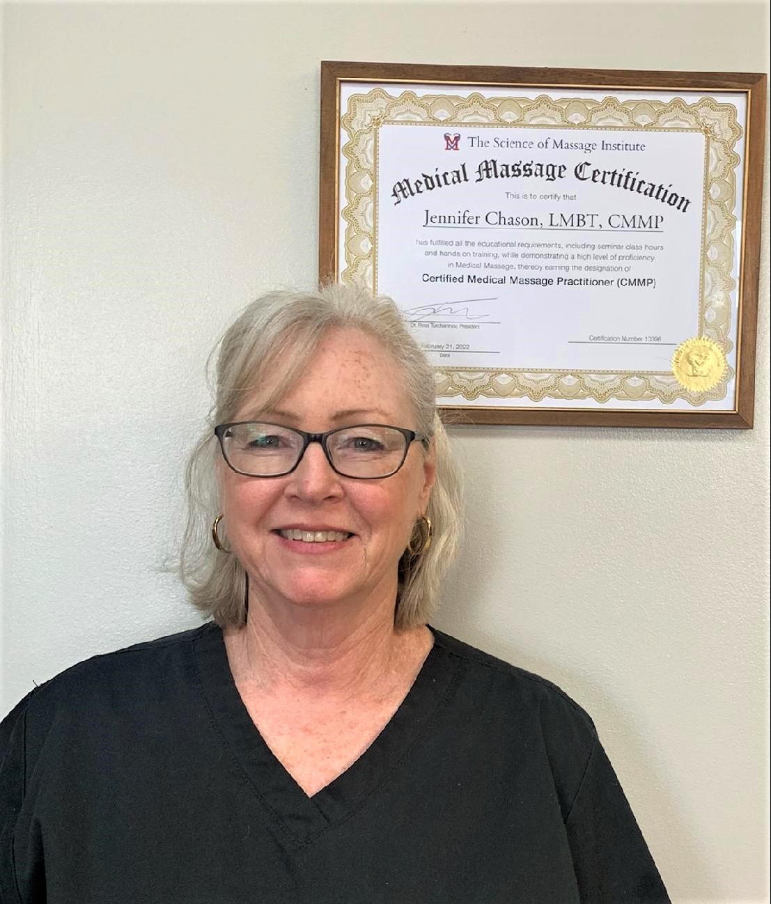
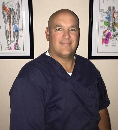

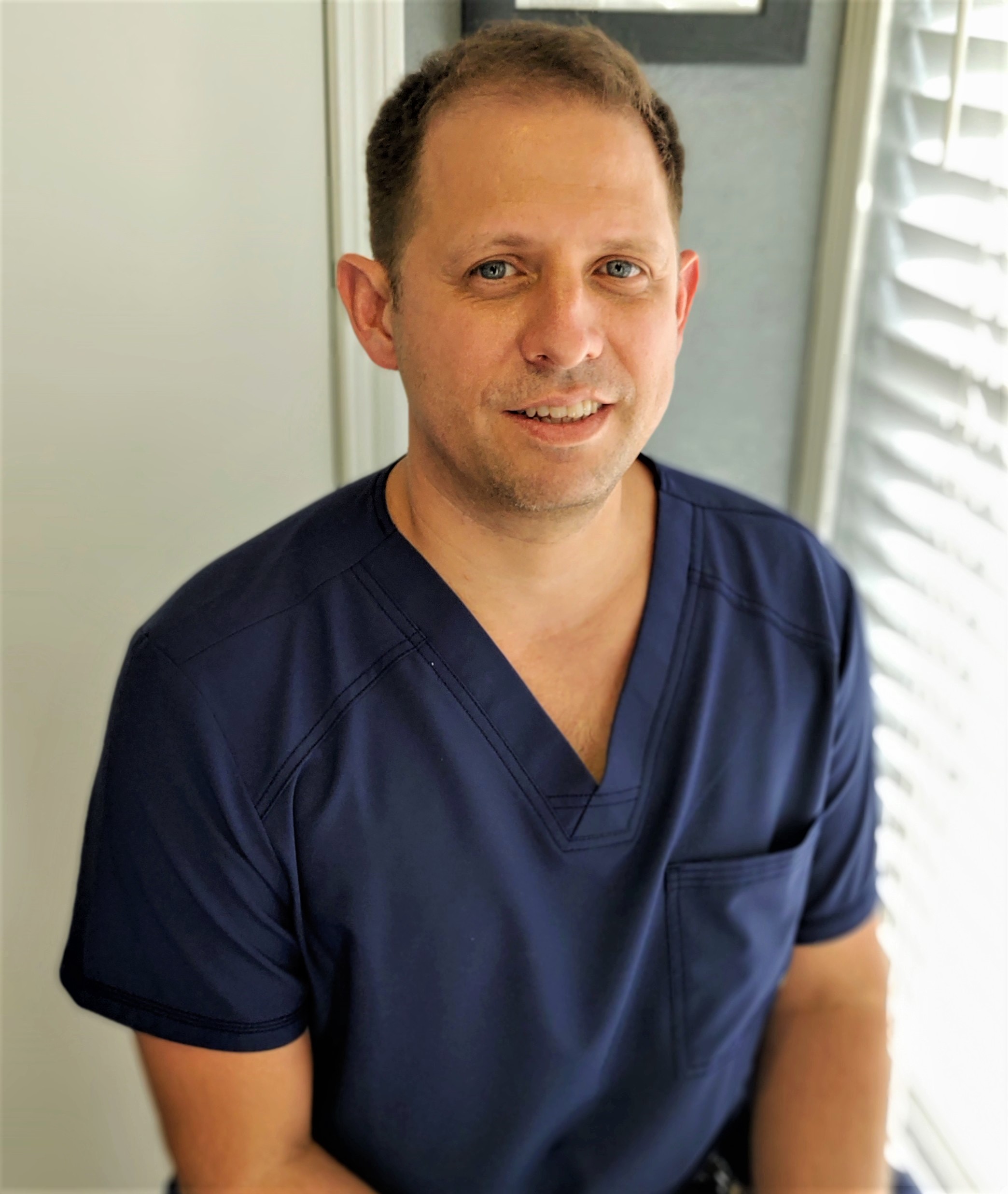

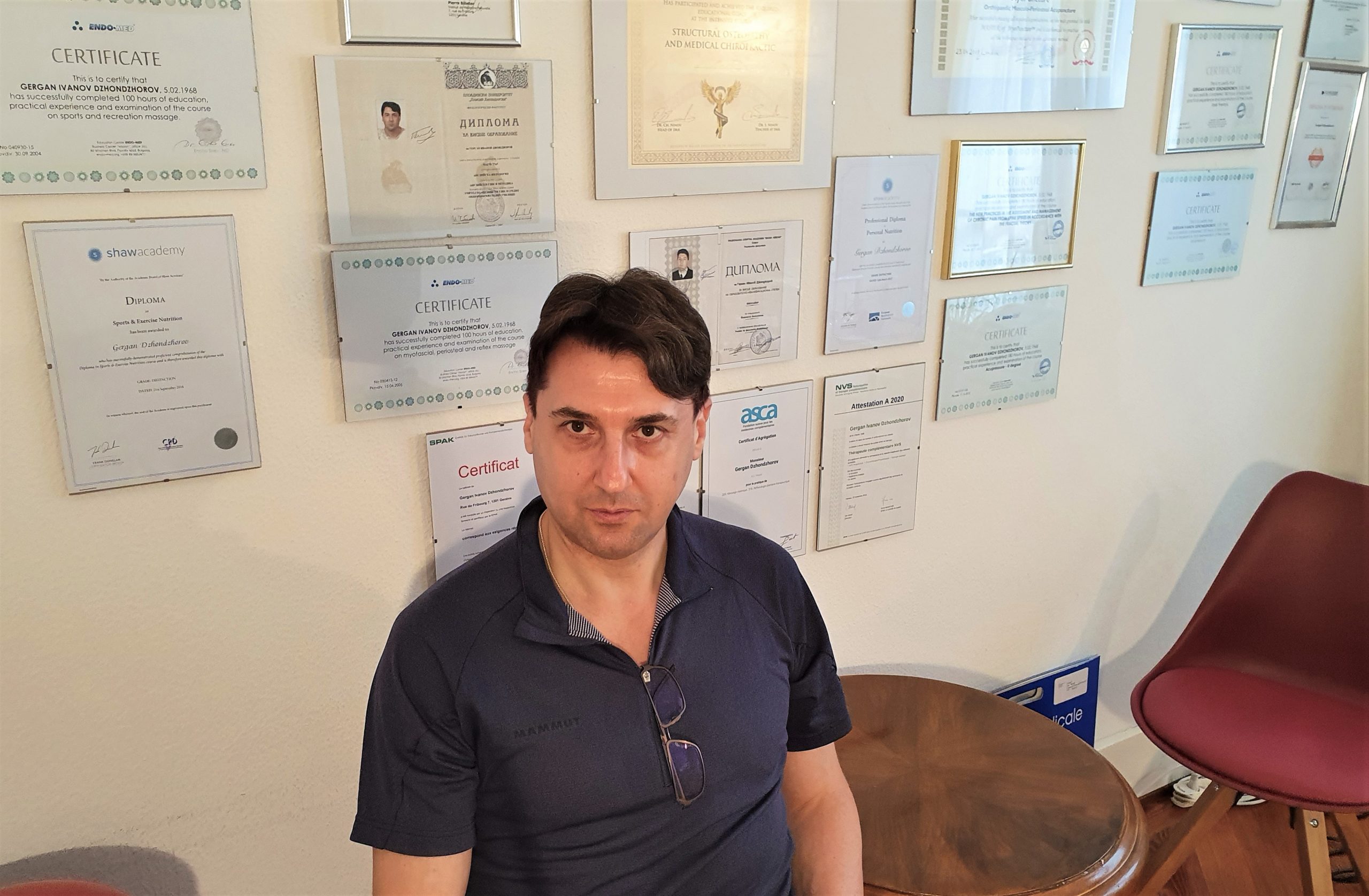


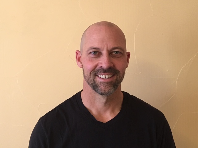


Register for a Free Membership Today
Three Free Introductory Lessons
Category: About Us